

Next Lesson - Diabetic Nephropathy
Core
Glomerular disease is a disease of the kidney affecting either the glomerulus itself or the tubules (the small tubes that carry filtered fluid), interstitium (the tissue between the tubules) or vascular structures (blood vessels) surrounding it. It mainly affects the glomerulus. However, disease in any part of a nephron can damage its other parts. For example, if the glomerulus is damaged, less blood reaches the rest of that nephron and it becomes ischaemic (short of blood).
In glomerular disease, the glomerulus can either become blocked, leading to renal failure and a decreased glomerular filtration rate (GFR), or it can leak, resulting in proteinuria and/or haematuria.
The pathology of the glomerular injury determines the clinical presentation.
Glomerulonephritis is inflammation of the glomeruli and it is caused by a number of different conditions. Four major structures are affected in glomerulonephritis: the capillary endothelium (the lining of the capillaries), the glomerular basement membrane, mesangial cells (supporting cells between the capillaries) and podocytes (cells that wrap around the capillaries). It often involves the immune system, with deposition of immune complexes (clumps of antibody bound to antigen) in the glomerulus. These are either subepithelial (beneath the podocytes) or subendothelial (beneath the capillary lining). They then initiate an immune response causing inflammation.
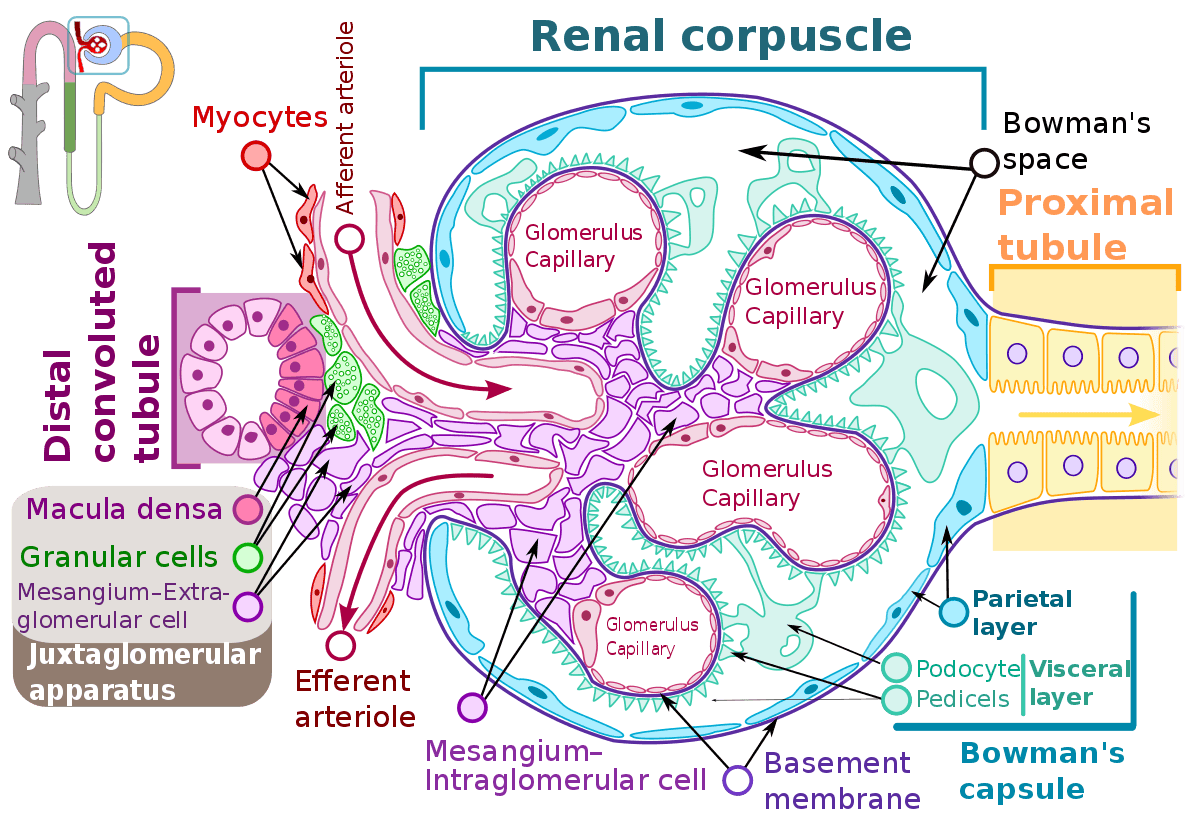
Diagram - The glomerulus within the renal corpuscle
Creative commons source by Shypoetess [CC BY-SA 4.0 (https://creativecommons.org/licenses/by-sa/4.0)]
The cause of glomerulonephritis is diagnosed based on the clinical presentation and histological appearance of the glomerulus, and can then be classified as either primary or secondary GN. Primary GN only affects the kidney, while secondary GN is either kidney disease that becomes systemic disease or originates outside the kidney and then affects the kidney.
We can decide which conditions are likely in a presenting patient by looking at the clinical presentation and seeing which features support each diagnosis. The following is a list of presentations and features for different diagnoses:
- Asymptomatic Proteinuria and/or Haematuria - usually caused by IgA nephropathy, systemic lupus erythematosus (SLE) and focal segmental glomerulosclerosis.
- Proteinuria is the presence of excess protein in the urine.
- Haematuria is the presence of blood in the urine.
- Chronic Glomerulonephritis - this is where there has been ongoing inflammation for a long period of time. IgA nephropathy, SLE and any previously treated glomerulonephritis can cause chronic glomerulonephritis.
- Acute Glomerulonephritis - in acute GN there is sudden inflammation of the glomerulus, and this usually presents as a nephritic syndrome (this will be discussed below). Causes include IgA nephropathy, SLE, post-infectious GN and ANCA-vasculitis.
- Rapidly Progressive Glomerulonephritis (RPGN) - in rapidly progressive GN there is a rapid loss of renal function which is associated with the formation of epithelial crescents (crescent-shaped collections of cells inside the glomerular capsule) in glomeruli when seen under the microscope. Causes of RPGN include ANCA-vasculitis, anti-glomerular basement membrane disease, post-infectious GN and SLE.
- Nephrotic Syndrome - this will be talked about further in this article. Causes of nephrotic syndrome include minimal change disease, focal segmental glomerulosclerosis and SLE.
Nephrotic vs Nephritic Syndrome
Glomerulonephritis presents mainly as two syndromes, nephrotic syndrome and nephritic syndrome. These are not diagnoses but help to determine the cause of GN.
Nephrotic syndrome is where there is damage to the podocytes, the subepithelial space or the glomerular basement membrane (GBM). This causes disruption to the glomerular filtration barrier, allowing proteins to leak through and be excreted with urine. This leads to a triad of:
- Proteinuria > 350 mg/mmol or > 3.5g per day. Proteinuria of > 350 mg/mmol alone is nephrotic range proteinuria.
- Hypoalbuminaemia (a low serum albumin)
- Oedema - hypoalbuminaemia from protein excretion leads to a lower oncotic pressure (the pull of albumin that holds fluid inside the blood vessels), so fluid leaks out into the extracellular space (the space between cells).
This triad can be accompanied by other features as well:
- High cholesterol
- Blood pressure may be normal or raised. High blood pressure (hypertension) is more likely when the kidneys retain sodium and the blood volume increases (hypervolaemia).
- Normal serum creatinine concentration (often preserved initially)
- Hypercoagulable state (a tendency to form blood clots) - there is a loss of antithrombin-III and proteins C and S (natural anticoagulants that help stop clots forming) through the damaged glomerulus. There is also a rise in fibrinogen levels (fibrinogen is a clotting protein). Together these cause a hypercoagulable state. This can predispose the patients to thrombosis.
- Immunocompromised - patients with nephrotic syndrome may become immunocompromised (more prone to infection) due to the loss of immune proteins (such as immunoglobulins) from the body.
- Low total thyroxine level - this is due to the loss of thyroid-binding globulin (the protein that carries thyroxine in the blood) through the damaged glomerulus. This results in a lower total thyroxine level but a normal free thyroxine level (the free hormone is the active part).
Nephrotic syndrome causes can be split into primary or secondary. Primary causes affect only the kidney. Secondary causes are either kidney diseases that become systemic (affect the whole body) or conditions that start outside the kidney and then affect it. This article will go into further depth on these conditions.
- Primary causes of nephrotic syndrome include minimal change disease, membranous glomerulonephritis and focal segmental glomerulosclerosis.
- Secondary causes of nephrotic syndrome include diabetes, SLE, amyloidosis, connective tissue disorders, and myeloma.
- Diabetes and amyloidosis can cause nephrotic syndrome without causing glomerulonephritis (inflammation of the glomeruli). Diabetes is one of the common secondary causes of nephrotic syndrome.
Nephrotic syndrome is managed by treating the underlying condition. Some features of nephrotic syndrome need to be treated to avoid complications while the definitive diagnosis is either being treated or investigated.
- Oedema should be treated using diuretics (e.g. furosemide), with doses adjusted carefully to avoid hypovolaemia (low blood volume) and renal impairment. Restricting salt and fluid can also help to treat oedema.
- Proteinuria can be reduced by giving a patient an ACE inhibitor (angiotensin-converting enzyme inhibitor, e.g. ramipril) which has an anti-proteinuric effect (it reduces the protein lost in the urine). Caution must be used if the patient is intravascularly depleted (has a low volume of blood in the vessels) or if the renal function deteriorates acutely.
- Hypoalbuminaemia - the lower the serum albumin level, the higher the risk of venous thromboembolism (a blood clot in a vein). In one large study the risk was highest when the serum albumin was below 25 g/L. The best threshold is uncertain for some causes of nephrotic syndrome. The decision to start anticoagulation (blood-thinning treatment) should be individualised after considering bleeding risk and other clinical factors.
- Hypercholesterolaemia (high cholesterol) - generally if the underlying disease is treated, the serum cholesterol levels will decrease. If the patient is suffering from long-term nephrotic syndrome then give the patient cholesterol-lowering drugs (e.g. atorvastatin) to protect them from atherosclerosis.
- If the patient has high blood pressure then this should be treated with either an ACE inhibitor or an angiotensin receptor blocker (ARB). NICE advises a clinic blood pressure below 130/80 mmHg for adults with chronic kidney disease and a urine albumin:creatinine ratio (ACR) of 70 mg/mmol or more, which includes nephrotic range proteinuria. For children and young people with the same ACR, it advises a clinic systolic pressure below the 50th percentile for height.
Complications of nephrotic syndrome include:
- Increased risk of infection
- Venous thromboembolism
- Progression of chronic kidney disease
- Hypertension
- Hyperlipidaemia (high levels of fats in the blood)
Nephritic syndrome is inflammation affecting the glomerulus and endothelium. Damage to the endothelium also causes an inflammatory response which further damages the glomerulus. Inflammation disrupts the glomerular basement membrane resulting in the loss of the filtration barrier and impairment of renal function. In nephritic syndrome, there is a triad of:
- Haematuria
- Decreased glomerular filtration rate - there is renal impairment and oliguria (decreased urine output)
- Hypertension
Other features of nephritic syndrome include:
- Proteinuria, usually less than in nephrotic syndrome (below 3.5 g/day). Some causes of nephritic syndrome, such as post-streptococcal glomerulonephritis, can also present as nephrotic syndrome.
- The onset of nephritic syndrome may be either acute or rapidly progressive (RPGN)
- Renal failure - nephritic syndrome can lead to renal failure which will present as increased serum urea and increased serum creatinine. This is also known as azotaemia.
- Red cell casts (tube-shaped clumps of red blood cells formed in the kidney tubules) - seen on urine microscopy.
Causes of nephritic syndrome include:
- Anti-GBM disease (Goodpasture’s disease)
- ANCA-associated vasculitis
- IgA nephropathy - this can also present as nephrotic syndrome.
- Post-infectious - rare in the UK. More common in children than in adults. Can present as nephrotic syndrome.
- Systemic lupus erythematosus - this can also present as nephrotic syndrome.
Management of nephritic syndrome is similar to that of nephrotic syndrome, as both are mainly about treating the underlying cause. While the underlying cause is being treated, clinical features and risk factors can also be treated:
- Blood pressure control - use an ACE inhibitor (e.g. ramipril) or an angiotensin receptor blocker (e.g. losartan) as long as the renal function is normal and does not decrease. Restrict salt and water intake as well.
- Proteinuria - use an ACE inhibitor (e.g. ramipril) to reduce the proteinuria if renal function allows.
- Treatment of oedema - the same as for nephrotic syndrome, i.e. large dose diuretics (e.g. furosemide), if there is adequate renal function.
- Reduce cardiovascular disease risk - stop smoking, diet changes (e.g. reduce salt). If the patient is at risk then a cholesterol-lowering drug, e.g. a statin (e.g. atorvastatin) can be given.
- Renal dialysis (a treatment that filters the blood when the kidneys cannot) - this can be used in the short term depending on the cause of nephritic disease. More information on renal dialysis can be found in our article about chronic kidney disease.
A useful way to remember the difference between nephrotic syndrome and nephritic syndrome is to look at the letter after ‘nephr’. Nephrotic has an ‘O’ there and so causes oedema, while nephritic has an ‘I’ there and so causes inflammation.
Minimal change disease (also called minimal change glomerulonephritis, although the glomerulus is not inflamed) is a condition that is common in childhood and adolescence. The disease is characterised by minimal changes to the histological appearance of the glomerulus. Examination through an electron microscope will reveal the loss of foot processes of podocytes resulting in the loss of the filtration barrier and nephrotic syndrome. It is thought to be caused by a circulating factor that damages the podocytes. This factor has not been identified (it is probably released by T cells, a type of white blood cell) and the idea remains unproven. There is no immune complex deposition.
Most cases are idiopathic (no cause is found) but other causes include non-steroidal anti-inflammatory drugs, rifampicin (an antibiotic), Hodgkin’s lymphoma and thymoma (a tumour of the thymus gland).
There is usually no progression to renal failure with minimal change disease, unlike most other glomerular diseases.
Features of minimal change disease include heavy proteinuria, nephrotic syndrome and ankle oedema. It is treated using corticosteroids and usually resolves but minimal change disease can recur if the steroids are stopped. If the disease does not respond to steroids, then an immunosuppressant (a drug that dampens the immune system) may be used.
Management will also include the general treatment for nephrotic syndrome.
- As minimal change disease is the most likely cause of nephrotic syndrome in children, there is no need to biopsy children unless treatment fails.
- With minimal change disease, 1/3rd will get better, 1/3rd will have infrequent relapses and 1/3rd will have frequent relapses until adulthood.
Focal Segmental Glomerulosclerosis (FSGS)
Focal segmental glomerulosclerosis is a glomerular disease caused by a circulating factor that damages podocytes. It doesn’t affect all glomeruli and does not damage all parts of the glomerulus. FSGS presents as nephrotic syndrome, e.g. ankle oedema and proteinuria, and mainly occurs in adults. These circulating factors remain even if the patient has a kidney transplant, meaning the transplanted kidney can also be affected by FSGS.
FSGS can be idiopathic or can have a cause, for example, malignancy, medication, HIV, heroin, Alport syndrome or infection.
Focal segmental glomerulosclerosis is treated using steroids and immunosuppressants, though it is less responsive to steroids than minimal change disease. The treatment will also include the general treatment for nephrotic syndrome.
FSGS causes glomerulosclerosis (scarring of the glomerulus). As the scarring builds up, less blood can be filtered and the GFR decreases. This means FSGS can progress to renal failure, especially if it does not respond to treatment.
Membranous Glomerulonephritis (MGN)
Membranous glomerulonephritis is an important primary cause of nephrotic syndrome in adults. It is caused by immune complex deposition in the glomerulus, either through an autoimmune process or secondary to another pathology. Secondary causes include SLE, hepatitis B virus and malignancy. Under the microscope (histologically), the glomerular basement membrane appears thickened.
The pathophysiology of membranous glomerulonephritis is as follows:
- An antigen is found in the glomerulus, either as a result of an autoimmune cause or another pathology. The antigen is typically found at the podocytes, so this is where the damage to the glomerulus occurs.
- IgG (immunoglobulin G, a type of antibody) is produced by the body against the antigen and binds to antigens at the level of the podocytes within the glomerulus.
- The immune deposits then activate complement (a group of blood proteins that form part of the immune system). This forms the membrane attack complex, which injures the podocytes. This is how the filtration barrier is damaged and nephrotic syndrome is caused.
- In primary membranous nephropathy, antibodies bind antigens on podocytes and immune deposits form subepithelially, beneath the podocytes. Complement-mediated injury damages the filtration barrier, allowing heavy protein loss into urine. The deposits are not explained simply by complexes being too large to filter.
The treatment of MGN is through immunosuppression and corticosteroids, including the general treatment of nephrotic syndrome. With the treatment of MGN, one-third of patients recover, one-third stay the same and one-third deteriorate.
IgA nephropathy is a common glomerulonephritis that can occur at any age, and classically presents with visible or invisible haematuria. It is caused by IgA deposition in the mesangium of the glomerulus resulting in an inflammatory response causing mesangial damage. It is the most common primary glomerulonephritis worldwide.
The histological features and the course of the disease can vary and the patient may have proteinuria instead of haematuria, so it can either present as nephritic syndrome or nephrotic syndrome.
IgA nephropathy is commonly triggered by mucosal infections (infections of the moist linings of the body, such as the throat), as IgA protects the mucous membranes. If a patient has an upper respiratory tract infection there may be over-activation of IgA causing it to be deposited in the glomerulus. This then results in an inflammatory response and damage.
IgA nephropathy presents with visible/invisible haematuria and/or proteinuria, an increased serum IgA concentration in about a third to a half of patients (it is not reliable enough to diagnose the disease), and a normal serum C3/C4 concentration (complement proteins, which are part of the immune system). There is typically a recent history of an upper respiratory tract infection within the past 1-2 days. The haematuria may be recurrent or IgA nephropathy may be picked up on the first presentation of haematuria.
There is no effective treatment for IgA nephropathy and 25-30% of patients will progress to end-stage renal failure within 20-25 years. However, it is managed using ACE inhibitors or angiotensin receptor blockers to control the proteinuria and hypertension, and reduce the progression of the disease.
Prognostic markers for the disease can help predict the course of the disease:
- Good Prognosis - frank (visible) haematuria.
- Poor Prognosis - male, proteinuria, hypertension, smoking, hyperlipidaemia.
This article will cover two main hereditary nephropathies: thin glomerular basement membrane nephropathy and Alport syndrome.
Thin Glomerular Basement Membrane Nephropathy
Thin glomerular basement membrane nephropathy is also known as benign familial nephropathy. In this hereditary condition the glomerular basement membrane is thin which causes the kidneys to leak a small amount of blood resulting in an isolated haematuria.
There are typically no symptoms with this condition.
Thin basement membrane nephropathy usually presents with microscopic haematuria and preserved kidney function. Its course is often favourable, but proteinuria, hypertension and progressive kidney impairment can occur, particularly in collagen IV-related disease. Continue follow-up of blood pressure, urine protein and renal function; the historical terms benign familial nephropathy and benign familial haematuria do not guarantee a harmless course.
Alport syndrome is a genetic disease most commonly caused by an X-linked mutation (a fault in a gene on the X chromosome). It can also be inherited in autosomal recessive or autosomal dominant forms (not linked to a sex chromosome). Mutations in each form lead to the production of abnormal collagen IV. In the kidneys it causes dysfunction of the glomerular basement membrane that results in haematuria and eventually renal failure.
Alport syndrome is associated with deafness as collagen IV is found in the cochlea (the hearing organ of the inner ear), which plays an important role in hearing. There are also visual disturbances as Alport syndrome affects the lens of the eye.
Alport syndrome typically presents with haematuria in childhood or adolescence, sensorineural hearing loss (hearing loss from damage to the inner ear or its nerve) and eye disorders.
Treatment includes supportive measures, but early use of ACE inhibitors or ARBs (angiotensin receptor blockers) is important as this can slow progression of renal disease. Renal replacement therapy (treatment that replaces kidney function, such as dialysis) is used for patients who develop renal failure. Patients can also undergo a renal transplant if they develop renal failure, but anti-glomerular basement membrane disease may develop in the transplanted kidney.
Granulomatosis with Polyangiitis
Granulomatosis with polyangiitis, formerly known as Wegener’s granulomatosis, is an immune vasculitis where there is necrotising granulomatous inflammation in small and medium-sized blood vessels around the body. It mainly affects small arterioles and so often affects the kidneys and lungs causing a pulmonary-renal syndrome (lung and kidney disease together).
Antibodies against proteins in neutrophils (a type of white cell), called anti-neutrophil cytoplasmic antibodies (ANCA), cause endothelial damage to blood vessels resulting in vasculitis and the inflammation seen in the disease. In this disease the renal lesion is typically pauci-immune, with little or no deposited immunoglobulin on biopsy.
Patients can present with: general symptoms such as fatigue, weight loss, arthralgia (joint pain) and myalgia (muscle pain); chest symptoms such as cough, haemoptysis (coughing up blood) and shortness of breath; kidney features such as haematuria and hypertension; and ear, nose and throat symptoms such as epistaxis (nosebleeds), hearing loss and a flat or saddled nose due to collapse of the nasal septum. Abdominal pain can also occur.
- Patients are most likely to present with systemic symptoms of the disease rather than solely renal symptoms.
- Patients can present after they develop pulmonary haemorrhage (bleeding into the lungs). This can be seen on a CT scan and causes haemoptysis (coughing up blood).
- This condition usually affects people aged 45-60 years.
- Investigations can reveal a positive result for anti-neutrophil antibodies in 90%, e.g. cANCA (cytoplasmic ANCA) and pANCA (perinuclear ANCA). These antibodies are directed against proteinase 3 (PR3) or myeloperoxidase (MPO).
- Renal biopsy will reveal the presence of epithelial crescents in Bowman’s capsule (the cup-shaped capsule around the glomerulus).
Treatment for granulomatosis with polyangiitis includes steroids together with an immunosuppressant, either rituximab or cyclophosphamide, to bring the disease into remission. Plasma exchange (removing and replacing the patient’s plasma) may be considered in selected severe cases, for example severe kidney involvement with a creatinine above 300 micromol/L. The condition has a good prognosis if caught early, with survival greatly improved since the introduction of modern immunosuppressive therapy. Granulomatosis with polyangiitis can cause a rapidly progressive glomerulonephritis, and kidney involvement occurs in 70-85% of patients. If it is not treated quickly, this rapidly results in renal failure.
ANCA-Associated Vasculitis is a term used to describe a group of conditions where there is destruction and inflammation of small blood vessels. This group includes granulomatosis with polyangiitis, microscopic polyangiitis and eosinophilic granulomatosis with polyangiitis.
Goodpasture’s Disease (Anti-Glomerular Basement Membrane Disease)
Goodpasture’s disease is an uncommon condition caused by antibodies targeting the alpha-3 chain of type IV collagen in the glomerular basement membrane. Type IV collagen is also present in the alveolar basement membrane (the thin layer between the air sacs and the blood), so Goodpasture’s disease can affect the lungs as well. However, it is more difficult for the antibodies to reach the alveolar basement membrane.
- The IgG antibodies are deposited in the basement membrane resulting in acute and rapid inflammation of the glomerulus.
- IgG antibodies also cause inflammation and damage to the alveolar membrane. This damage can result in pulmonary haemorrhage and haemoptysis.
- Patients are more at risk of pulmonary haemorrhage if they smoke, have a lower respiratory tract infection, have pulmonary oedema, are a young male or have inhaled hydrocarbons (chemical solvents).
Goodpasture’s disease can cause rapidly progressive glomerulonephritis and renal failure, so it needs to be recognised and treated quickly. It usually affects people aged 20-30 years or 60-70 years, and will present with an acute onset nephritic syndrome.
Signs and Symptoms:
- Shortness of breath
- Cough - may have haemoptysis (coughing up blood)
- Pallor (looking pale)
- Malaise (feeling generally unwell)
- Hypertension
- Abnormal renal function
Investigations will show haematuria and proteinuria on a urine dipstick (a test strip dipped into the urine sample). Blood tests may show the presence of anti-GBM antibodies. Renal biopsy will show linear IgG deposits along the glomerular basement membrane.
Treatment of Goodpasture’s disease is with immunosuppression, steroids and plasmapheresis. Plasmapheresis (plasma exchange) is where the patient’s plasma is separated from the blood and removed, then replaced with donor plasma or another replacement fluid. This removes the anti-GBM antibodies from the blood. The treatment will also include treating the nephritic syndrome to prevent the progression of the disease.
Vasculitis is the term used for a group of systemic disorders that cause inflammation of blood vessels. The different disorders are categorised depending on the size of the blood vessel they affect, so they will be classified as large vessel, medium vessel or small vessel vasculitis.
Some small vessel vasculitides are associated with anti-neutrophil cytoplasmic antibody (ANCA). ANCA activates neutrophils and leads to endothelial injury in small vessels, resulting in inflammation and damage to the glomerulus.
ANCA-associated vasculitis can cause pauci-immune necrotising and crescentic glomerulonephritis, with rapid loss of kidney function. Pauci-immune means little or no immunoglobulin deposition on biopsy, rather than an absolute absence of immune activity. Antibody-driven neutrophil activation and complement can contribute to the injury despite scant deposited immune material.
Besides granulomatosis with polyangiitis, vasculitides that affect the kidney include:
- Microscopic Polyangiitis - a small vessel vasculitis that commonly causes renal involvement, often presenting as a rapidly progressive glomerulonephritis, and may also cause respiratory symptoms, including pulmonary haemorrhage (bleeding into the lungs), which can be life-threatening.
- Blood tests usually show a raised serum ANCA, most often against myeloperoxidase (MPO-ANCA), and a renal biopsy will show segmental necrotising glomerulonephritis.
- Microscopic polyangiitis is treated with steroids together with an immunosuppressant, either rituximab or cyclophosphamide, to bring the disease into remission.
- Eosinophilic Granulomatosis with Polyangiitis (Churg-Strauss) - this is a small vessel vasculitis. These patients will typically have asthma and/or allergic rhinitis (hay fever) previously and present with purpura (purple spots on the skin from bleeding under the skin), peripheral neuropathy, sinusitis and dyspnoea (shortness of breath).
- Blood tests may show a raised serum ANCA and eosinophilia, and a renal biopsy will show focal segmental necrotising glomerulonephritis.
- It is treated with immunosuppression.
Systemic Lupus Erythematosus (SLE)
Systemic lupus erythematosus is an autoimmune systemic disease that affects multiple organ systems, including the kidney. There are many patterns of renal disease with SLE and it can cause either nephritic or nephrotic syndrome. It is an important diagnosis to look out for and to have as a differential if a patient presents with either nephrotic or nephritic syndrome.
Post-Streptococcal Glomerulonephritis
Post-streptococcal glomerulonephritis is a glomerular disease that occurs mainly in children aged 3-12 years, weeks after a group A β-haemolytic streptococcal infection. It mainly causes a nephritic syndrome.
It will typically present weeks after an infection, such as tonsillitis, pharyngitis, impetigo (a crusting skin infection) and cellulitis (infection of the deeper layers of the skin). Glomerular disease will be seen 1-2 weeks after tonsillitis or pharyngitis and 3-4 weeks after impetigo or cellulitis.
Investigations will show anti-streptococcal antibodies, a low C3/complement level and immune complex deposition (IgG, IgM and C3).
Post-streptococcal glomerulonephritis is usually self-limiting (it gets better without specific treatment), so the treatment is mainly supportive. This includes fluid restriction, diuretics and antihypertensive drugs. ACE inhibitors or angiotensin receptor blockers are usually avoided in the acute phase because they may worsen the fall in glomerular filtration and the high blood potassium (hyperkalaemia). Most patients recover, but a small number are left with persistent urinary abnormalities, hypertension or chronic kidney disease. If end-stage renal failure develops, then renal replacement therapy (dialysis or a transplant) is given to the patient.
Edited by: Dr. Tom Bradley
Reviewed by: Dr. Marcus Judge
- 991

