

Next Lesson - Micturition
Core
In a typical 70 kg adult the total body potassium content is about 3500 mmol, with the majority of K+ ions being found within the intracellular fluid (98%), and the remainder within the extracellular fluid (2%). This is important as even a slight shift in potassium from the intracellular fluid (ICF) into the extracellular fluid (ECF) can greatly increase the ECF potassium.
The difference between ECF and ICF potassium concentration is maintained by the Na-K-ATPase transporter (a pump in the cell membrane that uses energy) which moves Na+ ions out of the cell and K+ ions into the cell, helping to create the resting membrane potential. The serum K+ ion concentration must therefore be tightly controlled. If the resting membrane potential changes, the excitability of cardiac tissue changes, and this can lead to life-threatening arrhythmias (abnormal heart rhythms).
Role of Potassium:
- Cell-volume Maintenance - a net loss of potassium from a cell leads to cell shrinkage while a net gain will lead to cell swelling as water will follow K+ ion movement
- Intracellular and Extracellular pH Regulation - pH regulation is through the reciprocal cation shift (K+ ions and H+ ions swapping places across the cell membrane, explained under Internal Balance of Potassium)
- Cell Enzyme Functions
- DNA and Protein Synthesis
- Resting Cell Membrane Potential - an increased extracellular K+ ion concentration (hyperkalaemia) will lead to membrane depolarisation while a decreased extracellular K+ ion concentration (hypokalaemia) will cause membrane hyperpolarisation
- Neuromuscular Activity - hypokalaemia will lead to decreased muscle excitability while hyperkalaemia will cause partial depolarisation of the membrane, which can eventually stop muscle cells from responding. Both of these can lead to muscle weakness and paralysis
- Cardiac Activity - hypo- and hyperkalaemia will lead to arrhythmias
- Vascular Resistance - hypokalaemia will cause vasoconstriction while hyperkalaemia will cause vasodilation
Regulation of Extracellular Fluid Potassium
Control of extracellular fluid potassium is maintained by immediate and long-term control.
Immediate control is through the internal balance of potassium between the ICF and ECF. K+ ions move into and out of cells to help control serum potassium to prevent hypo- or hyperkalaemia. Immediate control occurs over minutes.
Long-term control is done by adjusting the renal excretion of K+ ions. This also helps to control the total potassium level in the body by increasing or decreasing the renal excretion of K+ ions. Long-term control happens over several hours.
The combination of immediate and long-term control can be seen following a meal where the intestine and colon absorb dietary potassium which raises the ECF K+ ion concentration. Within minutes, immediate control causes the majority of this potassium to move into cells to prevent a major rise in plasma potassium and hyperkalaemia. After a small delay, the kidneys start to excrete K+ ions from the body to maintain the total body K+ content at about 3500 mmol. Removal of potassium takes around 6-12 hours so the excretion rate of potassium can change depending on the body’s needs to ensure that not too little or too much is removed from the body.
The internal balance of K+ ions is the result of K+ ion movement into and out of cells. Movement into cells is mediated by Na-K-ATPase while movement out of cells is mediated by K+ ion channels, both of which are found in the cell surface membrane.
Factors that increase K+ ion uptake by cells
- Hormones which act via Na-K-ATPase:
- Insulin - insulin increases the activity of Na-K-ATPase in muscle and liver cells, increasing K+ ion uptake. A higher concentration of potassium in splanchnic blood (the blood draining the gut) stimulates insulin secretion to lower potassium levels in the blood. Insulin is used clinically to treat hyperkalaemia as it acts to shift K+ ions from the blood into cells and the ICF to lower ECF potassium
- Aldosterone - potassium in the blood stimulates aldosterone release which acts on cells to increase the activity of Na-K-ATPase causing increased K+ ion uptake into cells
- Catecholamines (hormones such as adrenaline) - these stimulate Na-K-ATPase and so increase cellular uptake of K+ ions
- Hyperkalaemia
- Alkalosis - a low serum H+ ion concentration drives K+ ions into cells as serum K+ ions are exchanged with cellular H+ ions to increase the serum H+ ion level and lower serum pH. This is known as the reciprocal cation shift. This will result in a low serum K+ ion concentration and hypokalaemia
Factors that decrease K+ ion uptake by cells:
- Exercise - there is a net release of potassium during the recovery phase of an action potential as K+ ions leave the cell. Skeletal muscle cells damaged during exercise release K+ ions
- Uptake of K+ by non-contracting tissues prevents dangerously high hyperkalaemia by counteracting the release of K+ from contracting tissues
- Catecholamine release during exercise helps to offset the rise in potassium by increasing uptake of K+ ions by non-contractile cells
- Cell Lysis - this releases potassium from the intracellular fluid to the extracellular fluid
- Increased ECF Osmolality (more dissolved particles in the extracellular fluid)
- Hypokalaemia
- Acidosis - a high serum H+ ion concentration reduces K+ ion uptake into cells as it causes K+ ions to leave the cell via the reciprocal cation shift. In acidosis serum H+ ions are exchanged with cellular K+ ions to decrease serum H+ ion level and raised serum pH. This will result in a high serum K+ ion concentration and hyperkalaemia
External balance of K+ ions is the long-term control of the total body K+ ion concentration. It is a slow process, taking around 6-12 hours, where the excretion of K+ ions from the body is changed to meet the body’s demands, and it occurs within the late distal convoluted tubule and the cortical collecting duct in the kidney nephron.
Potassium is freely filtered at the glomerulus and is then reabsorbed in the nephron. It is also secreted into the tubule late on in the distal convoluted tubule and the collecting duct.
- Potassium reabsorption occurs in:
- The proximal convoluted tubule - around 67% of filtered potassium is reabsorbed here
- The thick ascending limb of the Loop of Henle - around 20% of filtered potassium is reabsorbed here
- The distal convoluted tubule and the collecting duct by intercalated cells (a second cell type in the tubule wall) - around 10-12% of filtered potassium is reabsorbed here
- Potassium secretion occurs in:
- The distal convoluted tubule and the cortical collecting duct by principal cells (the main potassium-secreting cells of the tubule wall) - substantial secretion occurs with a normal or high potassium level but little secretion if the potassium is low
Potassium is secreted into the nephron tubule by principal cells in the distal convoluted tubule and collecting duct. A high intracellular K+ ion concentration creates a chemical gradient for secretion into the lumen, while an electrical gradient is created by Na+ ion reabsorption as it creates a negative luminal potential, promoting K+ ion secretion into the lumen. Together these create a favourable electrochemical gradient for K+ ion secretion via apical potassium channels (the apical membrane faces the tubule lumen, whereas the basolateral membrane faces the blood).
Factors that aim to increase potassium secretion will increase the electrical and/or chemical gradient across the cell membrane to promote potassium secretion. For example, by stimulating Na-K-ATPases in the basolateral cell membrane there is increased Na+ reabsorption into the cell, which enhances the electrochemical gradients that favour K+ ion secretion into the tubular lumen.
Factors that aim to decrease potassium secretion will decrease the electrical and/or chemical gradient across the cell membrane to reduce potassium secretion. For example, by blocking the epithelial sodium channels (ENaC) in the apical cell membrane, through which Na+ ions enter the cell, the electrical gradient is lost because fewer Na+ ions are being reabsorbed, and so there is no longer an electrical gradient promoting K+ ion secretion.
Factors that affect K+ ion secretion by principal cells:
- Extracellular Fluid Potassium Concentration - a high ECF K+ ion concentration stimulates the basolateral Na-K-ATPase and increases the permeability of the apical tubule cell membrane. It also stimulates aldosterone release to increase the number of apical potassium channels
- Aldosterone - aldosterone stimulates basolateral Na-K-ATPases, increases the number of apical potassium channels and increases the number of epithelial sodium channels in the apical membrane to promote potassium secretion
- The Acid-Base Balance of the Body:
- Acidosis inhibits Na-K-ATPases in principal cells and decreases potassium channel permeability to reduce K+ ion secretion
- Alkalosis stimulates Na-K-ATPases in principal cells and increases potassium channel permeability to promote K+ ion secretion
- Filtrate Flow Rate - a fast filtrate flow rate, e.g. caused by loop diuretics, means K+ ions are washed away more quickly, increasing the chemical gradient and promoting K+ ion secretion into the lumen
- Sodium Reabsorption by Principal Cells - more sodium reaching the principal cells means more Na+ ions are reabsorbed from the filtrate, and this creates a greater electrical gradient across the cell membrane (the tubule lumen becomes more negative). This increased electrical gradient promotes K+ ion secretion
Hyperkalaemia is a high plasma potassium concentration. The potassium concentration that defines hyperkalaemia changes depending on the lab testing, but it is usually either > 5.0 mmol/L or > 5.5 mmol/L. There are many causes of hyperkalaemia, and it is usually secondary to another problem. Some examples include:
- Increased Intake - this is unlikely and usually only happens if renal dysfunction is present or an inappropriate dose of IV potassium is given
- Decreased Renal Excretion - this can be due to acute kidney injury, chronic kidney disease, drugs that decrease K+ ion excretion (e.g. ACE (angiotensin-converting enzyme) inhibitors, and the potassium-sparing diuretics spironolactone and amiloride), Addison’s disease
- Metabolic Acidosis - this causes a reciprocal cation shift resulting in K+ ions leaving cells causing hyperkalaemia. The shift is clearest in acidosis caused by mineral (inorganic) acids, such as the acidosis of advanced kidney failure, and is small in organic acidosis such as lactic acidosis. Serum potassium still tends to be raised in diabetic ketoacidosis, even though the total body potassium is low
- Cell Lysis - rupture of cells releases potassium into the blood causing hyperkalaemia (e.g. tumour lysis, the breakdown of cancer cells after treatment, and rhabdomyolysis, the breakdown of damaged muscle)
- Nausea
- Tiredness
- Weakness
- Cardiovascular - hyperkalaemia can present with chest pain, arrhythmias, ECG changes and palpitations
- Changes on an ECG:
- Tall peaked T wave (‘tented’ T wave)
- Prolonged PR interval
- Flattened/absent P wave
- Widened QRS
- Sine-wave pattern
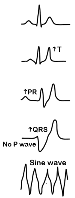
Diagram - The ECG changes seen in hyperkalaemia
SimpleMed original by Dr. Thomas Burnell
In the acute treatment of hyperkalaemia, the aims are to protect the heart from potassium, drive the potassium intracellularly and remove the excess potassium from the body. Acute treatment of hyperkalaemia mainly uses:
- IV Calcium Gluconate - this is given to protect the heart from potassium and prevent cardiac arrhythmias. It is given when there are ECG changes of hyperkalaemia. UK Kidney Association guidance reserves it for these patients, as there is no consensus on using it when the ECG is normal
- IV Insulin and IV Dextrose - insulin is given to shift the potassium intracellularly. The dextrose (a glucose solution) is given with the insulin to reduce the risk of hypoglycaemia (low blood glucose), which can still happen afterwards, so blood glucose is monitored
- Nebulised salbutamol (a beta agonist, the type of medicine used in asthma, breathed in as a mist) also shifts potassium intracellularly. It is an add-on to insulin and dextrose, not a replacement, and it should not be used on its own in severe hyperkalaemia
- Potassium binders (medicines that bind potassium in the gut so that it leaves in the stool) - sodium zirconium cyclosilicate or patiromer calcium are recommended to remove potassium from the body in the emergency management of severe hyperkalaemia. Calcium Resonium (calcium polystyrene sulfonate), an older resin, is no longer recommended for routine use in acute hyperkalaemia, and its effect is variable and unpredictable
- Renal Dialysis (a machine that filters the blood in place of the kidneys) - this is used to remove the potassium if other treatments do not work
- Furosemide - loop diuretics cause the kidneys to lose potassium (hypokalaemia), so they can be a useful add-on in hyperkalaemia, particularly chronic hyperkalaemia. They are only suitable for patients who are non-oliguric (still passing a reasonable amount of urine) and non-hypovolaemic (not short of body fluid)
- Sodium Bicarbonate - this is not recommended for routine use in acute hyperkalaemia, because there is insufficient evidence that it works (hyperkalaemic cardiac arrest is the exception). In patients with chronic kidney disease and a low serum bicarbonate (below 22 mmol/L) it is recommended even without hyperkalaemia, because treating the acidosis (too much acid in the blood) has benefits beyond lowering potassium
Chronic treatment is mainly to treat the underlying cause of hyperkalaemia, e.g. removing the offending drug, treating renal dysfunction, etc. On specialist initiation, oral potassium binders (sodium zirconium cyclosilicate for mild to moderate hyperkalaemia, patiromer calcium for moderate hyperkalaemia) may be used in certain patients who could not otherwise take an optimised dose of renin-angiotensin-aldosterone system (RAAS) inhibitors, such as ACE inhibitors, or take them at all, because of hyperkalaemia. They bind potassium in the gut and prevent its absorption.
Hypokalaemia is a low plasma potassium concentration. The potassium concentration that defines hypokalaemia changes depending on the lab, but it is usually < 3.5 mmol/L. There are many causes of hypokalaemia, and it is usually secondary to another problem. Some examples include:
- Excessive Loss - e.g. due to diarrhoea and/or vomiting
- Increased Renal Loss - e.g. potassium-losing diuretics, such as loop diuretics and thiazide diuretics, and hyperaldosteronism (too much of the hormone aldosterone, which makes the kidneys lose potassium)
- Metabolic Alkalosis - causes potassium to shift into the intracellular space, reducing the serum potassium concentration
- Inadequate Intake - this is an unlikely cause of hypokalaemia but it can happen
- Fatigue
- Constipation - this is caused by weakness of the smooth muscle in the bowel wall. Hypokalaemia results in membrane hyperpolarisation, making it more difficult for cells to depolarise. This leads to reduced bowel contractions (slow movement of the gut) and, in severe cases, to paralytic ileus (the bowel stops moving, so its contents do not pass along).
- Proximal Muscle Weakness - the membrane hyperpolarisation in hypokalaemia means it is more difficult for the muscle cells to depolarise and contract which presents as muscle weakness
- Cardiac Arrhythmias - hypokalaemia can present with cardiac arrhythmias
- ECG changes seen in hypokalaemia:
- Prolonged PR interval
- ST segment depression
- Low/absent T-wave
- High U wave
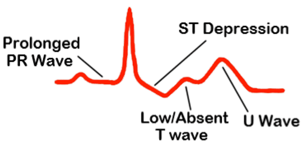
Diagram - The ECG changes seen in hypokalaemia
SimpleMed original by Dr. Thomas Burnell
The main treatment of hypokalaemia is through potassium replacement and treating the underlying cause of the low potassium. Potassium replacement can be oral or intravenous depending on the severity of the hypokalaemia, and in some cases, it may not be needed if the potassium returns to normal once the underlying cause is treated. Oral potassium replacement is for less severe cases while intravenous potassium is given if there is severe hypokalaemia.
Intravenous potassium replacement regimens vary according to severity, venous access and monitoring. Typical solutions use potassium chloride diluted in 0.9% sodium chloride (normal saline), with the concentration and infusion rate adjusted to reduce the risk of arrhythmia and vein irritation.
It is important to ensure that intravenous potassium replacement is not too quick, as rapid administration can lead to hyperkalaemia and arrhythmias. In general teaching, a maximum rate of 10 mmol/hour is used when cardiac monitoring is not in place, with closer monitoring required at higher rates.
Edited by: Dr. Maddie Swannack
Uploaded by: Dr. Thomas Burnell
Reviewed by: Dr. Marcus Judge
- 935
