

Next Lesson - Urinary Tract Obstruction
Core
Chronic kidney disease (CKD) refers to abnormalities of renal structure or function present for more than 3 months, which may be progressive. It is more common in older people, in people of black or south Asian family origin, in people with several other long-term conditions (co-morbidities) and in people who are socially deprived. Patients with CKD have an increased risk of developing acute kidney injury (AKI, a sudden fall in kidney function) because the kidneys are already damaged and so can be damaged more easily.
It is diagnosed using the glomerular filtration rate (GFR, how much fluid the kidneys filter from the blood each minute) and the urine albumin:creatinine ratio (ACR). A GFR of 90 ml/min/1.73m2 or more is normal or high (category G1). In CKD there is a lowered GFR because not enough fluid is being filtered by the kidneys due to the loss of renal function.
CKD is defined as a reduced GFR (below 60 ml/min/1.73m2 on at least 2 occasions 90 days apart) or other markers of kidney damage (for example protein or blood in the urine, or an abnormal kidney scan) that persist for more than 3 months.
In CKD, damaged renal tissue is replaced by an extracellular matrix (a network of proteins, such as collagen, that forms scar tissue), resulting in a smaller amount of functioning kidney tissue. In advanced CKD the kidneys become scarred. Under the microscope (histologically), a kidney biopsy shows four main features: a fibrotic cortex, sclerotic glomeruli, scattered inflammatory cells and thickened arteries.
Usually patients with CKD are asymptomatic (they have no symptoms). If a patient has risk factors, they are tested with blood and urine tests. The aim is to treat CKD early and prevent kidney failure. Any symptoms are usually non-specific and can include fatigue, nausea, pruritus (itching) and nocturia (passing urine at night).
Chronic kidney disease may progress over time to end-stage renal failure, and the main aim of treatment is to slow disease progression and reduce complications.
Stages of Chronic Kidney Disease
Chronic kidney disease is staged using the GFR and the urine albumin:creatinine ratio (ACR). The ACR is a urine test that measures how much albumin (a protein) is leaking into the urine. A higher ACR means more proteinuria. The lower the GFR and the higher the ACR, the worse the prognosis. The following table shows the GFR categories (G1 to G5) and the ACR categories (A1 to A3) used by NICE.
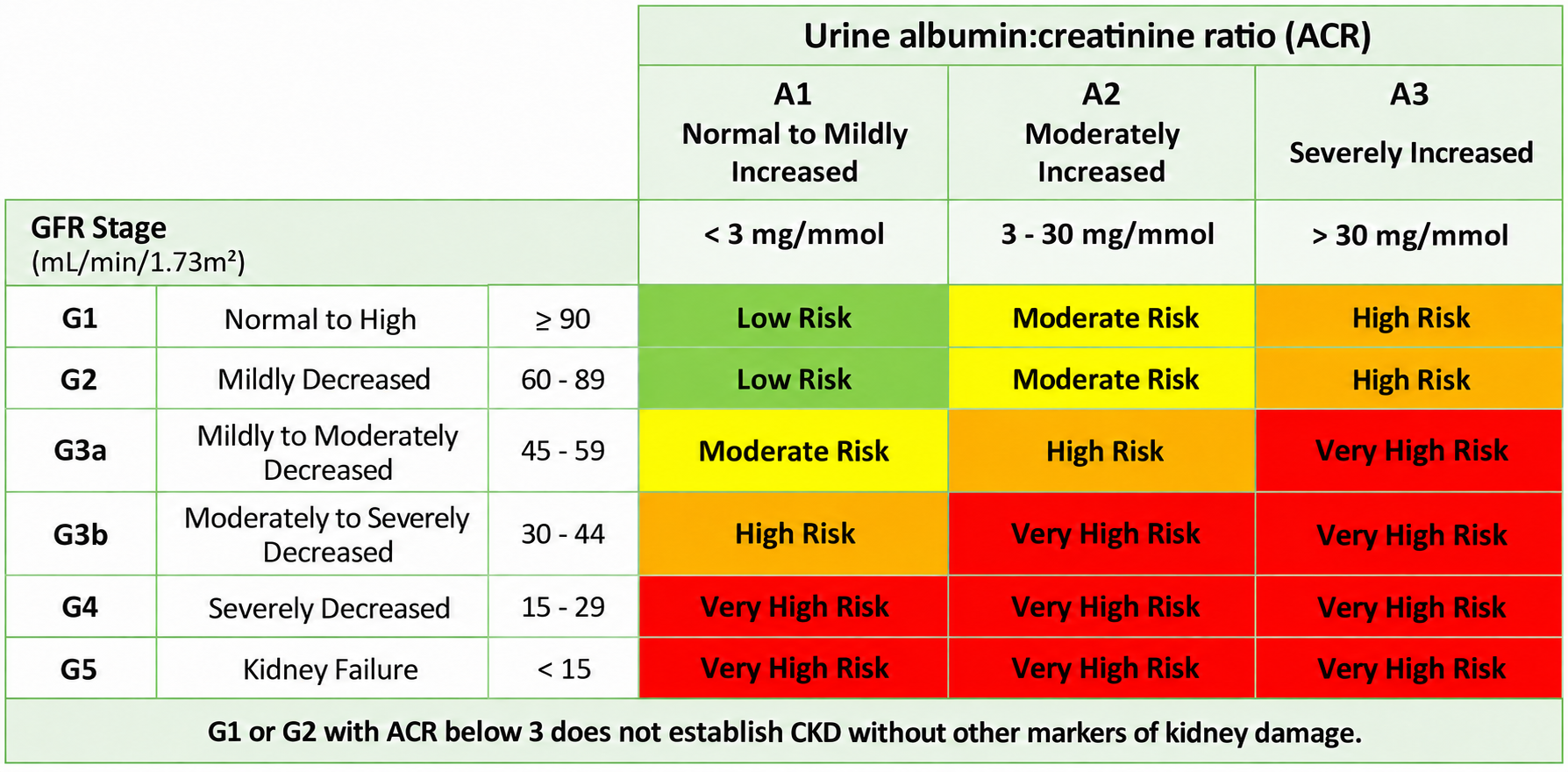
Table - NICE CKD risk categories by GFR and urine ACR. G1 or G2 with ACR below 3 mg/mmol does not establish CKD without other markers of kidney damage.
SimpleMed original by Dr. Thomas Burnell
Causes of Chronic Kidney Disease
CKD arises as a result of another condition damaging the kidneys over time and so there are many causes because a lot of conditions can affect the kidneys. Examples of these conditions include:
- Diabetes
- Hypertension
- Glomerulonephritis
- Infection - e.g. pyelonephritis (infection of the kidney)
- Genetic - e.g. autosomal dominant polycystic kidney disease (an inherited condition with many fluid-filled cysts in the kidneys), Alport’s syndrome (an inherited condition that damages the filters of the kidney and can also affect hearing and the eyes)
- Obstruction (a blockage to the flow of urine) and reflux nephropathy (kidney scarring caused by urine flowing back up towards the kidneys)
- Acute kidney injury - a current or previous episode of acute kidney injury (for example acute tubular injury, damage to the cells lining the kidney tubules) increases the risk of CKD
- Vascular disease - e.g. vasculitis (inflammation of the walls of the blood vessels)
- Medicines that can harm the kidneys (nephrotoxic drugs) - e.g. long-term use of non-steroidal anti-inflammatory drugs (NSAIDs, such as ibuprofen), lithium and calcineurin inhibitors (such as ciclosporin or tacrolimus)
- Idiopathic - CKD may just develop over time in an otherwise healthy patient with no known cause.
When investigating chronic kidney disease, it is important to identify the degree of renal impairment, the cause of renal impairment and to look for the complications of CKD.
The degree of renal impairment is identified using:
- Urine tests - the urine albumin:creatinine ratio (ACR) measures proteinuria, and a urine dipstick test (a test strip dipped in the urine) checks for haematuria (blood in the urine).
- Creatinine and eGFR (estimated GFR, calculated from the blood creatinine) - these help to define the stage of CKD.
- Other blood tests: urea and electrolytes (to check the potassium), full blood count (to look for anaemia), CRP (C-reactive protein, a marker of inflammation), liver function tests, iron status tests (to look for iron deficiency) and bone biochemistry (calcium, phosphate and parathyroid hormone)
The cause of renal impairment is identified using different techniques for different differential diagnosis:
- Autoimmune disease - an autoantibody screen (a blood test for antibodies that attack the body’s own tissues) and complement levels (complement is a group of immune proteins) can be used to look for autoimmune disease.
- Vasculitis - anti-neutrophil cytoplasmic antibody screen can be used to look for vasculitis.
- Myeloma (a cancer of the plasma cells in the bone marrow) - myeloma can cause CKD. The main tests are a serum immunoglobulin screen (a blood test for antibody levels), protein electrophoresis (a test that separates the proteins in blood or urine to find abnormal antibody proteins) and serum free light chain levels (small antibody fragments that are often raised in myeloma).
- Ultrasound scan - ultrasound scans can be used to see the kidney size and look for evidence of obstruction. Kidneys are often smaller in CKD, but they can be normal in size or enlarged in some conditions, such as diabetic kidney disease. NICE advises a renal ultrasound for some adults with CKD, for example if the GFR is below 30 ml/min/1.73m2, if there is visible or persistent invisible blood in the urine, or if there are symptoms of urinary tract obstruction.
- Kidney biopsy - this can be used if the cause of chronic kidney disease remains unknown after other investigations.
- Stones and masses - these can be investigated for using a CT scan or an MRI scan.
- Renal artery stenosis (narrowing of the artery that supplies a kidney) - an MR angiogram (an MRI scan of the blood vessels) can be used to look for it. Some gadolinium contrast agents (the dye sometimes used in MRI) must not be given in severe renal impairment, because of the risk of nephrogenic systemic fibrosis (a condition in which the skin and other tissues become thickened and hard). The choice of scan and contrast therefore depends on the GFR.
Identifying complications of CKD:
- Full blood count - this is used to look for anaemia.
- Cardiovascular risk factors - blood pressure, body mass index (BMI), HbA1c (a blood test that shows average blood glucose over the past 2 to 3 months) and a lipid profile (cholesterol levels) are checked to assess cardiovascular risk factors.
- Assessment of mineral and bone disorder (disordered calcium, phosphate and bone health) - this is done with blood tests of calcium, phosphate and parathyroid hormone. NICE advises measuring these in adults with a GFR below 30 ml/min/1.73m2, and not routinely if the GFR is 30 or more.
- Bone biochemistry - the usual pattern in advancing CKD is a low serum calcium, a high serum phosphate and a high parathyroid hormone, although results vary between patients.
The main aim in management of these patients is to prevent the development of CKD in the first place. Once a patient develops CKD, the aim of treatment is to prevent the progression of the disease and to treat any complications that develop. The main ways to prevent and treat CKD are to address modifiable risk factors, to control blood pressure and to reduce proteinuria (protein in the urine).
- Modifiable Risk Factors:
- Smoking - advise patients to stop smoking.
- Obesity - advise patients to lose weight and do more exercise.
- Hypertension Control:
- Anti-hypertensive medication - the target clinic blood pressure is below 140/90 mmHg if the ACR is under 70 mg/mmol, and below 130/80 mmHg if the ACR is 70 mg/mmol or more. An ACE inhibitor (angiotensin-converting enzyme inhibitor) or an angiotensin receptor blocker (ARB) is offered to people with CKD who have hypertension and an ACR above 30 mg/mmol, to people with CKD and diabetes if the ACR is 3 mg/mmol or more, and to people with CKD without diabetes if the ACR is 70 mg/mmol or more. They also reduce proteinuria.
- Diuretics
- Dietary advice on salt - advice on salt (and potassium and phosphate) intake is matched to the severity of CKD
- Proteinuria - This should be kept as low as possible in patients with CKD to prevent progression of the disease.
- ACE inhibitors and angiotensin receptor blockers are the main medications used to treat proteinuria. They reduce the pressure inside the glomeruli (the filtering units), so less protein is filtered into the urine.
- SGLT2 inhibitors (sodium-glucose cotransporter 2 inhibitors, such as dapagliflozin and empagliflozin) are recommended by NICE as an add-on to the highest tolerated dose of an ACE inhibitor or ARB for some adults with CKD. NICE recommends them if the eGFR is 20 to below 45 ml/min/1.73m2, or if the eGFR is 45 to 90 ml/min/1.73m2 and either the ACR is 22.6 mg/mmol or more or the patient has type 2 diabetes.
- Diabetes - people with diabetes are supported to optimise their blood glucose control, which is checked with the HbA1c blood test (the average blood glucose over the past 2 to 3 months).
- Lipid levels - people with CKD are offered atorvastatin 20 mg (a statin, a cholesterol-lowering medicine) to reduce their risk of cardiovascular disease, such as heart attacks and strokes. The aim is to prevent cardiovascular disease, not only to lower the cholesterol level.
There are many complications of chronic kidney disease and these must all be identified and treated to reduce the risk of further complications and improve patient’s quality of life. Some of the complications include hyperkalaemia, acidosis, anaemia, mineral bone disease and altered drug metabolism.
In chronic kidney disease there is reduced excretion of potassium. As less potassium is removed from the body, the level in the blood rises. This leads to hyperkalaemia (a high serum potassium level). To reduce the risk, the patient’s potassium intake and any medicines that raise potassium are reviewed, and the serum potassium is checked more often if they take such medicines.
If a patient’s serum potassium level is increasing, the patient may need the avoidance of drugs that increase potassium levels and a diet alteration to avoid foods high in potassium.
- ACE inhibitors and angiotensin receptor blockers used in the treatment of CKD can raise serum potassium, so it may need to be checked more often. NICE advises stopping them if the serum potassium reaches 6.0 mmol/litre or more and other medicines that raise potassium have already been stopped.
In chronic kidney disease the reduced kidney function leads to a decreased excretion of acid from the body and an increased loss of bicarbonate. This leads to a metabolic acidosis (a low blood pH caused by too much acid or too little bicarbonate, rather than by a lung problem) as the control of blood pH is lost. This acidosis will happen slowly and may not be noticed because the symptoms are quite non-specific.
Symptoms of Acidosis include:
- Headache
- Drowsiness
- Diarrhoea
- Muscle weakness
- Coughing
- Nausea and vomiting
- Shortness of breath, including fast, deep breathing
- Confusion
- Seizures
Acidosis may need treatment because it can contribute to worsening renal function and muscle breakdown. It can also cause bone demineralisation (loss of minerals such as calcium from the bone), which can further worsen mineral bone disease in CKD.
The main treatment of acidosis is oral sodium bicarbonate (NaHCO3), which will replace the bicarbonate that is being lost from the body. NICE advises considering it for adults with a GFR below 30 ml/min/1.73m2 and a serum bicarbonate below 20 mmol/litre. Bicarbonate supplements will help to prevent the pH from decreasing, and becoming acidotic, by neutralising the excess acid in the blood.
Anaemia is caused by multiple mechanisms in chronic kidney disease, all of which act to lower haemoglobin and cause anaemia. Some of the mechanisms that cause anaemia in CKD are:
- Erythropoietin deficiency - damaged kidneys make less erythropoietin (a hormone that tells the bone marrow to make red blood cells), so fewer red blood cells are made. This is one of the main causes of anaemia in CKD.
- Anaemia of chronic disease - inflammation causes a high level of hepcidin (a hormone, made by the liver, that reduces iron absorption and the release of iron from the body’s stores). This lowers the iron available to make red blood cells and causes anaemia.
- Shorter red blood cell lifespan - red blood cells survive for less time in CKD.
- Medication
- CKD mineral and bone disease
- Deficiency of Vitamin B12, folate, iron
The treatment of anaemia is very important as it improves the patient’s quality of life and reduces patient mortality. Treating anaemia helps to improve exercise capacity and cognitive function as well as slowing the progression of renal disease. It can also prevent left ventricular hypertrophy, as the heart has to pump harder to deliver enough oxygen around the body to counter the effects of anaemia, leading to strain on the heart, especially the left ventricle.
Before treating anaemia, iron status should be checked, as iron deficiency may be the cause of anaemia. NICE advises using the percentage of hypochromic red blood cells (red cells that look pale because they contain too little haemoglobin) or, if that is not possible, the reticulocyte haemoglobin content (the haemoglobin in young red cells). If neither is available, transferrin saturation (how full the iron-carrying protein transferrin is) and serum ferritin (a marker of the body’s iron stores) are used together, never alone. Folate and Vitamin B12 should also be checked to look for deficiencies. If any of these are found to be low then the appropriate treatment should be taken to increase the levels back to normal, e.g. oral folate supplements, Vitamin B12 injections.
If iron, folate and Vitamin B12 are all found to be fine but the patient is still anaemic. In that case, an erythropoietin stimulating agent (ESA, a medicine that acts like erythropoietin to stimulate red blood cell production) can be offered to patients who are likely to benefit. The aim is not to correct the haemoglobin to a normal level. In adults NICE advises typically keeping it between 100 and 120 g/litre and avoiding levels above 120 g/litre, because of the risk of death and serious cardiovascular events.
Mineral bone disease is seen in chronic kidney disease as the kidneys become unable to regulate calcium and phosphate levels within the body. There are many causes of mineral bone disease which all combine to cause a decreased calcium level and reduced bone mineralisation within the body.
Some of the causes of mineral bone disease include:
- Impaired renal function which decreases phosphate excretion from the body, resulting in an increased serum phosphate (hyperphosphataemia). Hyperphosphataemia stimulates the parathyroid glands leading to an increase in parathyroid hormone secretion. PTH acts on the bones, intestines and kidneys to raise the blood calcium. In the bones it causes increased bone resorption (breakdown of bone), which releases calcium into the blood. If this carries on for a long time, it is linked to disturbed bone turnover (the balance between bone breakdown and bone rebuilding).
- Decreased levels of activated Vitamin D due to a reduced ability of the kidneys to hydroxylate 25-hydroxyvitamin D (convert it into active vitamin D). This reduction in vitamin D levels impairs bone mineralisation and calcium absorption.
- Increased phosphate levels within the body cause phosphate in the blood to combine with free calcium. This forms calcium deposits throughout the body and decreases the concentration of free calcium in the blood. Less calcium is then available to mineralise bone, and the low calcium is a further trigger for the release of parathyroid hormone.
Mineral bone disease in CKD changes the structure of the bone, which is called renal osteodystrophy. The main patterns are osteitis fibrosa (high bone turnover, with bone broken down and rebuilt too quickly), adynamic bone disease (low bone turnover) and osteomalacia (soft bones from poor mineralisation). Osteosclerosis (abnormally dense bone) and osteoporosis (thin, weak bones) can also occur.
The blood results in mineral bone disease usually show a secondary hyperparathyroidism pattern, in which the parathyroid glands make too much parathyroid hormone because the calcium is low, the phosphate is high or the active vitamin D is low:
- Low serum Vitamin D
- Low serum Calcium
- High serum Phosphate
- High serum Alkaline phosphatase (ALP, an enzyme released by bone-forming cells)
- High serum Parathyroid Hormone
Mineral bone disease is managed through reduced phosphate intake (a renal dietitian can advise), the use of phosphate binders (medicines taken with food that bind phosphate in the gut, so less of it is absorbed) and, if the patient is deficient, vitamin D supplements (colecalciferol or ergocalciferol). If symptoms continue once the deficiency has been corrected, patients with a GFR below 30 ml/min/1.73m2 can be offered alfacalcidol (an active form of vitamin D, also called 1-alpha-calcidol) or calcitriol. Alfacalcidol is given by mouth or by injection, and the calcium level is monitored. The diet and phosphate binders lower the phosphate level, which helps to prevent calcium deposits and other complications of hyperphosphataemia. The vitamin D treatment aims to keep the calcium level within the normal range.
The kidneys are one of the main places in the body where drug excretion and metabolism occur. In CKD there can be reduced excretion of drugs and their metabolites (the substances made when the body breaks a drug down). The drugs can then accumulate, which gives an increased risk of toxicity.
Because of reduced drug excretion in CKD, many drugs require dose reduction to prevent their levels getting too high and causing toxicity. This is especially important in nephrotoxic drugs as these can further damage the kidney and worsen CKD.
Other Complications - some other complications that occur with CKD include uraemic syndrome, hyperparathyroidism, fluid overload, electrolyte disturbances, malnutrition and cardiovascular disease.
- Waste products can accumulate in the body because the kidneys excrete them less well. They contribute to uraemic syndrome (symptoms caused by waste products building up in the blood), reduced appetite, nausea and vomiting, and pruritus (itching).
End Stage Renal Disease/Failure
End-stage renal failure is the final stage of chronic kidney disease and is a result of extensive damage to the kidneys. Death is likely without renal replacement therapy. It is defined as an estimated GFR < 15 ml/min/1.73 m2, persisting chronically.
Non-specific symptoms of end-stage renal failure may not be recognised, so it is important to check the GFR of patients with CKD. Symptoms of end-stage renal failure include:
- Fatigue
- Symptoms of volume overload, e.g. peripheral oedema (swelling of the ankles)
- Nausea and vomiting
- Reduced appetite
- Restless legs and cramps
- Pruritus (itching)
- Sexual dysfunction and reduced fertility
- Increased risk of infections due to reduced cellular immunity (the immune cells) and humoral immunity (the antibodies)
Treatment of end-stage renal failure is through renal replacement therapy (RRT), which means dialysis or a kidney transplant. Dialysis is the usual first renal replacement therapy for end-stage renal failure: in the UK in 2024, only 6.3% of adults starting renal replacement therapy started with a transplant. Among all UK adults on renal replacement therapy at the end of 2024, a kidney transplant was the most common treatment (56.0%), followed by in-centre haemodialysis (haemodialysis given at a dialysis unit, 36.8%). Patients may also choose active conservative management if they decide not to have RRT, or if RRT is not suitable for them. The treatment options should be discussed with the patient before they reach renal failure, so that they understand the advantages and disadvantages of each option. NICE advises starting this assessment at least a year before treatment is likely to be needed.
Dialysis is the method by which waste products and excess fluid are removed from the body when the kidneys stop functioning. Dialysis filters the blood in one of two ways. In haemodialysis, the blood is diverted out of the body to be filtered by a machine. In peritoneal dialysis, a membrane in the body (the peritoneum, which lines the abdomen) is used to remove substances and fluid from the blood, with the help of a special dialysis fluid.
- There are two main types of dialysis, haemodialysis (HD) and peritoneal dialysis (PD) which are discussed below.
- Related methods include haemofiltration and haemodiafiltration (HDF), which are other ways of filtering the blood. NICE advises considering HDF rather than standard haemodialysis for people having treatment in a centre (a hospital or satellite unit). Intestinal dialysis (which uses the bowel to remove waste) is not used routinely. These methods are not discussed in this article.
Active Conservative Management
This treatment option for end-stage renal failure is used for patients who are either unsuitable for or choose not to have invasive therapy such as peritoneal dialysis, haemodialysis or kidney transplant. This complex decision is usually made after multiple discussions with the patient and their family.
In this option, patients are looked after by a multidisciplinary team (health professionals from different specialties working together). The team provides symptom control, advance care plans (written plans for future care and wishes) and supportive care for patients and their families. The team also aims to help the patient remain in their preferred place of care.
Haemodialysis involves pumping blood out of the body and into a machine, called a dialyser, that filters the blood to remove excess fluid and waste products. It is carried out in the hospital three times a week at designated slots, and lasts around 4 hours. Patients often take a lot of medication. They also need vascular access (a way of reaching the blood) so that the dialysis machine can be attached. The usual type is an arteriovenous fistula (AVF, a connection between an artery and a vein made by a small operation, usually in the arm). It is made some months before dialysis starts, to give it time to mature. Other types of access are a graft (an artificial tube placed under the skin to join an artery and a vein) or a central venous catheter (a tube placed in a large vein). The type is chosen after discussing the risks and benefits with the patient.
Patients also have large restrictions on their diet as they are restricted to a small volume of fluid per day and must avoid foods high in potassium and salt.
Haemodialysis can also be performed at home which allows for a little more freedom and less disturbance to daily life. Different types of home haemodialysis machine are available, and the dialysis team helps the patient to choose the best one. Usually patients on home haemodialysis can choose to have more dialysis than they would in hospital, for longer periods of time. This can improve their general wellbeing and symptoms such as tiredness and itching. However, home haemodialysis does require training and the ability to learn to use the machine, either alone or with the help of a family member, friend or carer.
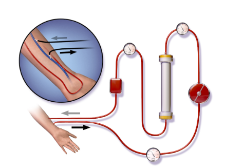
Diagram - The Process of Haemodialysis
Image by Blausen Medical Communications / Blausen.com staff (2014), source, licensed under CC BY 3.0.
- Failed vascular access - if no type of dialysis access (fistula, graft or central venous catheter) can be made. Being unable to have a fistula alone is not a contraindication, because a graft or catheter can be used instead.
- Heart failure - haemodialysis can cause rapid fluid and haemodynamic shifts (changes in blood flow and blood pressure) which may worsen cardiovascular instability. However, heart failure is not an absolute contraindication, and haemodialysis is commonly used with careful monitoring and adjustment of fluid removal.
- Coagulopathy (a disorder of blood clotting)
A high risk of bleeding, such as with a coagulopathy, does not by itself rule out haemodialysis. Heparin (a drug that stops blood clotting) is commonly given during haemodialysis to stop clots forming in the circuit outside the body, but it is contraindicated in patients at high risk of bleeding. These patients can still have haemodialysis without heparin, for example with intermittent saline flushes of the circuit.
- Infection - for example, a skin infection around the fistula
- Thrombosis
- Venous stenosis (narrowing of a vein)
- Haemorrhage
- Steal syndrome (the fistula diverts blood away from the hand)
- Anaemia
- Cardiovascular instability
- Cardiac arrhythmias (abnormal heart rhythms)
- Air embolism
- Nausea and vomiting
Peritoneal dialysis involves using the peritoneal cavity to exchange substances between dialysate and the blood. Dialysate is the fluid used in peritoneal dialysis. Waste products move from the blood into the dialysate by diffusion, and excess fluid is removed by ultrafiltration (fluid being drawn out of the blood across the peritoneal membrane). The dialysate contains dextrose (a glucose solution) which draws fluid out of the blood by osmosis. Once dialysis is complete, the dialysate is disposed of and a new bag is used the next time.
There are two main types, continuous ambulatory peritoneal dialysis (CAPD), in which the patient can move about and do their usual activities during treatment, and automated peritoneal dialysis (APD), in which a machine does the exchanges. Both of these involve pumping dialysate into the peritoneal cavity where substances can then be exchanged between the fluid and the blood.
Continuous ambulatory peritoneal dialysis involves one to four exchanges of dialysate per day where the patient’s abdominal cavity is filled with a bag of dialysate over around 30 to 40 minutes. The solution is then left in the abdomen for a few hours before being drained out. The process requires the patient to have a catheter inserted into their abdomen to fill and drain the abdominal cavity of dialysate. Once the used fluid is drained, the abdomen is filled with new fluid.
CAPD can be carried out anywhere, either at home or work, and requires the patient to be trained in how to perform CAPD. Exchanges can happen at any time of the day but most patients choose to do them before meals and before bed.
Automated peritoneal dialysis involves one session of peritoneal dialysis per day where the patient is attached to a machine for around 9 hours. This is usually carried out at home and requires the patient to be trained in how to use the machine. Most people choose to do their dialysis overnight due to the length of time required for APD.
When compared to haemodialysis there are fewer fluid and diet restrictions, but a dietitian still advises on any restrictions that the patient needs.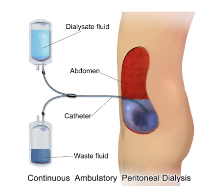
Diagram - The Process of Continuous Ambulatory Peritoneal Dialysis, with old dialysate fluid being exchanged for new fluid
Image by Blausen Medical Communications / Blausen.com staff (2014), source, licensed under CC BY 3.0.
- Failure of peritoneal membrane - peritoneal dialysis causes scarring of the peritoneal membrane which reduces diffusion of waste across the membrane.
- Intra-abdominal adhesions (bands of scar tissue inside the abdomen) - extensive adhesions can prevent peritoneal dialysis from working, but there are few absolute contraindications, so each patient is assessed individually.
- Previous abdominal surgery, hernia (a weakness in the abdominal wall, through which tissue can bulge) or stoma (an opening made from the bowel or urinary tract onto the skin of the abdomen) - these may be associated with peritoneal scarring and can make peritoneal dialysis more difficult, but suitability depends on individual assessment.
- Obesity or large muscle mass - these are relative contraindications. Peritoneal dialysis needs the peritoneal surface area to be large enough for the size of the body.
- Patient or carer unable to connect or disconnect the peritoneal dialysis
- Peritonitis (infection of the lining of the abdomen), which needs treatment straight away
- Catheter associated problems - e.g. infection, blockage, kinking and leaks.
- Constipation
- Fluid retention
- Hyperglycaemia
- Weight gain
- Ultrafiltration failure (the peritoneal membrane no longer removes enough fluid)
- Hernia
- Scarring of the peritoneum
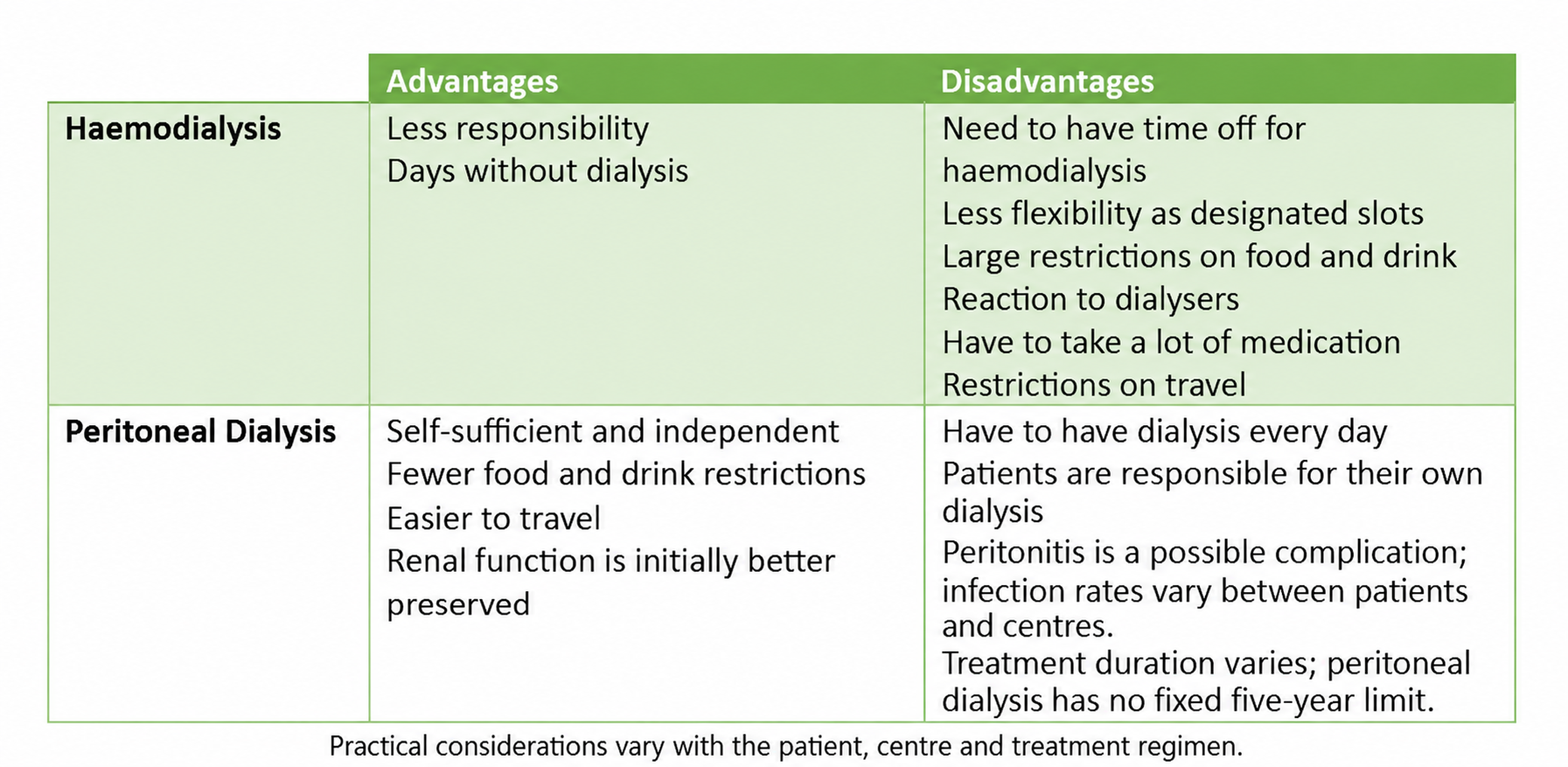
Table - Typical practical considerations for in-centre haemodialysis and self-managed peritoneal dialysis. Home or assisted treatments may differ. Diet, fluid limits, travel, medicines and residual kidney function depend on the patient, centre and regimen; peritoneal dialysis has no fixed five-year limit.
A kidney transplant is where a patient who has little or no kidney function (e.g. end-stage renal failure) receives a healthy kidney from another individual. This kidney can either come from a live or a deceased donor. Usually the donor and recipient are blood group compatible, and closer tissue matching improves transplant outcomes. Some living donor kidneys are transplanted across a blood group or tissue type (HLA) difference, which is called an antibody incompatible transplant. In a UK national registry study these made up around 10% of living donor kidney transplants. Pairs who are not compatible can also join the UK Living Kidney Sharing Scheme, which looks for compatible matches with other pairs. The replacement kidney is inserted into the right or left groin (lower abdomen), usually alongside the patient’s own kidneys. The renal vessels (the kidney artery and vein) and ureter of the new kidney are surgically attached so that it can work as normal.
Patients who undergo a renal transplant usually have both a greater life expectancy and better quality of life compared to patients who stay on dialysis. The individual risks and benefits, for example other health conditions, should be discussed with each patient. Transplantation means the patient can stop dialysis. It can also help to improve anaemia and mineral bone disease, although some problems, such as raised parathyroid hormone levels, often persist.
Contraindications for Transplantation
- Active infection or malignancy (cancer)
- Active viral hepatitis - hepatitis C, for example, can now be cured with direct-acting antiviral medicines, either before or after a transplant.
- Severe heart or lung disease
- Reversible kidney disease (the person’s own kidneys may recover)
- Uncontrolled substance abuse
- Psychiatric illness
- Short life expectancy
- Non-adherence to ongoing treatment
- Complications during operation - e.g. local infection and deep vein thrombosis.
- Immediate graft failure - where the transplanted kidney (the graft) does not function from the outset, for example due to vascular thrombosis (a blood clot in the vessels supplying the kidney) or primary non-function (the kidney never starts to work). Hyperacute rejection, in which antibodies the patient already has attack the kidney within minutes, is a rare cause.
- Infection
- Cancer
- Side-effects from immunosuppressive drugs
Renal transplants require lots of medication after the operation to prevent rejection of the transplanted kidney. Many of these medicines are immunosuppressants, which suppress the immune system so that it does not attack the new kidney. Examples are tacrolimus, mycophenolate mofetil and steroids such as prednisolone. The doses usually fall over time, but some are needed for the lifetime of the transplant. Each medicine has its own side effects. Tacrolimus can raise the blood sugar, cause tremors and, in high doses, impair kidney function. Steroids can raise the blood pressure and the blood sugar. Mycophenolate mofetil can cause nausea and diarrhoea, and can reduce the number of white cells in the blood. Side effects of these medicines include:
- Hypertension
- Gastrointestinal tract ulceration
- Nausea
- Diarrhoea
- Thrombocytopenia (a low platelet count)
- Leukopenia (a low white blood cell count)
- Diabetes mellitus
- Hypercholesterolaemia
The kidneys that are used in transplant come from three main donor sources:
- Living Related Donor - a relative may be a suitable living donor after assessment. Relatedness alone does not guarantee better graft survival than an unrelated living donation; donor health and the individual donor-recipient assessment matter.
- Living Unrelated Donor - a partner, friend or altruistic donor may provide a suitable living-donor kidney. Unrelated living donation is not inherently inferior to related donation; suitability is assessed individually.
- Deceased Donor - kidneys from deceased donors generally have shorter graft survival than living-donor kidneys, although outcomes vary between individuals. The transplant must also be done quickly, because kidneys do not survive long outside the body.
Patients who have had a kidney transplant require long term follow-up care at least once or twice a week at first, in the transplant clinic, to check that nothing has gone wrong. They then need to attend less often, and after a year most patients without serious problems attend every 3 to 4 months to check the function of the kidney. They are also screened for cancer, and are offered the hepatitis B, influenza and pneumococcal vaccines (the pneumococcal vaccine protects against infections such as pneumonia). Inactivated vaccines can be given, but many live vaccines are not suitable for people on immunosuppressants. They also have regular blood tests (e.g. calcium, phosphate and glucose levels) to monitor for complications (e.g. mineral bone disease and diabetes).
Complications of Transplant and Immunosuppressive Treatment
- Post-operative complications - e.g. bleeding, deep vein thrombosis, pulmonary embolism, local infection and pain.
- Infections - especially opportunistic infections (infections that take advantage of a weakened immune system) that can occur as a result of immunosuppressive treatment.
- Cancer
- Drug toxicity
- Bone marrow suppression - this can occur as a result of the immunosuppressive treatment.
- Urinary tract obstruction
- Graft rejection (the immune system attacking the transplanted kidney) can be hyperacute, accelerated, acute cellular or chronic. Hyperacute rejection happens within minutes of the transplant, because the patient already has antibodies against the donor kidney. It is now rarely seen because cross-match tests (tests for these antibodies) are done before the transplant. Many patients have some rejection later. It is usually mild and can be treated with different medicines, but rejection is still one of the leading causes of graft loss.
Edited by: Dr. Tom Bradley
Reviewed by: Adrian Judge and Dr. Marcus Judge
- 826


