

SimpleMedSimpleMed
Hit enter to search or ESC to close

Get in Touch

Contact Us

Use our contact form.

Pulmonary Embolism

Share:

By Dr. Jenny Hubball

Next Lesson - Asthma

Respiratory System


Contents

  1. Pathophysiology
  2. Presentation
  3. Investigations
  4. Management
  5. Quiz

Abstract

  • A pulmonary embolism (PE) is most commonly a blood clot (thromboembolism) that began as a deep vein thrombosis (DVT) in a leg or pelvic vein. The clot travels through the right side of the heart and lodges in the blood vessels of the lungs.
  • Risk factors for a PE include pregnancy, prolonged immobilisation, the combined oral contraceptive pill, and obesity.
  • PE can cause right ventricular strain (extra work for the right ventricle) and pulmonary vasoconstriction (narrowing of the blood vessels in the lungs). Hypoxaemia (low blood oxygen) and type 1 respiratory failure (low blood oxygen with normal or low carbon dioxide) may occur. The effects vary with the size of the clot (clot burden) and with how well the heart and lungs can cope (cardiopulmonary reserve).
  • Symptoms of a PE include dyspnoea (shortness of breath), pleuritic chest pain (sharp chest pain that is worse on breathing in), and a cough. Signs include tachypnoea (fast breathing), tachycardia (a fast heart rate), low BP, and a raised jugular venous pressure.
  • Investigations for PE start with a Wells score (a points score that estimates how likely a PE is). The next test is a d-dimer (a blood test for clot breakdown products) or a CT pulmonary angiogram (CTPA, a CT scan of the lung blood vessels). An ECG, chest x-ray and arterial blood gas (a blood test for oxygen and carbon dioxide levels) help to look for other causes and to show the effects of a PE.
  • Anticoagulation is used to treat PE; longer-term treatment requires reassessment of recurrence risk, bleeding risk and the patient's preferences.

Core

Pathophysiology

An embolus is material carried in the bloodstream from its site of origin to another site, where it may obstruct a vessel. A thrombus is a clot formed at its original site; a detached fragment travelling through the circulation is a thromboembolus.

A pulmonary embolism (PE) is therefore material that has moved from one part of the vasculature, through the right side of the heart, and lodges in the pulmonary arteries.

An embolus is usually made from a blood clot (thromboembolism), but can also be formed from tumour, air, fat, or amniotic fluid - note that the rest of this article will be focussing on thromboemboli. Pulmonary emboli are the third most common cause of cardiovascular death, after myocardial infarction and stroke, and are one of the most common causes of potentially preventable death in hospital patients. Venous thromboembolism (VTE), which includes deep vein thrombosis and pulmonary embolism, is also the third most common cardiovascular disease.

Most pulmonary thromboemboli arise from a deep vein thrombosis (DVT) in the legs or pelvis. A detached clot travels through the right heart before lodging in the pulmonary arteries. The main factors that contribute to thrombus formation are stasis of blood flow (slow or still blood), hypercoagulability of blood (blood that clots too easily), and endothelial injury (damage to the lining of the blood vessel). Together these are known as Virchow’s Triad. The risk factors for a DVT (and therefore PE) are factors that contribute to one or more parts of this triad:

  • Pregnancy
  • Prolonged immobilisation (e.g. post-operatively or on a long-haul flight)
  • Previous venous thromboembolism
  • Combined oral contraceptive pill
  • Cancer
  • Obesity
  • Hormone replacement therapy (HRT) taken by mouth. The risk is not increased with transdermal HRT (given through the skin, for example as patches or gel).
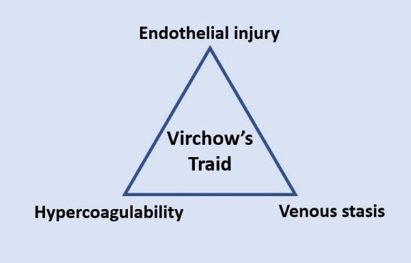


Virchow's Triad SimpleMed

Diagram - Virchow's Triad: the relationship between endothelial injury, hypercoagulability, and venous stasis 

Creative commons source by Dr.Vijaya chandar, MBBS [CC BY-SA 4.0 (https://creativecommons.org/licenses/by-sa/4.0)]


The above risk factors can be divided into two groups:

  • Temporary; for example, pregnancy, the pill, and immobilisation.
  • Persistent while present; for example, active cancer. Risk factors can change over time and should be reassessed.

About one-third to one-half of VTE episodes have no identifiable risk factor. The rest are caused by temporary (transient) or persistent risk factors. Prevention of a PE is worthwhile. For example, on a journey of 4 hours or more the NHS advises people to walk around whenever they can, to bend and straighten their legs, feet and toes every 30 minutes while seated, to drink water regularly and to wear flight socks.

Pulmonary emboli cause mechanical obstruction of pulmonary arteries. Obstruction and pulmonary vasoconstriction (narrowing of the lung blood vessels, driven by chemical signals including serotonin and thromboxane) increase pulmonary vascular resistance (the resistance to blood flow through the lungs) and right ventricular afterload (the pressure the right ventricle must pump against). The right ventricle may dilate and fail, reducing blood flow through the lungs and filling of the left ventricle. Cardiac output and systemic blood pressure can then fall. Acute right ventricular failure is the principal lethal mechanism in severe PE.

Ventilated lung supplied by an obstructed artery becomes poorly perfused, increasing alveolar dead space (air in the lung air sacs that reaches no blood flow, so no gas exchange takes place there). Redistribution of blood to other lung regions can produce low ventilation-perfusion ratios (areas where blood flow is high compared with air flow), contributing to hypoxaemia (low blood oxygen). Reduced cardiac output and atelectasis (collapse of part of the lung) can also contribute. Hyperventilation (fast, deep breathing) commonly lowers arterial CO2, so PE may cause type 1 respiratory failure: low arterial O2 with normal or low CO2. Normal oxygen saturation does not exclude PE.

The part of the lung that is poorly perfused may undergo infarction (death of tissue from loss of blood supply). The bronchial arteries, which supply the lung tissue itself, can often provide enough oxygen to prevent this, but infarction is not rare: one review reports an incidence of around 30%. A patient with pulmonary infarction may present with haemoptysis (coughing up blood) and pleuritic chest pain (sharp chest pain that is worse on breathing in).  

In a patient with a patent foramen ovale (a small hole between the two atria that stays open), PE-related elevation of right atrial pressure can permit right-to-left shunting (blood passing from the right side of the heart to the left without going through the lungs). A venous embolus may then cross into the arterial circulation, causing a paradoxical embolism (an embolus from a vein that reaches the arteries), including stroke. The risk depends on the presence of a shunt; PE does not make every patent foramen ovale shunt right to left.


Presentation

Symptoms of a PE include:

  • Shortness of breath
  • Pleuritic chest pain (sharp chest pain that is worse on breathing in)
  • Cough
  • Substernal chest pain (behind the breastbone)
  • Fever
  • Haemoptysis (coughing up blood)
  • Syncope (fainting)
  • Unilateral leg pain (DVT)  


Signs of a PE include:

  • Tachypnoea (fast breathing)
  • Tachycardia (fast heart rate, over 100 beats per minute)
  • Low BP
  • Raised jugular venous pressure
  • Pleural rub (a scratching sound heard through a stethoscope, from inflamed lung lining) in cases of pulmonary infarction
  • Evidence of DVT, e.g. erythema (redness), increased temperature and tenderness on palpation of the leg


The main differential diagnoses for a pulmonary embolism are pneumothorax (air in the space around the lung), pneumonia, musculoskeletal chest pain, and myocardial infarction.


Investigations

In a patient with a suspected PE, NICE advises that the first step is to estimate the clinical probability with the two-level Wells score (see below). The result decides the next test. The other investigations listed here help to look for other causes and to show the effects of a PE:

  • Arterial Blood Gas (ABG) - a blood test, taken from an artery, that measures oxygen, carbon dioxide and acidity. It may show respiratory alkalosis (low carbon dioxide, which makes the blood less acidic) with hyperventilation (fast, deep breathing), but findings are variable and a normal result does not exclude PE.
  • Chest X-ray - to exclude differentials such as pneumothorax or pleural effusion (fluid around the lung).
  • ECG:
    • The most common sign of a PE on an ECG is sinus tachycardia (a fast heart rhythm that starts in the heart's normal pacemaker).
    • An ECG could show an S1Q3T3 pattern: a large S wave in lead I, and a Q wave with an inverted T wave in lead III (waves are the parts of the heartbeat trace, and leads are the different views of the heart). These are classic signs of right heart strain. The pattern is a well-known textbook finding but is not common in clinical practice.
    • There could also be no signs on ECG - a normal ECG cannot rule out a PE.
  • D-dimers:
    • D-dimer is measured in a blood test. It is a fragment of fibrin (the protein that holds a clot together) that is released into the blood when the body breaks down a clot (a process called fibrinolysis).
    • In a patient in whom PE is unlikely (Wells score of 4 points or less), a d-dimer is the next test. A negative d-dimer rules PE out. A positive d-dimer does not confirm PE, so it is followed by a CTPA (or a ventilation-perfusion scan if CTPA is unsuitable). If the d-dimer result will take more than 4 hours, NICE advises interim therapeutic anticoagulation (a full treatment dose of an anticoagulant) while waiting.
    • In a patient in whom PE is likely (Wells score of more than 4 points), the pathway does not use a d-dimer. A CT pulmonary angiogram (CTPA, a CT scan of the lung blood vessels) is offered immediately if possible. If a scan cannot be done immediately, NICE advises interim therapeutic anticoagulation until it can.
  • A Wells score (a points score that estimates how likely a PE is) should be calculated for any patient in whom a PE is suspected. In pregnancy, the Royal College of Obstetricians and Gynaecologists (RCOG) advises that d-dimer testing should not be performed and that there is no evidence to support estimating the probability of PE before testing, so the pregnancy pathway in the Management section is used instead. The components of the two-level Wells score are:
    • Clinical signs and symptoms of a DVT (at least leg swelling and pain on palpation of the deep veins) - 3 points
    • PE is the most likely clinical diagnosis - 3 points
    • Heart rate >100 beats per minute - 1.5 points
    • The patient has been immobilised for more than 3 days or has had surgery in the past 4 weeks - 1.5 points
    • Previous PE or DVT - 1.5 points
    • Haemoptysis - 1 point
    • Malignancy (on treatment, treated in the last 6 months, or palliative) - 1 point
    • A score of >4 indicates a PE is likely 
    • A score of <= 4 indicates a PE is unlikely
  • Imaging:
    • CT Pulmonary Angiogram (CTPA): a CT scan of the lung blood vessels, using contrast dye injected into a vein
    • Ventilation Perfusion Lung Scintigraphy (VQ scan): a scan that compares air flow (ventilation, V) with blood flow (perfusion, Q) in the lungs, using a small amount of radioactive tracer


Management

Pulmonary emboli can be imaged with a CT pulmonary angiogram (CTPA), which is offered first. Ventilation-perfusion lung scintigraphy (a V/Q scan) is an alternative for people who cannot have CTPA, for example because of an allergy to contrast dye, severe kidney impairment or a high risk from radiation. In pregnancy, the RCOG advises a chest X-ray first, then either a V/Q scan or CTPA (CTPA is preferred if the chest X-ray is abnormal). Scan results should be interpreted together with the clinical history.

A patient with a suspected PE should be given O2 if they are hypoxic (have low blood oxygen), and anticoagulation (a medicine that stops clots growing, for example a direct oral anticoagulant (DOAC) or heparin, an injected anticoagulant) where clinically appropriate. Anticoagulation reduces mortality by preventing the thrombus from propagating (growing) in the pulmonary artery and allowing the body’s fibrinolytic system (its own clot-dissolving system) to break the thrombus down. It reduces the frequency of further embolism but doesn’t dissolve the clot already present.

For a confirmed PE, NICE advises apixaban or rivaroxaban (both DOACs) as the first choice. If neither is suitable, the options are low molecular weight heparin (LMWH) for at least 5 days followed by dabigatran or edoxaban (other DOACs), or LMWH together with a vitamin K antagonist (an anticoagulant tablet such as warfarin). Other choices apply in some groups, for example people with active cancer or kidney impairment, and for a PE with haemodynamic instability NICE advises a continuous infusion of unfractionated heparin (see below). In pregnancy, LMWH is used (RCOG).

Patients with a confirmed PE and haemodynamic instability (a dangerously low blood pressure or poor circulation caused by the PE) may also require haemodynamic support (treatment to support the circulation), respiratory support, and thrombolytic therapy (a drug that dissolves the clot). NICE advises a continuous infusion of unfractionated heparin (UFH, a form of heparin given into a vein) for these patients and that thrombolysis is considered. It advises against systemic thrombolysis in patients with a PE who are haemodynamically stable. Catheter-directed techniques are reserved for selected cases in specialist settings. Procedures used to remove the embolus include percutaneous catheter directed thrombectomy (removal of the clot through a thin tube passed into a blood vessel through the skin), or surgical pulmonary embolectomy (removal of the clot by open surgery). 

After 3 months of anticoagulation for PE (3 to 6 months in people with active cancer), reassess the benefits and risks of continuing, stopping or changing treatment with the patient.

  • For a provoked PE (one with a clear temporary trigger, such as surgery), consider stopping after the recommended initial course if the provoking factor has resolved and the clinical course has been uncomplicated.
  • For an unprovoked PE (one with no identifiable trigger) or an ongoing risk factor, consider longer-term treatment according to the balance between recurrent VTE and bleeding risk, taking the patient's preferences into account. Continuing indefinitely is not automatic.


Edited by: Dr. Marcus Judge

Reviewed by: Dr. Maddie Swannack

Free account

Keep reading for free

Create a free account to continue reading Pulmonary Embolism.

Your account includes:

  • Thousands of free practice questions
  • Tools to track your revision progress
Sign up free Already have an account? Log in

No payment. No spam. Just free medical revision.

Quiz

Preview the Pulmonary Embolism quiz

Practice this topic

Free single best answer questions written by UK doctors, with an explanation after every answer.
  • Pulmonary Embolism: free SBA questionsQuestions on this article→
  • Respiratory System question bankEvery question in this subject→
  • Free medical question bankAll subjects, with full explanations→
SimpleMed+ EncyclopaediaShort and simple explanations of hard topics, with memory aids→
SimpleMed+

Want More?

Access more on SimpleMed+

Lock it in with practice questions, the full question bank and revision tools that track what you know.

Practice now
6475
simplemedlonglogo.png
  • Home
  • Terms & Conditions
  • Privacy Policy
  • Contact

The medical information on this site is provided as an information resource only, and is not to be used or relied on for any diagnostic or treatment purposes. This information is intended for medical education, specifically to biological/medical learning inclined individuals, and does not create any doctor-patient relationship, and should not be used as a substitute for professional diagnosis and treatment.

©2026 Lili's Ark Limited, UK Company #15424953
S+

Search

  • Pre-Clinical
    • Cardiovascular System
      • 1. Anatomy of the Cardiovascular System
      • 2. The Cardiac Cycle
      • 3. Pulses and Pressures
      • 4. Control of Cardiac Output
      • 5. Cardiac Conduction and Contraction
      • 6. Embryology of the CVS
      • 7. Congenital Heart Defects
      • 8. Hypertension
      • 9. Electrocardiograms
      • 10. Electrocardiogram Pathology
      • 11. Cardiovascular Drugs
      • 12. Murmurs
      • 13. Special Circulations
      • 14. Heart Failure
      • 15. Artery and Vein Disease
      • 16. Acute Coronary Syndromes
      • 17. Haemodynamic Shock
    • Musculoskeletal System
      • The Human Body
        • 1. Anatomical Terms
        • 2. Structure of the Muscles
        • 3. Muscles in Action
        • 4. Connective Tissue
        • 5. Cartilage: Structure, Growth and Repair
        • 6. Structure of Bones
        • 7. Basics of Bone Anatomy
        • 8. Joints
        • 9. The Spine
        • 10. Dermatomes and Myotomes
        • 11. The Brachial Plexus
        • 12. The Lumbar Plexus
        • 13. The Sacral Plexus
      • Clinical Conditions
        • 1. Conditions of the Foot
        • 2. Conditions of the Ankle
        • 3. Conditions of the Lower Leg
        • 4. Conditions of the Knee
        • 5. Conditions of the Thigh
        • 6. Conditions of the Hip
        • 7. Conditions of the Lower Back
        • 8. Conditions of the Back and Neck
        • 9. Conditions of the Shoulder and Clavicle
        • 10. Conditions of the Arm
        • 11. Conditions of the Elbow
        • 12. Conditions of the Forearm and Wrist
        • 13. Conditions of the Hand
      • Musculoskeletal Anatomy
        • 1. Muscles of the Gluteal Region
        • 2. Muscles of the Thigh
        • 3. Key Areas of the Lower Limb
        • 4. Bones of the Lower Limb
        • 5. Bones of the Foot
        • 6. Muscles of the Lower Leg
        • 7. Bones of the Upper Limb
        • 8. Muscles of the Shoulder
        • 9. Muscles of the Upper Arm
        • 10. Bones of the Hand
        • 11. Muscles of the Forearm
        • 12. Key Areas of the Upper Limb
    • Cell Physiology & Biology
      • 1. An Introduction to the Cell
      • 2. Introduction to DNA
      • 3. The Cell Cycle, Checkpoints and Apoptosis
      • 4. Structure of Proteins
      • 5. Protein Function in Oxygen Transport
      • 6. Enzymes and Regulation of Protein Function
      • 7. Molecular Techniques for Medics
      • 8. Post-translational Modification and Collagen Biosynthesis
      • 9. Protein Targeting
      • 10. Biological Membranes
      • 11. G-Protein Coupled Receptor
      • 12. Enzyme-linked and Intracellular Receptor Signalling
      • 13. Membrane Transport and Intracellular Calcium Regulation
      • 14. Membrane Potentials
      • 15. Action Potentials
      • 16. Autonomic Nervous System Introduction
    • Metabolism
      • 1. Nutrition, Diet and Body Weight
      • 2. Energy Reactions in Cells
      • 3. Dietary Energy Production
      • 4. Vitamins, Cofactors, Minerals and Trace Elements
      • 5. Glycolysis
      • 6. Oxygen-dependent Pathway
      • 7. Lipid Metabolism and Transport
      • 8. Protein and amino acid metabolism
      • 9. Purine and Pyrimidine Metabolism
      • 10. Inborn Errors of Metabolism and Newborn Screening
      • 11. Alcohol Metabolism
      • 12. Oxidative Stress
      • 13. Energy Storage
      • 14. Temperature Regulation
      • 15. Metabolic and Endocrine Regulation
    • Endocrinology
      • 1. An Introduction to Endocrinology
      • 2. Control of Appetite
      • 3. The Adrenal Glands and Adrenal Disorders
      • 4. HPA and Growth Hormone
      • 5. Pituitary Disorders
      • 6. The Thyroid Gland
      • 7. The Endocrine Pancreas
      • 8. Diabetes Mellitus
      • 9. Calcium Metabolism
    • Haematology
      • 1. Haematopoiesis and the Spleen
      • 2. Full Blood Count
      • 3. Blood Groups, Crossmatching and Transfusion Reactions
      • 4. Anaemia
      • 5. Vitamin B12 and Folate
      • 6. Iron Metabolism
      • 7. Haemoglobinopathies
      • 8. When Haematopoiesis goes wrong
      • 9. Haematology in Systemic Disease
    • Genetics
      • 1. Mitosis and Meiosis
      • 2. Genotype, Phenotype and Inheritance
      • 3. Gene Expression
      • 4. Epigenetics, Genomic Imprinting and Mitochondrial Inheritance
      • 5. Introduction to Gene Mutation
      • 6. DNA Repair and Cancer
      • 7. Genetic Testing and Counselling
    • Infection
      • 1. An Introduction To Infection
      • 2. The Infection Model
      • 3. Bacterial Structure, Classification and Growth
      • 4. Biofilms
      • 5. Innate Immunity
      • 6. Adaptive Immunity
      • 7. Gram Staining and Infection Investigations
      • 8. Staphylococci and Other Gram-positive Bacteria
      • 9. Streptococci
      • 10. Gram-negative Bacteria: Structure, Entry Routes and Endotoxin
      • 11. The Organisms Gram Staining Misses
      • 12. Sepsis
      • 13. Antibiotics
      • 14. Principles of Medical Virology
      • 15. Hepatitis
      • 16. Human Immunodeficiency Virus
      • 17. Fungi and Parasites: The Eukaryotic Pathogens
      • 18. The Immunocompromised Host
      • 19. Travel-related Infections
    • Pathology
      • 1. An Introduction to Pathology
      • 2. How to Read a Histology Slide
      • 3. The Basics of Pathology
      • 4. Epithelium, Skin and Exocrine Glands
      • 5. Acute Inflammation
      • 6. Chronic Inflammation
      • 7. Regeneration and Repair of Tissues
      • 8. The Lymphatic System
      • 9. Haemostasis and Clotting
      • 10. Thrombosis and Embolism
      • 11. Atherosclerosis
      • 12. Cellular Adaptations
      • 13. Introduction to Neoplasia
      • 14. Pathogenesis of Neoplasms
      • 15. Behaviour of Neoplasms
    • Population & Social Science
      • 1. Introduction to Population Science
      • 2. Describing Data and Testing Hypotheses
      • 3. Epidemiological Studies
      • 4. Models of Health
      • 5. Beliefs of the Public
      • 6. Health Economics
      • 7. Evidence-Based Practice
      • 8. Patient Safety and Quality Control
      • 9. Summary and Glossary
    • Patient-Centred Care
      • 1. Lifestyle Medicine
      • 2. Childhood Immunisations
      • 3. Sepsis in Paediatrics
      • 4. Child Safeguarding
      • 5. Ageing
      • 6. Falls
      • 7. Men's Health
      • 8. Palliative Care
    • Reproductive System
      • 1. Anatomy of the Man
      • 2. Anatomy of the Woman
      • 3. The Pelvic Floor
      • 4. Development of the Reproductive System
      • 5. Puberty
      • 6. The Menstrual Cycle and Menstrual Abnormalities
      • 7. Gametogenesis
      • 8. Sex, Infertility and Contraception
      • 9. Physiology of the Mother
      • 10. Diseases of the Mother in Pregnancy
      • 11. Physiology of the Fetus
      • 12. The Placenta
      • 13. Labour and Delivery
      • 14. The Postpartum Period
      • 15. Infections of the Reproductive Tract
      • 16. Pelvic Inflammatory Disease
      • 17. The Menopause
      • 18. Malignancies of the Reproductive Tract
      • 19. Introduction to the Breast
      • 20. Benign Conditions of the Breast
      • 21. Malignant Breast Disease
    • Respiratory System
      • 1. Anatomy of the Respiratory System
      • 2. Development of the Respiratory System
      • 3. Ventilation and Lung Mechanics
      • 4. Properties of Gas Exchange
      • 5. Carbon Dioxide Transport
      • 6. Chemical Control of Breathing and Plasma pH
      • 7. Lung Function Testing
      • 8. Pulmonary Embolism
      • 9. Asthma
      • 10. Chronic Obstructive Pulmonary Disease
      • 11. Pneumonia and other Lower Respiratory Tract Infections
      • 12. Bronchiectasis and Cystic Fibrosis
      • 13. Tuberculosis
      • 14. Pneumothorax and Pleural Effusion
      • 15. Lung Cancer
      • 16. Respiratory Failure
    • Urinary System
      • 1. Anatomy of the Kidneys, Bladder and Urethra
      • 2. Development of the Urinary System
      • 3. The Nephron
      • 4. The Glomerulus – Tubular Reabsorption and Secretion
      • 5. Control of Plasma Volume
      • 6. Control of Plasma Osmolarity
      • 7. Control of Potassium
      • 8. Micturition
      • 9. Diuretics
      • 10. Glomerular Filtration Rate and the Measurement of Kidney Function
      • 11. Hyponatraemia
      • 12. Urinary Incontinence
      • 13. Glomerular Disease
      • 14. Diabetic Nephropathy
      • 15. Acute Kidney Injury
      • 16. Chronic Kidney Disease
      • 17. Urinary Tract Obstruction
      • 18. Urinary Tract Infections
      • 19. Prostate Pathology
      • 20. Male Urological Conditions
      • 21. Urological Malignancy
    • Gastrointestinal System
      • 1. Development of the Gastrointestinal Tract
      • 2. Abdominal Wall and Inguinal Canal
      • 3. Anatomy and Physiology of the Foregut
      • 4. Anatomy and Physiology of the Midgut
      • 5. Anatomy and Physiology of the Hindgut
      • 6. Gastrointestinal Motility and the Enteric Nervous System
      • 7. Liver, Biliary and Exocrine Pancreatic Physiology
      • 8. Gastrointestinal Secretion, Digestion and Absorption
      • 9. Upper Gastrointestinal Pathology
      • 10. Hepatic, Biliary and Pancreatic Pathology
      • 11. Intestinal Pathology
      • 12. Distal Gastrointestinal Tract Pathology
      • 13. Gastrointestinal Malignancy
      • 14. Gastroenteritis and Enterocolitis
      • 15. Gastrointestinal Emergencies
    • Head & Neck
      • 1. Introduction to Head & Neck Anatomy
      • 2. Blood Supply of the Head and Neck
      • 3. Lymph Nodes and Neck Lumps
      • 4. The Skull
      • 5. The Temporomandibular Joint
      • 6. The Meninges
      • 7. Cranial Nerves I-VI
      • 8. Cranial Nerves VII-XII
      • 9. Development of the Head and Neck
      • 10. Autonomics in the Head and Neck
      • 11. Anatomy of the Ear
      • 12. Ear Pathology
      • 13. The Nose
      • 14. The Oral Cavity and Pharynx
      • 15. The Eye
      • 16. The Larynx
      • 17. Head and Neck Cancers
      • 18. Imaging of the Head and Neck
      • 19. Fascial Planes, Muscles and Triangles
    • Immunology
      • 1. An Introduction to Hypersensitivity
      • 2. Type 1 Hypersensitivity
      • 3. Food Allergy
      • 4. Introduction to Autoimmunity
      • 5. Rheumatoid Arthritis
      • 6. Systemic Lupus Erythematosus (SLE)
      • 7. Immunological Therapies | Monoclonal Antibodies
    • Neuroanatomy & Psychiatry
      • 1. Basics of Neuroanatomy
      • 2. Cells and Components of the Nervous System
      • 3. Arterial Supply to the Brain
      • 4. Development of the Nervous System
      • 5. Production and Circulation of CSF and Related Disorders
      • 6. Somatosensory System
      • 7. Motor System
      • 8. The Cerebellum: Circuits, Coordination and Clinical Signs
      • 9. Visual System
      • 10. Auditory and Vestibular Systems
      • 11. The Hypothalamus and Limbic System
      • 12. Higher Cortical Function
      • 13. Reticular Formation
      • 14. Movement Disorders
      • 15. Infections of the Central Nervous System
      • 16. Dementia
      • 17. Delirium
      • 18. Headaches
      • 19. Stroke
      • 20. Raised Intracranial Pressure (ICP)
      • 21. Subarachnoid Haemorrhage
      • 22. Anxiety
      • 23. Mood Disorders
      • 24. Psychosis
    • Embryology
      • 1. The Basics of Embryology
      • 2. Development of the Limbs and Body Wall
      • 3. Teratology and Congenital Malformations
      • 4. Development of the Gastrointestinal Tract
      • 5. Development of the Reproductive Systems
      • 6. Development of the Urinary System
      • 7. Development of the Respiratory System
      • 8. Development of the Head and Neck
      • 9. Development of the Nervous System
      • 10. Embryology of the Cardiovascular System
    • Pharmacology
      • 1. Research Methods in Pharmacology
      • 2. Pharmacokinetics
      • 3. Pharmacodynamics
      • 4. Pharmacovigilance and Pharmacogenetics
      • 5. Safe Prescribing
      • 6. Cholinergic Pharmacology
      • 7. Adrenergic Pharmacology
      • 8. Hypertension and Heart Failure
      • 9. Cardiac Arrhythmias
      • 10. Antiplatelets, Anticoagulants and Thrombolysis
      • 11. Hyperlipidaemias
      • 12. Diuretics and Renal Pharmacology
      • 13. Pharmacology of Insulin
      • 14. Reproductive and Post-reproductive Health
      • 15. Antibiotics
      • 16. Antivirals
      • 17. Cancer Chemotherapy
      • 18. Anaesthetics
      • 19. Opioids
      • 20. Introduction to Psychopharmacology
      • 21. Epilepsy and Anti-epileptic Drugs
      • 22. Parkinson's Disease and Myasthenia Gravis
      • 23. NSAIDs
      • 24. Immunosuppression and Disease-modifying Therapy
      • 25. Pharmacology of Airway Control
      • 26. Drugs Affecting Acid Secretion
      • 27. Antiemetics and Laxatives
      • 28. Poisoning and STOPP/START
  • OSCE
    • Index Conditions for OSCE exams
    • Cardiovascular System Examination
    • Shoulder Examination
    • Hand Examination
    • Venepuncture
    • Blood Pressure
    • Respiratory System OSCE Examination
    • Peripheral Vascular OSCE Examination
    • Varicose Vein OSCE Examination
    • Deep Vein Thrombosis (DVT) OSCE Examination
    • Abdominal OSCE Examination
    • Rectal (PR) OSCE Examination
    • Stoma OSCE Examination
    • Hernia OSCE Examination
    • Upper Limb Neurological OSCE Examination
    • Lower Limb Neurological OSCE Examination
    • Cranial Nerve OSCE Examination
    • Cerebellar OSCE Examination
    • GALS Screening OSCE Examination
    • Elbow OSCE Examination
    • Hip OSCE Examination
    • Knee OSCE Examination
    • Ankle and Foot OSCE Examination
    • Spine OSCE Examination
    • Thyroid Status OSCE Examination
    • Diabetic Foot OSCE Examination
    • Cushing's Syndrome OSCE Examination
    • Acromegaly OSCE Examination
    • Ear, Otoscopy and Hearing OSCE Examination
    • Nasal OSCE Examination
    • Eye and Vision OSCE Examination
    • Fundoscopy (Ophthalmoscopy) OSCE Examination
    • Visual Acuity OSCE Assessment
    • Anterior Segment Eye OSCE Examination
    • Pigmented Skin Lesion OSCE Examination
    • Rash and Non-Pigmented Skin Lesion OSCE Examination
    • Breast OSCE Examination
    • Testicular OSCE Examination
    • Neck Lump OSCE Examination
    • Lymphoreticular OSCE Examination
  • Question Bank
  • About
  • How to Use SimpleMed
  • SimpleMed Lecture Series
  • DON'T PANIC - Guide to the Wards
  • SimpleMed Plus
Your privacy We use optional cookies for analytics and advertising, to improve SimpleMed and measure our ads. Privacy policy
