
Next Lesson - Carbon Dioxide Transport
Core
Gases are molecules that move randomly in a space, and pressure is generated when these molecules collide with a surface. If these collisions occur more frequently, then the pressure is higher.
Boyle’s Law states that the pressure of a fixed quantity of gas at a constant temperature (e.g. body temperature) is inversely proportional to its volume (when one goes up, the other goes down). By decreasing the volume, the number of collisions between gas molecules increases, and so does the pressure.
In the lungs, as the volume of the thoracic cavity increases during inspiration, the number of collisions of molecules of gases decreases, meaning the pressure inside the lungs decreases below the atmospheric pressure. This allows air from the atmosphere to move into the lungs down a pressure gradient.

Diagram - Demonstration of Boyle’s Law
Creative commons source by OpenStax College [CC BY-SA 4.0 (https://creativecommons.org/licenses/by-sa/4.0)]
Partial Pressure of an Individual Gas in a Gas Mixture
When there is a mixture of gases, such as in the atmosphere, each individual gas exerts a partial pressure. This is the pressure that the gas would exert if it were the only gas present. The partial pressure is in proportion to the percentage of the mixture that the gas makes up.
Therefore, the total pressure is equal to the sum of the partial pressures of the individual gases.
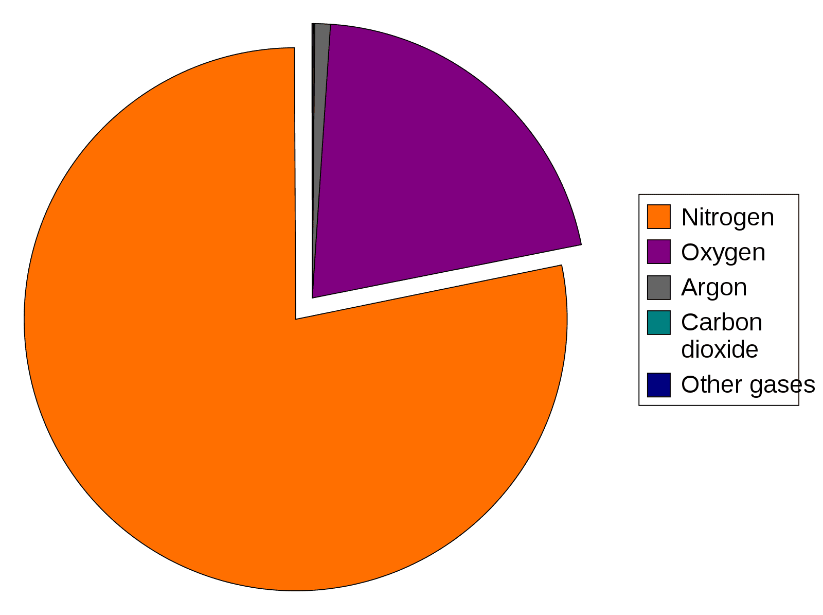
Image - The composition of Earth's atmosphere
Public Domain Source by Dbc334 [Public domain]
The atmospheric pressure is 101 kilopascals (kPa) and air contains 20.9% of oxygen. So the partial pressure of oxygen (pO2) in atmospheric air is 101 x 0.209 = 21.1 kPa. The other ~80 kPa is made up of the partial pressures of other gases in the atmosphere, as shown in the diagram above.
At higher altitudes, the atmospheric pressure is lower, meaning the molecules of gases are further apart, so there are fewer oxygen molecules available for us to breathe. This helps explain why many climbers use supplemental oxygen when ascending very high altitudes such as Mount Everest.
Gases move down a pressure gradient from high partial pressure to low partial pressure; this explains why oxygen moves from the alveolus (high) into the blood (low).
Partial Pressure of a Gas in a Liquid
When a gas comes into contact with a liquid, the gas molecules dissolve into the liquid. Some of the molecules already in the liquid will evaporate, becoming gases. When the rate of the gas entering the liquid is equal to the rate of gas leaving the liquid, the system is said to be in equilibrium.
The partial pressure of a gas in a liquid is the partial pressure that the same gas would have in the air that is in equilibrium with the liquid, at the same temperature.
The partial pressure of a gas in blood matters in medicine. In the alveoli, the partial pressure of oxygen is about 13.3 kPa. This ability of oxygen to diffuse into a liquid and form an equilibrium means that the partial pressure of oxygen in blood leaving the lungs approaches this value.
The amount of a gas dissolved in the liquid and the partial pressure of a gas are two different measurements.
The content of a gas in blood is the amount carried per unit volume: the amount physically dissolved plus the amount bound to components such as haemoglobin. Dissolved concentration and partial pressure are different quantities; a pressure in kPa cannot be added directly to a concentration in mmol/L.
At equilibrium, dissolved gas concentration = partial pressure × solubility coefficient (Henry’s law). The solubility coefficient shows how much gas dissolves per unit of pressure. For a specified gas, solvent and temperature, the coefficient relates concentration to pressure; when concentration is in mmol/L and pressure is in kPa, its units are mmol L−1 kPa−1.
At 37 °C, a commonly used oxygen solubility coefficient is approximately 0.003 mL O2 dL−1 mmHg−1. Expressing the gas volume at standard dry conditions (0 °C and 101.325 kPa), this corresponds to approximately 0.01 mmol L−1 kPa−1. At a pO2 of 13.3 kPa (about 100 mmHg), the dissolved oxygen concentration is therefore approximately 0.3 mL O2/dL, or 0.13 mmol/L. Only a small proportion of blood oxygen is carried in this dissolved form.
Oxygen can also be bound to haemoglobin in the blood to be transported around the body. The content of oxygen in blood is therefore the amount of oxygen bound to haemoglobin plus the amount of oxygen dissolved in the blood.
Normal haemoglobin (Hb) concentration is around 2.2 mmol/L. Each molecule of haemoglobin can bind to four oxygen molecules. So the most oxygen that can be bound to haemoglobin is 8.8 mmol/L (4 x 2.2), when the haemoglobin is fully saturated. Overall, the total oxygen content is then 8.93 mmol/L (8.8 + 0.13). This shows why it is important to have haemoglobin molecules in the blood, as they are responsible for most of the oxygen transport in the blood.
Once oxygen has dissolved in the blood, it can enter red blood cells to bind with haemoglobin molecules. After the haemoglobin molecule is fully saturated with four oxygen molecules, oxygen can continue to dissolve into the blood until the pO2 of plasma equals pO2 of alveolar air.
When gas molecules encounter water in the airways they dissolve, and water molecules evaporate to enter the air. An equilibrium is set up when the rate of evaporation is equal to the rate of water vapour returning to the liquid phase. Once equilibrium has been reached, the gas mixture inside the airways is saturated with water vapour and there is a pressure exerted by the water vapour. This pressure is called saturated vapour pressure and it is dependent on body temperature.
The total pressure in the airways is still equal to atmospheric pressure (101kPa). However, in the airways, water is evaporating and condensing at equal rates (equilibrium), and the water vapour contributes 6.28 kPa of pressure. This means that the other gases make up the remainder: 94.7 kPa (101 - 6.28). The other gases are in the same proportion in the lungs as in the atmosphere meaning the pO2 of the humidified air in the airways = 19.8kPa (94.7 x 0.209).
This means that the partial pressure of oxygen changes as it moves from the nostrils to the alveoli. In the nostrils and upper respiratory tract, the inhaled air becomes humidified.

Diagram - The distribution of gas and water partial pressure
SimpleMed original by Dr. Peter Parkinson
Partial Pressure of Gases in the Alveoli
The partial pressure of oxygen (pO2) in the alveolus is set by two factors. These are the rate at which oxygen diffuses into the blood and the rate at which ventilation replaces it. The ideal pO2 in the alveoli is around 13.3 kPa and it is the balance between perfusion and ventilation that maintains this value. If a person hyperventilates (breathes faster and deeper than the body needs) or hypoventilates (breathes less than the body needs), the pO2 in the alveolar gas will change.
The normal value for the partial pressure of carbon dioxide (pCO2) in the alveoli is 5.3 kPa. This is determined by the rate at which CO2 enters the alveoli from the blood and the rate at which CO2 is breathed out. Again, if a person hyper- or hypoventilates, the pCO2 will change.
Partial Pressure of Gases in the Venous System
Venous blood that reaches the pulmonary capillaries has a pO2 of 6kPa and pCO2 of 6kPa. At the alveolar capillary membrane, gas exchange takes place and the blood equilibrates with the alveolar air, meaning the blood leaving the lungs for the heart has a pO2 of 13.3kPa and a pCO2 of 5.3kPa. This change is what allows the lungs to supply the body with oxygen and to get rid of the carbon dioxide produced.

Diagram - The partial pressures in the respiratory tract and the pulmonary capillaries
SimpleMed original by Dr. Peter Parkinson
The rate at which oxygen and carbon dioxide diffuse between the blood and the alveolar space is determined by three factors:
1. Area Available for Gaseous Exchange
The area for gas exchange is around 70 m2 and is formed by the large alveolar surface and the huge number of alveoli in the lungs.
This can be affected by lung diseases like emphysema as there is destruction of the alveolar walls and this reduces the total surface area available for gas exchange.
2. Diffusion Barriers
Even though the distance between the alveolar gas and the alveolar capillary blood is short, there are a number of structures between the two. Gas must cross every one of them, and a thicker barrier slows diffusion. The structures are:
- Alveolar epithelial cell
- Interstitial fluid (the thin layer of tissue fluid between the cells)
- Capillary endothelial cell (the cell that lines the capillary)
- Plasma
- Red cell membrane
Gas exchange occurs mainly across the thin side of the alveolar-capillary membrane. This has three layers: a single layer of alveolar epithelium, a basement membrane (a thin sheet of supporting material) and a single layer of capillary endothelium. The plasma and the red cell membrane lie beyond it, in the blood.
The resistance to diffusion is not the same for oxygen and carbon dioxide; the rate of diffusion for the gases is affected by the solubility of the gases in water. Carbon dioxide is more soluble than oxygen and therefore diffuses across the barriers at a faster rate. As a result, oxygen diffusion is affected more in diseased states such as interstitial lung disease, where there is excessive deposition of collagen and extracellular matrix (the supporting material between cells) in the interstitium (the thin layer of tissue between the alveoli and the capillaries). This process increases the distance of the diffusion pathway and thickens the walls of alveoli. Conditions like pulmonary oedema also increase the diffusion pathway because there is fluid in the alveolus.
3. Gradient of Partial Pressure
Each gas diffuses down its own gradient of partial pressure, not down a gradient of concentration. To allow oxygen to diffuse into the blood and carbon dioxide to diffuse out, a steep partial pressure gradient is beneficial. For oxygen, the gradient is large: the alveolar pO2 is 13.3 kPa and the blood arriving at the lungs has a much lower pO2. For carbon dioxide, the gradient is small: the alveolar pCO2 is 5.3 kPa and the blood arriving at the lungs has a slightly higher pCO2. A small gradient is enough for carbon dioxide because it is much more soluble than oxygen (about 23 times more soluble in water), so it diffuses across the barriers faster.
Oxygen is not soluble enough in blood for this to be the only mechanism of transport around the body. This means that an alternative mechanism of oxygen transport is needed. The protein, haemoglobin, found in red blood cells reversibly loads (picks up) and unloads (releases) oxygen within a very narrow range of partial pressures of oxygen. Myoglobin is only found in muscle tissue and acts as a storage molecule by only unloading oxygen at very low partial pressures of oxygen e.g. anaerobic conditions.
Myoglobin is a haem protein (a protein that contains haem, an iron-containing group) and is monomeric (single polypeptide chain). One myoglobin molecule comprises one haem group that contains an iron ion (Fe2+), which binds with an oxygen molecule. This lets myoglobin store oxygen in muscle.
Myoglobin is only found in muscle tissue and acts as a storage molecule for oxygen, which then can be released in times of oxygen deprivation. When oxygen is in high demand (like when exercising), the oxygen can be released from myoglobin and can allow the muscle cells to carry on generating energy.
Each myoglobin molecule has only one oxygen-binding site, so there is no cooperative binding (explained below in the haemoglobin section). This gives a hyperbolic curve (one that rises steeply, then flattens out) on an oxygen dissociation curve. This is a graph of the percentage saturation of myoglobin against the partial pressure of oxygen.
The graph below shows this hyperbolic curve when plotting the saturation of myoglobin against increasing partial pressures of oxygen. It shows that myoglobin has a high affinity for oxygen (it holds on to oxygen tightly). This is because the partial pressure of oxygen needed to occupy 50% of the myoglobin molecules is very low. This pressure is called the P50 and is shown in red on this graph.

Diagram - Myoglobin oxygen dissociation curve and the P50 of myoglobin (red line)
SimpleMed original by Dr. Peter Parkinson
Haemoglobin like myoglobin is a haem protein but is made up of four subunits. Each subunit contains a haem group containing an iron ion, which binds with the oxygen molecules. This means that each haemoglobin molecule can bind with a maximum of four oxygen molecules.
Because haemoglobin has four polypeptides (protein chains) joined together, which is called its quaternary structure, the binding of oxygen can cause changes within the structure of the molecule. Each haemoglobin molecule can exist in a tense (T) or a relaxed (R) state. When no oxygen molecules are bound to haemoglobin, it exists in the T state. This makes it difficult for the first oxygen molecule to bind. After one oxygen molecule binds to a haem group, the whole haemoglobin molecule settles into the R state (the R conformation, or shape). This makes it easier for the subsequent oxygen molecules to bind.
The relationship between oxygen and haemoglobin can be described using the haemoglobin-oxygen dissociation curve. This is a graph of haemoglobin saturation against the partial pressure of oxygen. The curve is sigmoidal (S-shaped), due to the cooperative binding of oxygen. Each oxygen molecule that binds makes it easier for the next one to bind. The first oxygen molecule binds with difficulty because of the low affinity for oxygen in the T state. Binding becomes easier for subsequent oxygen molecules because the haemoglobin molecule settles into the R state.
The oxygen–haemoglobin dissociation curve plateaus at high pO2 because haemoglobin has a finite number of oxygen-binding sites, most of which are already occupied. Further increases in pO2 therefore produce only a small increase in haemoglobin saturation. The plateau reflects near-complete site occupancy at equilibrium.
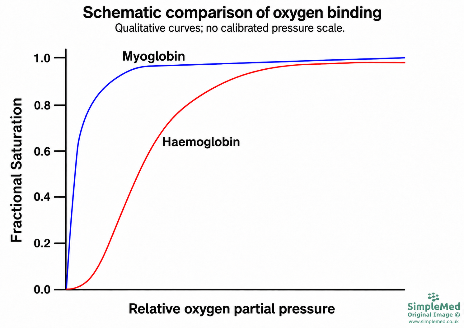
Diagram - Schematic oxygen-binding curves for myoglobin and haemoglobin. The horizontal axis is qualitative and is not a calibrated pressure scale; values cannot be read from this illustration.
SimpleMed original by Dr. Peter Parkinson

Diagram - P50 of myoglobin in comparison to haemoglobin
SimpleMed original by Dr. Peter Parkinson
Once the blood leaves the lungs, it is carrying nearly all the oxygen it can and has the pO2 of 13.3kPa (equal to the pO2 of the alveolar gas). Once it gets to the tissues, there is an increased amount of CO2 diffusing into the blood. This causes a fall in the pH of the blood (because CO2 reacts with H2O to form H+ and HCO3-). This change in pH encourages haemoglobin into the T state. As explained earlier, the T state lowers the affinity of haemoglobin for oxygen (haemoglobin holds on to oxygen less tightly). The haemoglobin molecule therefore releases the oxygen it carries into the blood, so that the oxygen can diffuse into the tissues.
In actively respiring tissues, there is a large production of carbon dioxide through increased metabolism and this will lower the pH of the local blood. This causes haemoglobin’s affinity for oxygen to decrease, meaning more oxygen will be provided to actively respiring tissues through increased unloading.
The overall physiological effect of carbon dioxide acting to decrease the pH and cause a decreased affinity of haemoglobin for oxygen is called the Bohr effect. This effect enhances the unloading of oxygen through metabolically active tissues and ensures the delivery of oxygen meets the demands of the tissues.
The Bohr effect will shift the oxygen dissociation curve to the right, increasing the P50 of haemoglobin, therefore, reducing the affinity of haemoglobin for oxygen. A good way to remember the direction of the shift is that the BohR shift moves the curve Right.

Diagram - The Bohr effect
Public Domain Source by Ratznium, edited by Dr. Peter Parkinson [Public domain]
Increased temperature can also shift the oxygen-haemoglobin curve to the right, because heat encourages haemoglobin into the T state. Metabolically active tissues are slightly warmer, so more oxygen is dissociated from haemoglobin there.
2,3-bisphosphoglycerate (2,3-BPG)
2,3-BPG is a molecule in the blood that acts as an allosteric effector (a molecule that binds away from the oxygen-binding site and changes how well haemoglobin binds oxygen) on haemoglobin. It can bind to the positively charged amino acids in the centre of the haemoglobin structure, encouraging stabilisation of haemoglobin in the T state. By binding to haemoglobin, 2,3-BPG lowers the affinity haemoglobin has for oxygen, meaning high concentrations of 2,3-BPG increase the amount of oxygen released in peripheral tissues.
There are situations where a person may have a higher than expected level of 2,3-BPG due to low tissue oxygen concentrations. This is beneficial because it encourages more oxygen to dissociate at the peripheral tissues. This may occur if the person has acclimatised (adapted) to living at high altitude, where the inspired pO2 is lower anyway. It may also occur in heart failure, where blood flow to the peripheral tissues is reduced.
Measuring Adequacy of Oxygenation
Two common ways to assess oxygenation are a pulse oximeter and an arterial blood gas.
The patient’s oxygen saturation can be measured by using a pulse oximeter, a non-invasive (it does not break the skin), quick technique that can monitor the percentage of haemoglobin saturated with oxygen. The instrument works because oxygenated haemoglobin and deoxygenated haemoglobin absorb different wavelengths of light.
It does have limits. The reading can be less reliable if a patient has poor circulation to their peripheries (the fingers, toes and earlobes where the probe is placed), for example when they are very unwell. Skin pigmentation, nail varnish, and the presence of methaemoglobin (an abnormal form of haemoglobin) or carboxyhaemoglobin (haemoglobin bound to carbon monoxide) can also affect the reading. In carbon monoxide poisoning the saturation may appear normal, because most pulse oximeters measure carboxyhaemoglobin together with oxyhaemoglobin. Pulse oximeters can also over-estimate the true oxygen level in people with darker skin tones. The reading can also be misleading in severe anaemia, as oxygen saturation may appear normal despite a reduced oxygen content due to low haemoglobin.
A sample of blood from an artery is taken, usually from the radial artery in the forearm. A machine then analyses the sample to measure the pO2, pCO2 and pH, and the concentration of certain ions. These measurements are very helpful when assessing how well the lungs are working and the acid-base balance of the blood, which depends on the lungs and kidneys. However, taking a blood sample from an artery can be painful and difficult to perform. It is used when the clinician needs information that a pulse oximeter cannot give, such as the pCO2 and the acid-base balance. One example is a patient at risk of hypercapnic respiratory failure (a raised pCO2 because the lungs cannot clear enough carbon dioxide).
To get enough oxygen to the peripheral tissues and to exhale all the waste carbon dioxide, the ventilation and the blood supply of the alveoli need to be well matched.
To calculate minute ventilation, multiply the tidal volume (the volume of each breath) by the respiratory rate. For the average adult (70 kg), minute ventilation is around 6 L/min. Some of each breath fills the dead space, which is the part of the airways that holds air but takes no part in gas exchange. The ventilation (V) used in the V/Q ratio is the alveolar ventilation, the air that reaches the alveoli. This is lower than minute ventilation, at approximately 4.2 L/min.
Perfusion (Q) of the lungs is defined as the total volume of blood that reaches the pulmonary capillaries in a given period of time.
The V/Q ratio ideally would be 1 because this would mean that the most efficient diffusion of oxygen and carbon dioxide was taking place. In reality, the V/Q ratio varies between regions of the lung. When a person stands upright, the V/Q ratio is high at the apex (the top of the lung) and low at the base (the bottom of the lung), because gravity increases perfusion towards the base. The overall V/Q ratio of the average lung is therefore closer to 0.8.
Ventilation-perfusion mismatch is the most common cause of hypoxaemia (a low level of oxygen in the arterial blood).
V/Q mismatch can occur due to reduced ventilation of regions of the lungs. There are some disorders that can result in poor ventilation to a region of alveoli, and these include:
- Early stages of asthma: airway narrowing secondary to smooth muscle contraction makes it more difficult to ventilate the alveoli.
- Early stages of chronic obstructive pulmonary disease (COPD): airway narrowing due to inflammation and loss of radial traction (the outward pull of the surrounding lung tissue that helps to hold the airways open) makes it harder to ventilate alveoli.
- Pneumonia: exudate (inflammatory fluid and cells) found in the alveoli increases diffusion distance and impairs delivery of oxygen to the alveolar membrane.
- Respiratory distress syndrome in neonates (newborn babies): alveoli are less compliant (stiffer and harder to inflate), meaning ventilation takes a lot of effort.
The poorly ventilated alveoli will have a V/Q ratio of < 1, which results in a decrease in pO2 of the affected alveoli. This low pO2 prompts hyperventilation (breathing faster and deeper than the body needs). The extra carbon dioxide is breathed out, so the pCO2 usually stays normal or falls. If the ventilation problem is severe enough, the pCO2 can rise instead.

Diagram - Ventilation-perfusion mismatch due to poor ventilation
SimpleMed original by Dr. Peter Parkinson
V/Q mismatch can also occur due to reduced perfusion to regions of the lungs. Perfusion to areas of the lungs can be reduced by a pulmonary embolus, a blood clot blocking a branch of the pulmonary artery. Ventilation to the region of the lung affected by the embolus is wasted as no gas exchange can occur there. This region is called alveolar dead space (alveoli that are ventilated but not perfused), and its V/Q ratio is greater than 1.
In poorly ventilated regions of the lungs, the body diverts blood to better ventilated areas through a process called ‘hypoxic vasoconstriction’ (narrowing of the small pulmonary arteries around alveoli with a low pO2). In pulmonary embolism, the embolus itself blocks blood flow to part of the lung, so blood is redirected to the unaffected regions. These regions receive more blood than their ventilation can match, so their V/Q ratio falls below 1. However, the blood that is supplying the functional regions is already saturated with oxygen (as is normal for any area of the lungs) meaning it cannot saturate further. The low pO2 of the blood leaving the lungs drives hyperventilation, which results in a low or normal pCO2 of the blood leaving the lungs.
Edited by: Dr. Maddie Swannack
Reviewed by: Dr. Thomas Burnell and Dr. Marcus Judge
- 10062