
Next Lesson - Pulmonary Embolism
Core
Lung function can be assessed without any needles or surgery (non-invasively) through spirometry, a breathing test in which the patient blows into a machine that records the volume and speed of the air. These tests are useful in the diagnosis and management of diseases of the respiratory system.
On a spirometry tracing, tidal volume (VT) is the volume of air entering and leaving the lungs during each normal breath. Quiet breathing is usually automatic and requires no deliberate maximal effort, but quiet inspiration still requires respiratory muscle activity. Breathing in and out in this way is called quiet inspiration and quiet expiration.
If a patient takes a deep breath in, the extra air they can breathe in above a normal tidal breath is known as the inspiratory reserve volume (IRV). It can be measured by asking the patient to breathe in normally (as for tidal volume), but then take a very big breath in. This action is called forceful inspiration, because the patient has to consciously move air into the lungs.
If the patient then breathes out more than normal, the extra air breathed out below a normal tidal breath is called the expiratory reserve volume (ERV). It is measured by asking the patient to breathe out normally (as for tidal volume), but then breathe out all of the air they can. This action is called forceful expiration, because the patient has to consciously move air out of the lungs.
After maximal forced expiration, gas remains in the lungs. This is the residual volume (RV). Simple spirometry cannot measure residual volume, because it records gas moving into or out of the lungs rather than the gas remaining inside.

Diagram - Schematic of lung volumes and capacities. Simple spirometry records moving volumes; residual volume, functional residual capacity and total lung capacity require additional measurement.
SimpleMed original by Dr. Peter Parkinson
A lung capacity is the sum of two or more lung volumes. Capacities do not change with the pattern of breathing, so two patients can have the same lung capacity but different values for breathing volumes like tidal volume (see above). Capacities can still change in disease.
The inspiratory capacity is defined from the end of quiet expiration to the maximum inspiration, and this can be calculated by the addition of inspiratory reserve volume and tidal volume.
The functional residual capacity (FRC) is the volume of air in the lungs at the end of quiet expiration. It can be calculated by the addition of expiratory reserve volume and residual volume.
Vital capacity (VC) is defined as the maximal volume of air that can be expelled from a patient’s lung after maximal inspiration. It is the maximum amount of air that a patient is capable of moving into or out of their lungs. The same value can be calculated in three equivalent ways, because each capacity is built from the same lung volumes:
- Inspiratory capacity plus expiratory reserve volume.
- Inspiratory reserve volume plus tidal volume plus expiratory reserve volume.
- Total lung capacity minus residual volume.
Total lung capacity (TLC) is the total volume of air present in the lungs after maximal inspiration. It can be calculated by adding the vital capacity and the residual volume.
Vital capacity is one of the most important measurements of the lungs from a spirometry tracing. A patient’s vital capacity can be predicted based on sex, height and age, and any deviation from this predicted value can indicate pathology.
Vital capacity requires a patient to inhale maximally then exhale maximally. This means that any pathology of the lungs (e.g. stiffness in fibrosis), muscles of breathing (e.g. neuromuscular disorders, where nerve or muscle disease causes weakness) or of the chest wall (e.g. kyphoscoliosis, a curve of the spine that bends the chest sideways and forwards) can affect the vital capacity. All of these conditions can reduce the vital capacity and give a restrictive pattern on spirometry (even if the cause is outside the lungs) because the lungs cannot fill effectively.
In obstructive lung disease, expiratory airflow is reduced. Vital capacity may remain near normal, particularly in mild disease, but air trapping (air that cannot be breathed out) can reduce the forced vital capacity (FVC). A reduced vital capacity therefore does not by itself establish restriction: a total lung capacity below the lower limit of normal (the lowest value expected in a healthy person of the same age, sex and height) is needed to confirm a restrictive ventilatory impairment, a pattern in which the lungs cannot expand fully.
To separate patients with restrictive lung disease from those with an obstructive lung disease, the patient takes a maximal inspiration and then exhales rapidly and forcefully into a single breath spirometer. The result can then be plotted on a time-volume graph, with the volume expired plotted against the time in seconds.

Diagram - Time-volume graph for a healthy individual
SimpleMed original by Dr. Peter Parkinson
To distinguish between restrictive and obstructive lung disease, measure the forced expiratory volume in one second (FEV1) and the FVC (the total volume blown out in that forced breath), then compare them as the FEV1/FVC ratio.
Interpret FEV1/FVC against an appropriate reference range: the ratio normally falls with age. A value below the lower limit of normal indicates airflow obstruction. For diagnosing COPD (chronic obstructive pulmonary disease), a post-bronchodilator FEV1/FVC below 0.70 is a commonly used fixed criterion. Post-bronchodilator means measured after an inhaled medicine that opens the airways. Interpret the result alongside symptoms and clinical context. It is not a universal rule for all ages or lung diseases.
In obstructive lung disease, such as COPD, FEV1 falls disproportionately to FVC, producing a ratio below the age-appropriate lower limit of normal. In asthma the airway obstruction is variable and reversible, so the ratio is low when the airways are narrowed and can return towards normal after a bronchodilator. FVC may remain near normal in mild disease, but can also fall because of air trapping.

Diagram - Time-volume graph for a patient with an obstructive lung disease
SimpleMed original by Dr. Peter Parkinson
In restrictive lung disease, such as pulmonary fibrosis, FVC and FEV1 may both fall, with a normal or increased FEV1/FVC ratio. Low FVC with a normal ratio suggests but does not confirm restriction; confirmation requires TLC below the lower limit of normal. A low FVC can also reflect poor effort or air trapping.
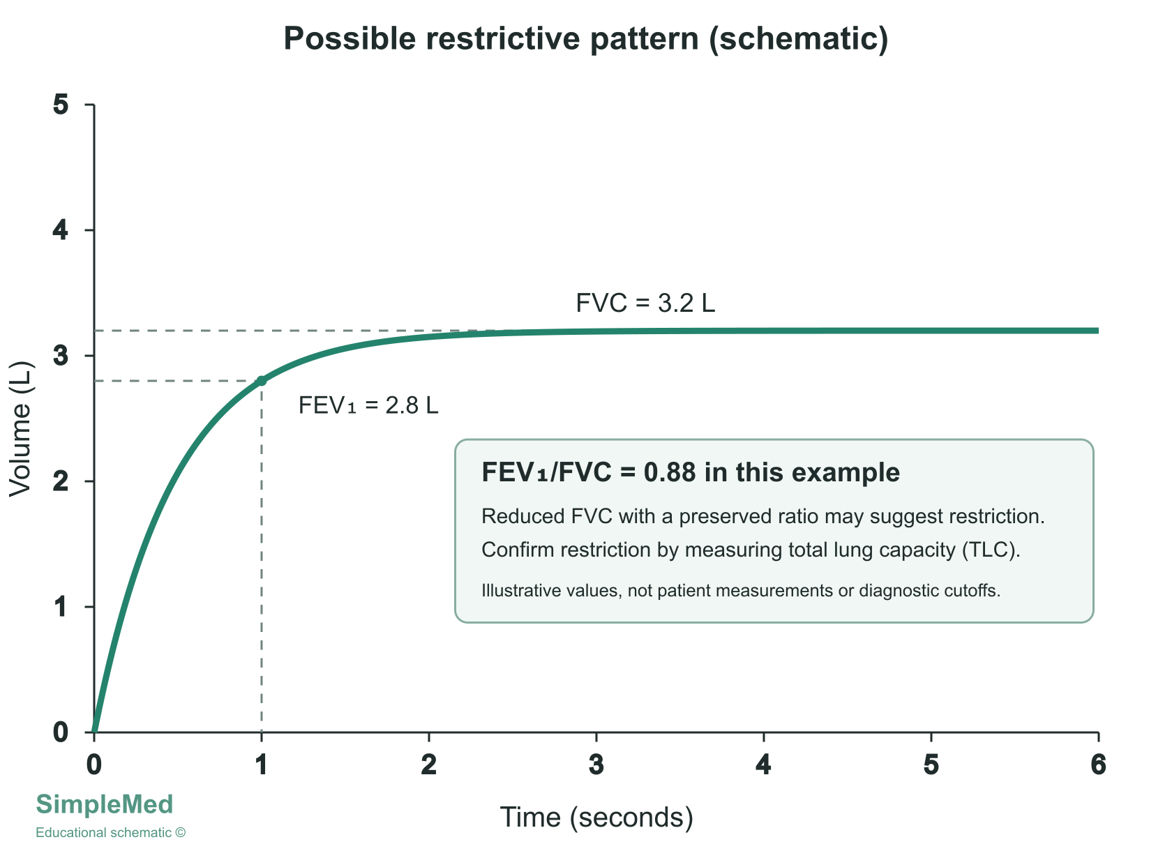
Diagram - Illustrative time-volume curve with FEV1 approximately 2.8 L and FVC 3.2 L (FEV1/FVC approximately 0.88). A reduced FVC with a normal ratio suggests restriction; low total lung capacity is required to confirm it. These illustrative values are not patient measurements or diagnostic cutoffs.
SimpleMed original by Dr. Peter Parkinson
Flow volume curves (also called flow-volume loops) plot airflow in litres per second against lung volume. The curve tracks the patient through full cycles of breaths, from complete exhalation through inhalation and back to exhalation again. They are useful in identifying obstructive lung diseases, because the shape of the curve shows how airflow changes as the lungs empty. They can also be used to assess whether airflow is appropriate for the patient’s lung volume.
For example, patients with a restrictive disease such as pulmonary fibrosis have a reduced lung volume. If airflow is measured alone (as on a time-volume graph), it appears decreased. When airflow is measured together with lung volume (as on a flow-volume curve), it is higher than normal for that lung volume.
When the lungs are expanded at the start of expiration, the airways are stretched fully open and the peak expiratory flow can be seen. During expiration, the radial traction (the outward pull on the airways that keeps them open) decreases. The small airways are then compressed, causing a decrease in the flow of air.
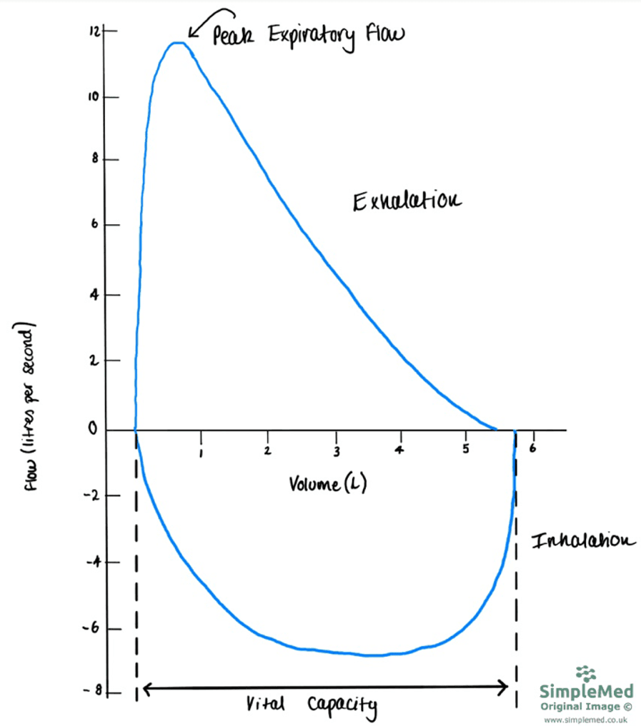
Diagram - Flow-volume curve for a healthy individual. Note how inhalation has a negative flow, and exhalation has a positive flow
SimpleMed original by Dr. Peter Parkinson
When there is an obstruction of the small airways, an early fall in the flow rate is seen. Mild obstruction of the small airways (as seen in asthma) can lead to scalloping (also called spooning: a scooped-out, concave downslope) on the flow-volume curve. In severe cases, a reduction in the peak expiratory flow rate is also seen (the loop is lower).
In patients with an obstructive lung disease, reversibility testing can be conducted. This involves giving the patient an inhaled bronchodilator (a medicine that opens the airways) to assess how responsive the patient’s airway is to the medication. In asthma the obstruction is reversible. For adults, NICE (the National Institute for Health and Care Excellence) advises diagnosing asthma if the FEV1 rises by 12% or more and by 200 ml or more after a bronchodilator, but this is only one of several tests. Reversibility testing is not routinely needed to diagnose COPD.

Diagram - The differences in the flow volume curve of a healthy individual (blue) vs a patient with obstructive lung disease (green)
SimpleMed original by Dr. Peter Parkinson
In patients with restrictive lung disease, the flow volume curve will be narrower because the lungs struggle to fill completely. The width of the curve is the vital capacity, so a narrow curve means a lower vital capacity.

Diagram - Flow-volume curve for patient with a restrictive lung disease (pink) compared to a healthy patient (blue). Note the narrowed vital capacity in comparison to the normal flow-volume curve
SimpleMed original by Dr. Peter Parkinson
A patient’s peak expiratory flow rate (PEFR) can be measured in clinical practice with a cheap, simple device called a peak flow meter. Doctors in general practice can use these devices when they suspect a patient has asthma. If spirometry is not available or is delayed, NICE advises measuring peak flow twice daily for 2 weeks, and variation in the readings of 20% or more supports a diagnosis of asthma. In suspected COPD, serial home peak flow readings can help to exclude asthma if there is still doubt. Patients can also use these devices at home to monitor their condition. The patient’s PEFR value can be plotted on a graph to compare against their predicted value based on sex, age and height to assess the severity of the condition. In a patient who already has asthma, their own personal best (their highest usual reading) is also used to judge control and to set up an action plan.
It is important that healthcare professionals properly educate patients in how to use the peak flow meters so that the readings are accurate. To get an accurate measurement of the PEFR, the patient needs to blow a short sharp breath into the mouthpiece of the peak flow meter. It is important for clinicians to emphasise that it is the speed of the air that is important, not the volume.

Image - Peak flow meter
Public Domain Source [Public domain]
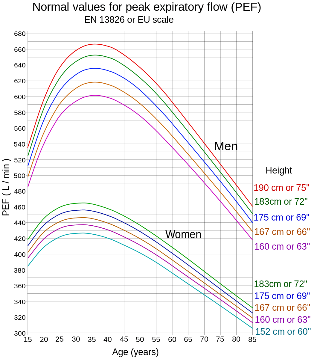
Image - Normal values for peak flow meter readings
Public Domain Source [Public domain]
The following tests are not performed routinely but can be useful when pathology is difficult to identify on other tests like spirometry.
A patient’s lung volumes, such as functional residual capacity, can be estimated using the helium dilution test, although values may be underestimated in obstructive lung disease. This test requires a closed-circuit system: the patient’s mouth is secured to the mouthpiece of the device so that no air can leak. The spirometer is filled with a mixture of oxygen and helium. There is a known concentration and volume of helium in the spirometer before the test begins. The patient breathes normally into the machine, and the helium spreads out into the air in their lungs, so its concentration falls (it is diluted). The lung volume is then calculated from how much the helium has been diluted.
The helium dilution test can be used to measure lung volumes when the airways are not obstructed, as in restrictive lung disease. In obstructive lung disease it can underestimate lung volumes, because helium may not reach air trapped behind narrowed airways. Body plethysmography (measuring lung volumes inside a sealed booth) is then preferred, because it can also assess trapped air.
Single-breath nitrogen analysis can estimate anatomical dead space (the part of each breath that fills the airways and takes no part in gas exchange) using Fowler’s method. After one breath of 100% oxygen, nitrogen concentration is plotted against expired volume. Oxygen-rich gas from the conducting airways (the tubes that carry air down to the alveoli) comes out first, followed by a transition to nitrogen-containing alveolar gas. This is different from multiple-breath nitrogen washout, in which the patient breathes oxygen over successive normal breaths while resident lung nitrogen is progressively cleared. Multiple-breath washout estimates functional residual capacity and assesses ventilation inhomogeneity (uneven ventilation between different regions of the lung); slowly ventilated regions can prolong washout. Nitrogen is found throughout the lungs, not only in the conducting airways, so washout clears it from every ventilated region.
To assess how well gas moves from the air sacs (alveoli) across the alveolar membrane into the blood, carbon monoxide transfer factor (TLCO) can be used. The assessment measures the overall lung’s ability to transfer gas. Carbon monoxide (CO) is used because it binds very strongly to haemoglobin in red blood cells, so its uptake mainly reflects how easily gas crosses the alveolar membrane. The result is adjusted for the patient’s haemoglobin level and for any CO already in the blood (carboxyhaemoglobin). The patient breathes in a test gas that contains a small, fixed concentration of carbon monoxide, holds their breath for about 10 seconds, and then breathes out. The fall in CO concentration between the gas breathed in and the gas breathed out is used to estimate how quickly CO is transferred into the blood.
The TLCO test is used to monitor lung conditions such as pulmonary fibrosis and COPD (because these conditions affect the amount of oxygen absorbed) and can be used to assess how well a patient’s lungs will react to chemotherapy. A raised value (i.e. more gas transfer) can be seen in obesity (usually a small rise) and in asthma. It can also be seen when more blood flows through the lungs, when there are more red blood cells than normal (erythrocytosis), or when there is bleeding into the lungs (alveolar haemorrhage), because blood in the airways also binds CO. A decreased value can be seen in pulmonary fibrosis (a thickened membrane), a smaller gas-exchange surface (for example in emphysema, a type of COPD), anaemia and pulmonary embolism (a blockage in the lung’s blood vessels).
Edited by: Dr. Maddie Swannack
Reviewed by: Dr. Thomas Burnell and Dr. Marcus Judge
- 9064