

Next Lesson - Adrenergic Pharmacology
Core
Acetylcholine Life Cycle
Cholinergic pharmacology becomes manageable when every drug is placed on one transmitter cycle. In the nerve terminal, choline acetyltransferase joins choline to acetyl-CoA to make acetylcholine (ACh). The vesicular acetylcholine transporter, VAChT, concentrates ACh inside synaptic vesicles. An arriving action potential opens voltage-gated calcium channels; calcium then triggers SNARE-dependent vesicle fusion and quantal exocytosis. Released ACh crosses only a tiny cleft before binding nicotinic or muscarinic receptors.
Termination is unusually fast because acetylcholinesterase (AChE) hydrolyses ACh to acetate and choline within the cleft. The transmitter itself is not simply taken back intact. High-affinity choline transport returns choline to the terminal for reuse. This sequence creates five conceptual intervention points: choline uptake, synthesis, storage, release and breakdown. Clinically familiar drugs concentrate on receptors and AChE, but knowing the whole cycle explains why inhibiting storage or release decreases every downstream cholinergic response, whereas inhibiting AChE increases ACh wherever the inhibitor can reach an active cholinergic synapse.
Experimental tools make the sequence tangible. Hemicholinium limits high-affinity choline uptake, vesamicol blocks VAChT, and botulinum toxins cleave selected SNARE proteins and prevent vesicle fusion. These are not interchangeable “anticholinergics”: one restricts precursor recycling, one empties the future vesicle pool, and one prevents already filled vesicles from releasing. Their effects emerge at different speeds because terminals can temporarily draw on existing choline, cytosolic ACh or stored vesicles. A receptor agonist can still activate a postsynaptic cell despite an upstream release lesion, which is a useful way to localise a defect experimentally.
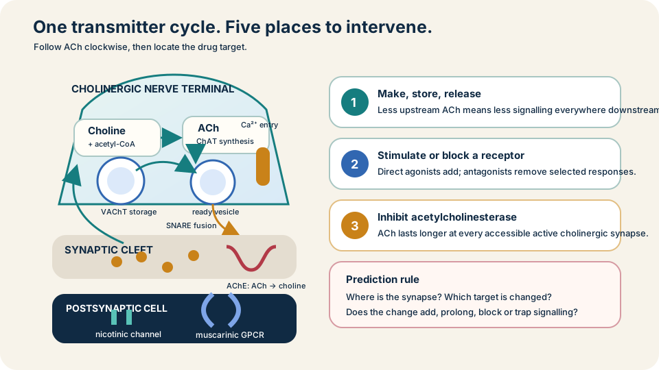
Follow ACh from recycled choline to receptor activation, then place each drug at its point of action.
SimpleMed original educational diagram
Locate the Receptor
Nicotinic receptors are ligand-gated cation channels, so they produce rapid depolarisation. Neuronal Nn receptors sit on postganglionic neurons in both sympathetic and parasympathetic ganglia and on adrenal-medullary chromaffin cells. Muscle Nm receptors sit at the skeletal neuromuscular junction. A drug at Nn can therefore disturb both autonomic divisions; a drug selective for Nm changes skeletal-muscle transmission without directly blocking muscarinic organ responses.
Muscarinic M1-M5 receptors are G-protein-coupled receptors. M1 is prominent in neural and enteric signalling; M4 and M5 are mainly emphasised in central pathways. At preclinical level, M2 and M3 do most of the peripheral predictive work. Preganglionic autonomic fibres release ACh onto Nn in both divisions. Most postganglionic parasympathetic fibres release ACh onto muscarinic receptors at the organ. Somatic motor fibres release ACh onto Nm. The memorable exception is the sympathetic postganglionic supply to eccrine sweat glands: it is cholinergic and acts mainly through M3. This lesson uses that map pharmacologically; the pathway anatomy remains in Autonomic Nervous System: Introduction.
Location also separates speed from direction. Nicotinic channel opening is always a fast excitatory event at the receptor, although the downstream pathway can later increase or decrease an organ's activity. Muscarinic receptors signal more slowly and can either excite or inhibit according to subtype and cell machinery. Calling ACh simply “excitatory” or “parasympathetic” therefore loses the information needed to predict a drug response.
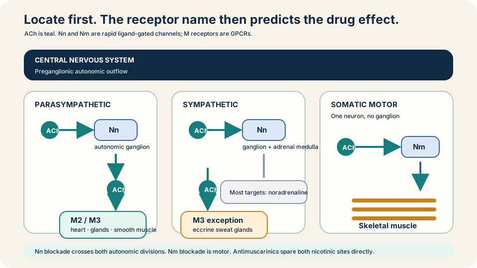
Location predicts consequence: Nn links autonomic neurons, Nm drives skeletal muscle, and muscarinic receptors control target organs.
SimpleMed original educational diagram
M2 and M3 Signalling
M2 and M4 preferentially couple to Gi/o. In sinoatrial and atrioventricular nodal cells, M2 activation reduces adenylyl cyclase activity and cAMP, and G-protein subunits open GIRK potassium channels. Hyperpolarisation slows pacemaker firing and AV conduction. The useful prediction is not that every muscarinic receptor excites: cardiac M2 activation restrains electrical activity.
M1, M3 and M5 preferentially couple to Gq/11. M3 activates phospholipase C, producing IP3 and raising intracellular calcium. In most target tissues that means glandular secretion or smooth-muscle contraction: miosis and near accommodation in the eye, bronchoconstriction and airway secretion, increased gastrointestinal motility and secretion, and detrusor contraction that favours bladder emptying. The vascular endothelium is the high-yield exception. Endothelial M3 calcium activates nitric oxide synthase; nitric oxide diffuses to adjacent vascular smooth muscle and relaxes it through cGMP. The receptor is still Gq-coupled, but the observed tissue response is relaxation because the signalling cell and responding muscle are different.
This pathway logic is more reliable than memorising “odd receptors excite, even receptors inhibit”. M2 can also regulate transmitter release and smooth muscle outside the heart, while M3 expression and tissue architecture shape the final response. The coupling rule is a first prediction, not a claim that every receptor has one effect in every cell.
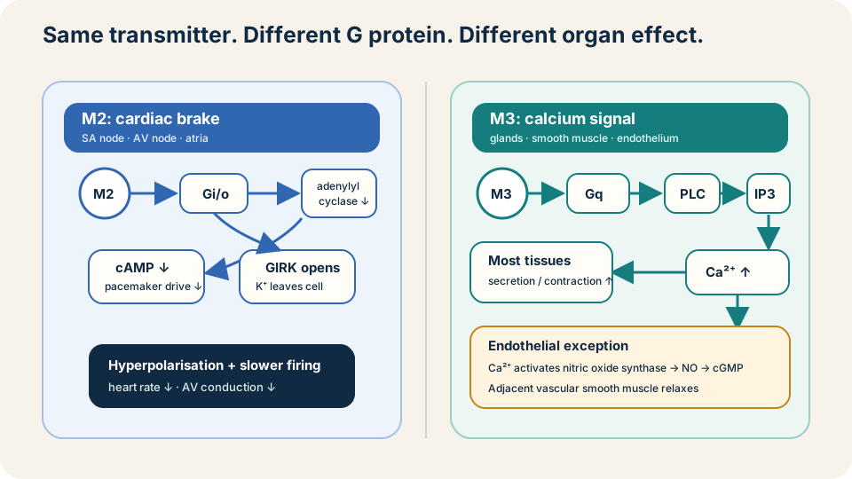
M2 usually slows through Gi and potassium current; M3 raises calcium, with endothelial nitric oxide creating a vascular exception.
SimpleMed original educational diagram
Direct Agonists and Enzyme Inhibitors
Direct muscarinic agonists bind the receptor themselves. Bethanechol favours muscarinic effects and can promote detrusor and gastrointestinal smooth-muscle activity; pilocarpine drives secretion and, in the eye, miosis with ciliary-muscle contraction. Their adverse effects are extensions of the same receptor map: bradycardia, sweating, salivation, abdominal cramping, diarrhoea, urinary urgency, bronchoconstriction and secretions. Obstruction, marked bradycardia or reactive airway disease can therefore make added muscarinic drive hazardous. These are mechanism predictions, not a complete prescribing checklist.
Indirect agonists inhibit AChE rather than stimulating one receptor subtype. Neostigmine and pyridostigmine prolong endogenous ACh at accessible cholinergic synapses, so both muscarinic and nicotinic responses can increase. That explains useful strengthening of neuromuscular transmission alongside unwanted bradycardia, secretions or gut activity. Physostigmine enters the central nervous system more readily than quaternary agents such as neostigmine; distribution therefore changes which synapses are amplified. The existing Parkinson's Disease and Myasthenia Gravis lesson retains disease-specific treatment detail.
Direct and indirect agonism also differ in dependence on nerve activity. A receptor agonist supplies a signal even when little ACh has just been released. An AChE inhibitor mainly magnifies transmitter present after endogenous impulses, so its pattern depends on active synapses, tissue access and whether the molecule reaches the brain. Neither drug class is automatically selective simply because one clinical use dominates its name.
Antimuscarinics
Antimuscarinics competitively prevent ACh from activating muscarinic receptors while leaving Nm and Nn transmission directly intact. Atropine removes vagal M2 restraint from the heart and reduces muscarinic secretions. Ipratropium and tiotropium reduce airway M3-mediated bronchoconstriction; tropicamide relaxes the iris sphincter and ciliary muscle; oxybutynin reduces detrusor drive. The same logic predicts class effects: dry mouth, reduced sweating, mydriasis with blurred near vision, tachycardia, reduced gut motility and urinary retention. Central penetration can add confusion, especially in susceptible older people.
Risk follows physiology. Blocking detrusor contraction can worsen urinary retention; reducing gut movement can worsen obstruction; mydriasis can be dangerous in susceptible narrow angles; reducing sweating can impair heat loss. A drug may be selected for one organ, but receptor distribution creates effects elsewhere. “Anticholinergic” is sometimes used loosely, so state the actual target: an antimuscarinic does not directly block nicotinic weakness at the neuromuscular junction or autonomic ganglion.
Exposure and distribution change the balance. An inhaled antagonist is delivered toward the airway, a short-acting ocular preparation is used locally, and a lipid-soluble systemic drug can reach central receptors. Local delivery reduces but never makes off-target effects conceptually impossible. Always combine receptor selectivity with route, concentration and access to the relevant compartment.
Neuromuscular Blockers
At the neuromuscular junction, motor-neuron ACh opens postsynaptic Nm channels. The endplate depolarises, reaches threshold and triggers a muscle action potential. Non-depolarising blockers such as rocuronium competitively occupy Nm receptors without opening them. Fewer channels respond to ACh, the endplate potential fails to reach threshold, and flaccid paralysis follows. Raising cleft ACh with an AChE inhibitor can oppose suitable non-depolarising blockade, but also raises muscarinic signalling elsewhere.
Suxamethonium (succinylcholine) is different: it is an Nm agonist that depolarises the endplate. Initial activation can cause fasciculation, but persistent depolarisation prevents the membrane from resetting; with continued exposure, desensitisation contributes to transmission failure. Both classes produce paralysis without unconsciousness, analgesia or amnesia. Practical selection, monitoring and reversal belong in Anaesthetics; the pharmacological distinction here is competitive silence versus depolarising activation followed by failure.
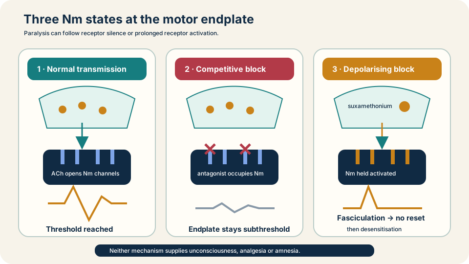
Non-depolarising drugs prevent Nm activation; suxamethonium activates first, then traps the endplate in transmission failure.
SimpleMed original educational diagram
Organophosphate Poisoning
Organophosphates phosphorylate AChE, allowing ACh to accumulate at muscarinic, nicotinic and central synapses. Muscarinic excess produces salivation, lacrimation, sweating, bronchorrhoea, bronchoconstriction, bradycardia, gastrointestinal hyperactivity, urination and miosis. Nicotinic excess can cause fasciculation followed by weakness and respiratory-muscle paralysis. Central effects can include agitation, confusion, seizures and depressed consciousness. Respiratory failure can therefore combine secretions and bronchospasm with weak ventilation and central impairment.
The treatment logic has separate jobs. Immediate decontamination, airway support, oxygenation and ventilation address exposure and physiology. Atropine competitively blocks muscarinic receptors, so it treats bronchial secretions, bronchoconstriction and other muscarinic effects; it does not restore Nm transmission directly. Pralidoxime can remove the phosphate group and reactivate AChE before the inhibited enzyme undergoes ageing, a chemical stabilisation that makes reactivation much harder or impossible. Timing matters mechanistically, but this is not a dose protocol. Suspected poisoning needs urgent specialist toxicology and emergency care.
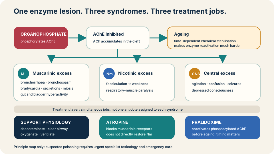
Support breathing first; atropine blocks muscarinic excess, while pralidoxime targets unaged phosphorylated AChE.
SimpleMed original educational diagram
The Three-Question Method
For any unfamiliar cholinergic problem, ask three questions in order. First, where is the synapse: autonomic ganglion, parasympathetic target, sympathetic sweat gland, neuromuscular junction or central pathway? Second, which receptor or enzyme is involved: Nn, Nm, muscarinic subtype or AChE? Third, is ACh signalling being added, prolonged, blocked or converted into persistent depolarisation? The phenotype then follows from ordinary physiology.
This method prevents common category errors. AChE inhibition is broader than a selective muscarinic agonist. Atropine can dry secretions while nicotinic weakness persists. An Nm blocker paralyses skeletal muscle without directly removing consciousness. M3 activation usually contracts smooth muscle but relaxes vessels indirectly through endothelial nitric oxide. Locate, name, predict: that sequence turns a long drug list into a small set of reusable mechanisms and prepares the contrast with Adrenergic Pharmacology.
Reviewed by: Dr. Marcus Judge
- 131
