

Next Lesson - The Basics of Pathology
Core
Magnification and Resolution
An unfamiliar histology slide is read as a map, not as a random zoom into colour. Begin at low power so the whole section is visible. Ask first what organ fragment or tissue plane you have been given, then where free surfaces, lumina, capsules and large vessels lie. Only after that frame is stable should the objective be increased. High power without orientation produces confident descriptions of the wrong structure.
Magnification and resolution are related but not identical. Magnification is how much larger the image appears than the object. Resolution is the smallest distance at which two points remain distinguishable. Empty magnification enlarges blur without revealing new detail. Work low to high: survey architecture, choose a relevant region, then use higher power to confirm nuclei, cell borders, fibres or inflammatory cells.
A reliable sequence follows that optical logic. Start wide. Find an edge or lumen. Identify the lining cells. Inspect the supporting tissue. Only then name the tissue or organ region. The order works across organs because every section is a two-dimensional cut through a three-dimensional structure. Fixation, embedding and microtomy are covered in An Introduction to Pathology; here the task is to read a stained slide without inventing disease from colour alone.
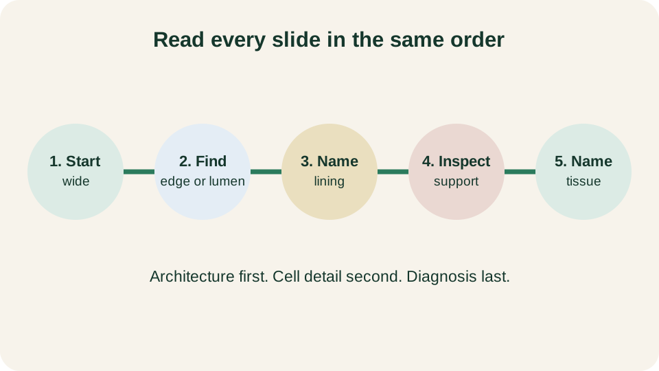
Low-to-high workflow: survey the section, locate an edge or lumen, identify the lining, inspect the support, then name the tissue.
SimpleMed original educational diagram
Haematoxylin and Eosin
Haematoxylin and eosin (H&E) is the default stain of routine histology because it separates acidic and basic tissue chemistry into readable colours. Haematoxylin behaves as a basic dye and binds acidic components, especially DNA and RNA. Nuclei therefore appear blue to purple, a property called basophilia; RNA-rich cytoplasm can also be basophilic.
Eosin is an acidic dye that binds basic, protein-rich material. Cytoplasm, collagen, muscle fibres and many extracellular matrix proteins stain pink to red and are described as eosinophilic. Intensity is not a diagnosis: processing and tissue composition both affect it. Read colour as chemistry first, then architecture, comparing structures within the same slide.
H&E shows nuclei and protein bulk well, making it ideal for first-pass morphology. It is weaker for some carbohydrates, lipids, fine reticulin frameworks and organisms; special stains can target these later. Ask which structure is basophilic, which is eosinophilic, and whether that distribution matches the tissue plan.
Orientation and Artefact
Every slide is a plane of section through a solid object. A tube cut longitudinally shows elongated lumina and long walls; the same tube cut transversely shows rings. Folded mucosa can look multilayered, while tangentially cut glands may appear solid or incomplete. Before calling a pattern abnormal, reconstruct the likely three-dimensional shape and knife angle.
Processing introduces artefacts that can mimic pathology. Folds produce dark bands where the section doubles. Chatter creates regular parallel ridges. Shrinkage can pull epithelium from stroma, leaving false clefts. Tears lack a biological border, while crush flattens nuclei and smears chromatin. A finding confined to one edge, fold or poorly fixed strip should be checked in well-preserved fields before it is accepted as biological.
Describe conservatively: record what is present, where it sits and whether it is consistent across the section. Do not upgrade a processing defect into inflammation, necrosis or neoplasia. Preparation is summarised in An Introduction to Pathology; here the skill is not to confuse laboratory effects with disease.
Four Tissue Families
Adult histology is organised into four tissue families: epithelium, connective tissue, muscle and nervous tissue. Most organs are assemblies of these families in repeating architectural patterns. Epithelium covers surfaces and lines cavities and forms many glands. Connective tissue provides support, packing, vessels and specialised matrices such as bone, cartilage and blood. Muscle generates force through contractile proteins. Nervous tissue conducts and integrates signals through neurons and their supporting glia.
On a new slide, assign large regions to families before naming an organ. A continuous polarised sheet on a free surface is epithelium. Fibrous tissue with vessels and scattered cells is connective tissue, often called stroma when it supports epithelium. Elongated eosinophilic fibres suggest muscle; neuropil, axons or ganglion cells point to nervous tissue. This family-level sort prevents premature diagnoses.
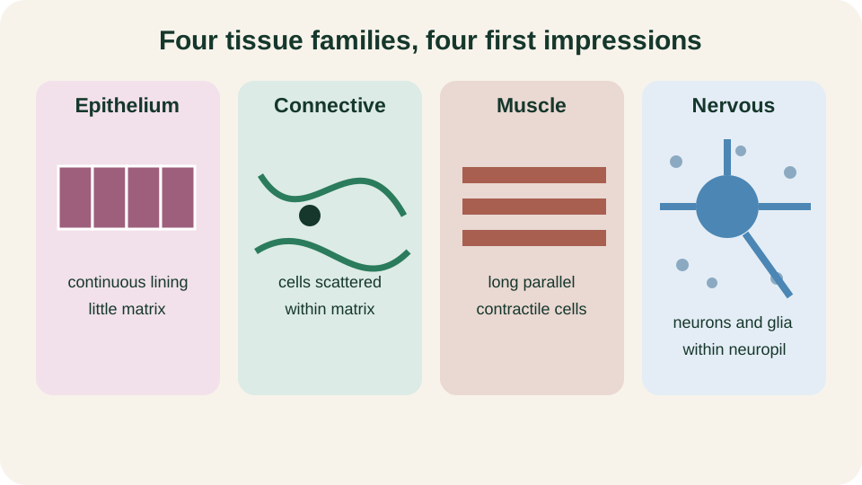
Comparative patterns of the four tissue families: polarised epithelium, fibrous stroma, contractile muscle and nervous tissue in context.
SimpleMed original educational diagram
Epithelium and Stroma
Epithelium is defined by cell-to-cell cohesion, polarity and attachment to a basement membrane rather than by a single cell shape. The apical surface faces a lumen or free exterior; the basal surface rests on basement membrane; lateral borders form junctions with neighbours. Shape terms such as squamous, cuboidal and columnar, and layering terms such as simple and stratified, refine the description once polarity is clear. Detailed epithelial taxonomy belongs with surface specialisations and glands in later lessons; the slide-reading task is to recognise a lining or covering sheet and to state how many layers and what surface features are visible.
Stroma is an organ's supporting connective tissue framework. Beneath epithelium it commonly contains basement membrane, loose connective tissue, vessels and fibroblasts; deeper scaffolding may be dense connective tissue, cartilage or bone. Epithelium is avascular and depends on diffusion from stromal vessels. After identifying a lining, ask whether its stroma is loose or dense, vascular or fibrotic, inflamed or quiet, and whether the interface is sharp. Claims of breach or invasion require more than one suspicious field.
Muscle and Nervous Tissue
Muscle is recognised by abundant eosinophilic contractile cytoplasm and nuclei whose position and shape fit the subtype. Skeletal muscle fibres are long, cylindrical and multinucleate, with peripheral nuclei and cross-striations visible when the plane of section and stain quality allow. Cardiac muscle shows striations too, but fibres branch, nuclei are usually central, and intercalated discs may be seen as transverse junctions between cells. Smooth muscle lacks striations; cells are spindle-shaped with single central nuclei and form sheets in viscera and vessel walls. If nuclei are crushed or the section is oblique, use neighbouring context: attachment to bone or dermis favours skeletal muscle; myocardium favours cardiac muscle; muscularis of gut or media of artery favours smooth muscle.
Nervous tissue is read in context more than by a single nuclear shape. In the central nervous system, neuropil forms a finely textured eosinophilic background with scattered glial and neuronal nuclei; large neurons may show prominent nucleoli and Nissl-rich cytoplasm. In the peripheral nervous system, nerves appear as bundled axons with Schwann-cell nuclei and variable myelin, often beside vessels and connective-tissue sheaths. Ganglia contain large neuronal cell bodies with satellite cells. Do not label every elongated nucleus as a nerve; fibroblasts and smooth muscle can look similar until the bundle pattern, myelin tinctorial quality or organ location is considered. Family assignment first, subtype second, clinical diagnosis later.
Special Stains
Special stains answer targeted questions after H&E has framed the anatomy. They are not a second random colour atlas. Periodic acid-Schiff (PAS) highlights neutral polysaccharides and some mucins, so basement membranes, glycogen-rich cytoplasm and certain fungal walls become clearer. Trichrome methods contrast collagen with muscle and other elements, helping separate fibrosis from residual parenchyma. Reticulin stains outline fine connective-tissue frameworks, useful where architecture rather than bulk collagen is the issue. Immunohistochemistry uses antibodies to detect specific antigens and can support cell lineage or protein expression, but the brown or red chromogen is only as meaningful as the antibody, controls and morphological context.
Colour alone is never the diagnosis. A PAS-positive blob could be glycogen, mucin, basement membrane or contaminant depending on digestion steps and location. Trichrome-blue fibrosis must still be mapped to an anatomical compartment. An immunohistochemical label that does not match the H&E cell of interest is a laboratory or interpretation problem, not a new disease. Use special stains to test a hypothesis generated at low and high power on H&E: Is this matrix collagen-rich? Is a basement membrane intact? Does this cell express a marker expected for its family? Keep the question narrow and the morphology primary.
A Repeatable Description Method
A neutral description sequence prevents both silence and overcalling. First state the specimen type and overall architecture at low power: solid, cystic, mucosal, encapsulated or fragmented. Second, locate landmarks: free surfaces, lumina, vessels, septa and any obvious tissue interfaces. Third, identify linings and name their epithelial pattern only as far as the image supports. Fourth, describe the supporting stroma or specialised connective tissue, including vessels and any infiltrate, without forcing an aetiology. Fifth, assign non-epithelial regions to muscle or nervous tissue when the criteria above are met. Sixth, note artefacts explicitly so they are not recycled as findings. Seventh, only after that scaffold is written, add measurements, special-stain results or a differential if the teaching task requires them.
Keep language observational. Prefer "stratified squamous epithelium with underlying dense irregular connective tissue" to disease labels borrowed from later modules. Prefer "fold with double thickness of section" to "hyperplasia" when the dark band tracks a crease. Prefer "eosinophilic fibrillar cytoplasm with central nuclei in a vessel wall" to an unsupported claim of myocyte injury. The method is repeatable because the order does not change when the organ changes: wide view, edge or lumen, lining, support, tissue name, then cautious interpretation. Mastery of this sequence is the practical goal of the lesson. Organ-specific catalogues and diagnostic histopathology build on it; they do not replace it.
Reviewed by: Dr. Marcus Judge
- 151
