

Next Lesson - Higher Cortical Function
Core
Position and Usable Map
The hypothalamus forms the ventral diencephalon. It floors and walls the third ventricle, sits above the optic chiasm and pituitary stalk, and ends caudally at the mammillary bodies. The infundibulum links the median eminence to the pituitary. This small volume samples or acts on almost every major homeostatic variable.
Two simple grids keep the map usable. Rostrocaudally, preoptic and anterior regions lie near the chiasm, tuberal (middle) regions surround the infundibulum, and posterior regions hold the posterior hypothalamic area, with mammillary bodies at the caudal pole. Mediolaterally, a thin periventricular zone hugs the third ventricle, a medial zone packs many named nuclei, and a lateral zone is more loosely arranged and continuous with lateral hypothalamic and basal forebrain territory. Real nuclei straddle these labels; the grid is a reading frame, not a set of sealed boxes.
Inputs arrive from blood-borne signals, visceral and somatic afferents relayed through brainstem, and extensive forebrain and limbic projections. Outputs leave as neuroendocrine signals, autonomic preganglionic drive via brainstem and spinal relays, and organised behavioural programmes. Teach the system as linked sensors, integrators and effectors rather than as a disconnected nucleus list.
Major Nuclei and First Rules
Nucleus names matter when they predict a rule. The suprachiasmatic nucleus (SCN) sits above the optic chiasm and receives direct retinal input; it is the master circadian pacemaker that times other hypothalamic and pituitary rhythms. Preoptic and anterior regions sense core temperature and drive heat-loss or heat-conservation responses, linking to the broader account in Temperature Regulation.
The paraventricular nucleus (PVN) and supraoptic nucleus (SON) are the principal magnocellular sources of oxytocin and vasopressin for the posterior pituitary. PVN also houses parvocellular neurons that control anterior-pituitary releasing and inhibiting factors and that contribute to autonomic outflow. The arcuate (infundibular) nucleus sits near the median eminence and integrates energy and metabolic signals that shape appetite and growth-axis tone; detailed peptide and receptor physiology belongs with Control of Appetite.
Ventromedial and dorsomedial nuclei, with the lateral hypothalamic area, organise feeding, energy expenditure and defensive or exploratory behaviour. Mammillary bodies receive fornix input and project via the mammillothalamic tract in memory-linked Papez traffic. The neighbouring posterior hypothalamic area supports autonomic tone, arousal and heat conservation. Memory aid: light to SCN; salt and water near PVN and SON; heat near preoptic-anterior; hunger near arcuate and medial-lateral partners; stress via PVN.
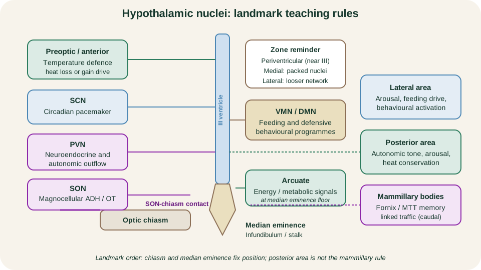
Usable hypothalamic map: each labelled region carries a first teaching rule rather than an exhaustive eponym list.
SimpleMed original educational diagram
Three Coordinated Output Channels
Hypothalamic control is coordinated, not single-track. Three main channels leave the same integrator and often fire together when a set point is threatened.
The first channel is endocrine. Parvocellular neurons release factors into the hypophyseal portal circulation at the median eminence, so the anterior pituitary secretes or withholds its hormones under hypothalamic permission. Magnocellular axons of PVN and SON project through the infundibulum into the posterior pituitary and release oxytocin or vasopressin directly into systemic blood. Portal control and axonal neurosecretion are therefore two endocrine strategies, not synonyms.
The second channel is autonomic. Descending projections reach brainstem and spinal autonomic networks that adjust heart rate, vascular tone, sweating, gut motility and adrenal medullary drive. The third channel is behavioural and somatic: thirst-seeking, feeding, thermoregulatory posture, defensive freezing or flight, and sleep-wake organisation recruit cortex, basal ganglia and brainstem motor patterns under hypothalamic bias.
When plasma osmolality rises, the same neighbourhood can increase vasopressin release, promote renal water retention, generate thirst behaviour and shift autonomic tone. That is one set-point problem solved through all three channels rather than three separate organs inventing independent answers.
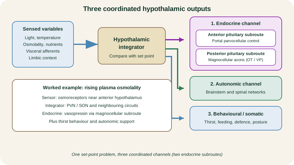
Output map: three channels (endocrine, autonomic, behavioural/somatic) restore set points; endocrine has two pituitary subroutes.
SimpleMed original educational diagram
Set-Point Examples
Each example uses the same skeleton: sensed variable, integrator, coordinated output. Detail lives in linked lessons; the map lives here.
Circadian timing. Light intensity and spectrum reach the SCN through the retinohypothalamic tract. The SCN times melatonin-related and other endocrine rhythms and synchronises peripheral clocks so physiology anticipates day and night rather than only reacting after the fact.
Osmolality and thirst. Osmoreceptors and related circuits sense effective plasma tonicity. Integration near the anterior hypothalamus and PVN-SON complex raises vasopressin when water is scarce, conserves free water at the kidney, and drives thirst and water-seeking behaviour. The renal and receptor detail is developed in Control of Plasma Osmolarity.
Temperature. Preoptic and anterior sensors compare core temperature with a defended range. Heat-loss responses (vasodilation, sweating, behavioural cooling) or heat-gain responses (vasoconstriction, shivering, behavioural warming) follow. Full effector physiology sits in Temperature Regulation; here the rule is that the same region both measures and commands.
Appetite and energy balance. Arcuate and neighbouring medial-lateral circuits integrate gut, adipose and nutrient signals with taste, circadian phase and experience. Outputs adjust feeding behaviour, energy expenditure and pituitary axes. Peptide catalogues and receptor lists remain in Control of Appetite.
Stress. Salient threat or physiological challenge recruits PVN parvocellular neurons that drive corticotrophin-releasing hormone into the portal system, activating the HPA cascade described in HPA and Growth Hormone. Parallel autonomic and behavioural outputs raise arousal and mobilise resources. Stress is therefore a multi-channel set-point shift, not a single hormone pulse.
| Set point | Key integrator | Typical coordinated outputs |
|---|---|---|
| Circadian phase | SCN | Timed endocrine and behavioural programmes |
| Plasma osmolality | Anterior / PVN-SON | Vasopressin, thirst, autonomic support |
| Core temperature | Preoptic-anterior | Heat loss or gain, posture and preference |
| Energy balance | Arcuate and medial-lateral | Feeding, expenditure, pituitary tone |
| Stress mobilisation | PVN with limbic partners | HPA drive, autonomic arousal, defence |
Limbic Anatomy on the Medial Brain
On a midline sagittal view the limbic landscape is a ring of cortex and subcortical nodes around the diencephalon and corpus callosum. Cingulate gyrus arches above the corpus callosum. Parahippocampal and entorhinal cortex occupy the medial temporal surface and gate much of the traffic into the hippocampal formation. The hippocampus and dentate gyrus roll into the temporal lobe; their major output axon bundle, the fornix, arches under the corpus callosum to the mammillary bodies and septal region.
Anterior thalamic nuclei sit in the limbic loop between mammillary bodies and cingulate cortex. The amygdala lies in the medial temporal lobe anterior to the hippocampus and links sensory association cortex with hypothalamic and brainstem effectors. Septal nuclei and ventral forebrain regions connect to hippocampus and hypothalamus. Medial prefrontal cortex evaluates and regulates affective responses rather than merely registering them.
The phrase limbic system is a useful historical map for these interconnected structures. It is not one sharply bounded organ with a single modern definition. Authors vary on orbitofrontal cortex, insula or ventral striatum. For first-year teaching, keep the medial-ring inventory and insist on connectivity.
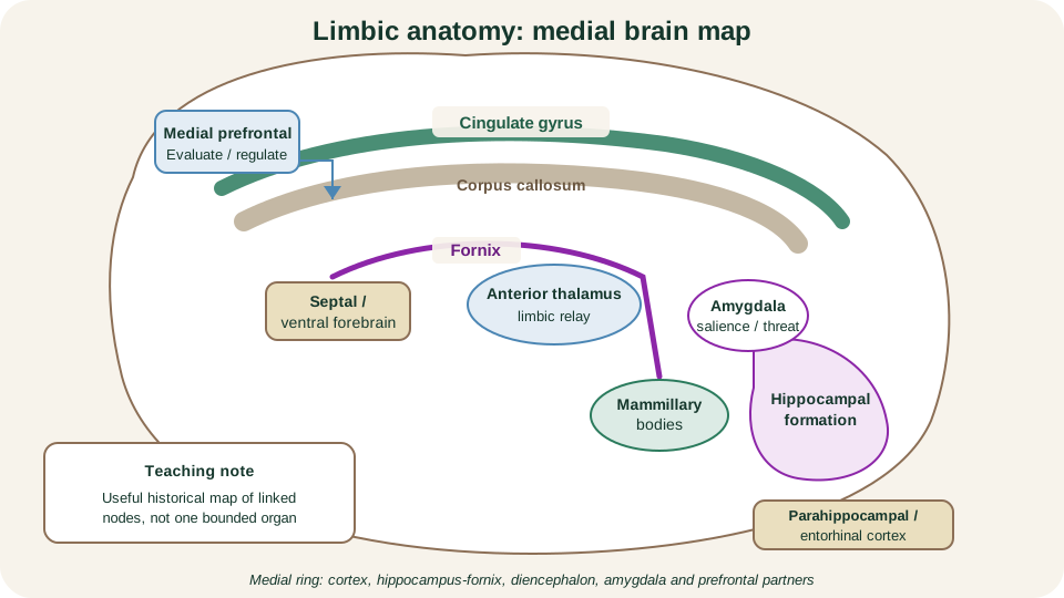
Medial limbic map: cortical rim, hippocampal-fornix path, diencephalic nodes and amygdala-prefrontal partners form a connected network, not a single organ.
SimpleMed original educational diagram
Papez Circuit and Memory Context
The classical Papez circuit has a fixed teaching direction that students should be able to recite and draw: hippocampal formation to fornix to mammillary bodies to mammillothalamic tract to anterior thalamic nucleus to cingulate gyrus to cingulum to parahippocampal and entorhinal cortex, then back into the hippocampal formation. That closed loop is the examination core.
What the loop mainly does, on modern reading, is support memory and contextual integration. Hippocampal processing binds episodic and spatial detail; diencephalic and cingulate stages help consolidate and re-enter that information into wider cortical networks. Emotion is influenced because the same anatomy talks to hypothalamus, amygdala and prefrontal evaluators, but Papez is not a complete modern emotion circuit. Broader cortico-limbo-thalamo-cortical models add amygdala, prefrontal, striatal and brainstem partners that the 1930s diagram omitted.
Detailed taxonomies of memory systems and cortical localisation continue in Higher Cortical Function. This lesson owns the anatomical ring and its interface with hypothalamic set-point control.
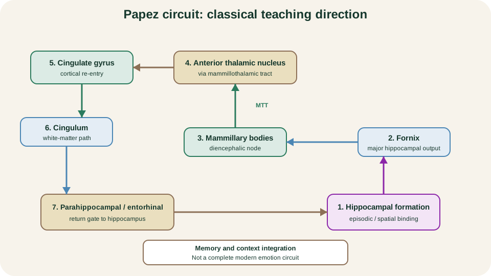
Papez direction: hippocampus-fornix-mammillary-anterior thalamus-cingulate-cingulum-parahippocampal-entorhinal return; a memory-context loop, not a full modern emotion map.
SimpleMed original educational diagram
Amygdala, Reward and Lesion Logic
The amygdala tags sensory and interoceptive events with salience, especially threat and social value. Through dense links to hypothalamus and brainstem, it can trigger coordinated autonomic, endocrine and defensive behavioural packages rapidly. Through links to hippocampus and prefrontal cortex, it also biases what is encoded and how strongly it is later retrieved. Hippocampus supplies episodic and spatial context and supports consolidation of declarative detail. Medial prefrontal and cingulate regions help evaluate whether a response remains appropriate and can damp or redirect amygdala-driven output when the situation changes.
Reward learning at first-year depth uses a short midbrain-striatal story. Ventral tegmental dopamine neurons project to nucleus accumbens in the ventral striatum and to prefrontal and other limbic partners. Phasic dopamine signals help stamp in which actions predicted valuable outcomes, so future choice is biased. That is action learning and incentive salience, not a simple equation that dopamine equals pleasure. Pleasure and liking can dissociate; keep the claim modest.
Lesion logic follows the map. Loss of magnocellular PVN-SON output or their tracts impairs vasopressin or oxytocin release and water balance behaviour. SCN damage disorders circadian organisation. Preoptic injury disturbs temperature defence. Arcuate and medial hypothalamic lesions can produce coherent feeding and endocrine failures without primary gut disease. Fornix, mammillary or anterior thalamic damage interrupts the Papez path and undermines new episodic memory formation. Amygdala disruption blunts threat tagging and coordinated defence while sparing basic sensory detection. In each case the pattern is predictable from the integrator and its channels: endocrine, autonomic, behavioural and memory-salience failures travel together when their shared anatomy is lost. Disease catalogues sit outside this lesson; read failures back to the map.
References and Further Reading
- Neuroanatomy, Hypothalamus, Bear, Reddy and Bollu.
- Functional Anatomy of the Hypothalamus and Pituitary, Lechan and Toni, Endotext.
- The Limbic System, Purves et al., Neuroscience.
- Neuroanatomy, Limbic System, Torrico and Abdijadid.
- The Cortico-Limbo-Thalamo-Cortical Circuits: An Update to the Original Papez Circuit, Kamali et al.
Reviewed by: Dr. Marcus Judge
- 129
