

Next Lesson - Visual System
Core
Gross Form and Peduncles
The cerebellum occupies the posterior cranial fossa beneath the tentorium cerebelli, behind the fourth ventricle, pons and medulla. Two hemispheres meet at a midline vermis. Superficially it is divided into anterior, posterior and flocculonodular lobes by the primary and posterolateral fissures. Those lobe names describe anatomy; functional teaching uses longitudinal zones and connection patterns instead.
A thin, highly folded cortex covers white matter that embeds paired deep nuclei. The surface area is large for the organ's volume, so a short rostrocaudal length still supports dense processing. The cerebellum does not initiate voluntary contraction in the way corticospinal upper motor neurons do. It receives copies of motor plans and sensory reports of what the body is doing, then shapes the commands that leave brainstem and cortical motor centres.
Three peduncles attach each half of the cerebellum to the brainstem: superior to midbrain, middle to pons and inferior to medulla. The superior cerebellar peduncle carries mainly deep-nuclear axons that decussate in the midbrain to reach contralateral red nucleus and thalamus, with thalamic relays continuing to motor and premotor cortex. Some afferent fibres also travel in this peduncle, so "mainly output" is the first-pass rule rather than an absolute law. The middle cerebellar peduncle is dominated by crossed pontocerebellar axons that relay cerebral cortical information into the cerebellar hemispheres. The inferior cerebellar peduncle is mixed: it admits spinal, vestibular and inferior olivary afferents and returns some fibres to vestibular nuclei and reticular formation.
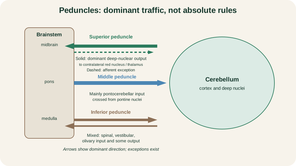
Peduncle traffic: superior mainly deep-nuclear output, middle mainly pontocerebellar input, inferior mixed; arrows show dominant directions, not exclusive one-way rules.
SimpleMed original educational diagram
Spatial memory rule: superior out, middle in from pons, inferior mixed. Keep the exceptions in mind when a tract name seems to break the slogan.
Functional Divisions
Connection-based divisions predict clinical territory better than lobe names alone. The vestibulocerebellum, largely the flocculonodular lobe, receives vestibular nuclear and labyrinth-related input and helps stabilise posture, balance and eye-head coordination. Much of its output returns to vestibular nuclei, either directly from cortex or via the fastigial nucleus.
The spinocerebellum occupies the vermis and intermediate (paravermal) zones. It is the only major division that receives substantial direct spinal proprioceptive and somatosensory copy. Vermis circuits favour axial and proximal musculature and gait posture; intermediate zones favour distal limb adjustment during ongoing movement. Outputs leave mainly through fastigial and interposed (globose and emboliform) nuclei towards brainstem and thalamic targets.
The cerebrocerebellum occupies the large lateral hemispheres. Pontine nuclei collect wide cortical input, including motor, premotor and association areas, and send massive crossed fibres through the middle peduncle. This division supports planning and timing of skilled, sequential movements, including aspects of speech motor control. Its principal deep nuclear partner is the dentate nucleus, which projects via the superior peduncle towards thalamus and then motor and premotor cortex.
| Division | Territory | Dominant input | Typical concern |
|---|---|---|---|
| Vestibulocerebellum | Flocculonodular | Vestibular | Balance, eye-head control |
| Spinocerebellum | Vermis, intermediate | Spinal sensory copy | Posture, limb execution |
| Cerebrocerebellum | Lateral hemisphere | Cortex via pons | Skilled timing and sequences |
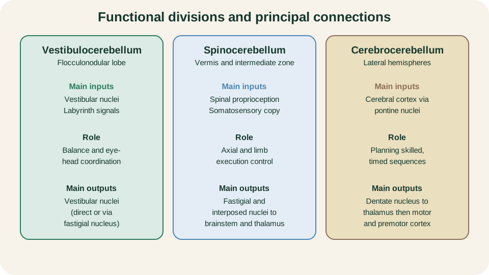
Functional divisions: vestibulocerebellum for balance and gaze, spinocerebellum for axial and limb execution, cerebrocerebellum for skilled cortical sequences.
SimpleMed original educational diagram
Cortical Microcircuit and Polarity
Cerebellar cortex has three layers from outside in: molecular, Purkinje and granule. Granule cells are tiny and numerous. Their axons ascend and bifurcate as parallel fibres that run along the molecular layer, contacting many Purkinje dendritic trees. Purkinje cells form a single sheet of large inhibitory neurons; their axons are the sole cortical output line. The molecular layer also contains inhibitory interneurons that refine local timing, but the examination core is the excitatory-inhibitory sequence that reaches the deep nuclei.
Two afferent fibre systems enter the cortex. Mossy fibres arise from many sources, including pontine nuclei, spinal pathways and vestibular relays. They excite granule cells (and provide collaterals to deep nuclei). Granule parallel fibres then excite Purkinje cells with relatively weak individual synapses that become powerful when many fibres coincide. Climbing fibres arise from the inferior olivary complex. Each climbing fibre wraps a Purkinje dendritic tree and delivers a powerful excitatory complex spike. Mossy and climbing pathways therefore both excite, but they do so with different source logic and different postsynaptic impact.
Polarity must stay exact. Mossy fibres excite. Climbing fibres excite. Parallel fibres excite Purkinje cells. Purkinje cells inhibit deep cerebellar nuclei or, for some vestibulocerebellar targets, vestibular nuclei. Deep nuclei are not simple slaves of that inhibition: they also receive excitatory collaterals from the same major afferent streams, so nuclear output is a balance between direct excitation and Purkinje brake. That balance is what leaves the cerebellum as its major output.
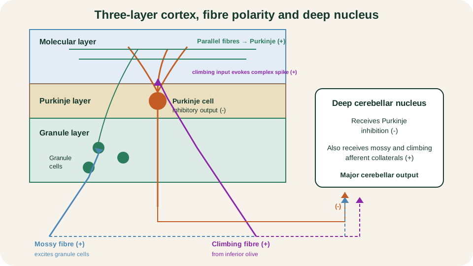
Microcircuit polarity: mossy and climbing fibres excite, parallel fibres excite Purkinje cells, Purkinje axons inhibit deep nuclei that also receive excitatory collaterals.
SimpleMed original educational diagram
Deep Nuclei and the Comparator
From medial to lateral the deep nuclei are fastigial, interposed and dentate. Roughly, fastigial partners the vermis and axial control, interposed partners intermediate limb zones, and dentate partners the lateral hemispheres. Nuclear axons form most of the superior peduncle traffic and some inferior peduncle returns to vestibular and reticular targets. Because Purkinje cells are inhibitory, a rise in Purkinje firing can suppress nuclear output, while reduced Purkinje tone can release it. Afferent collaterals keep a tonic excitatory drive under that cortical control.
The teaching spine is a comparator. Cerebral and brainstem centres broadcast intended movement through mossy routes such as the cortico-ponto-cerebellar path. Sensory systems report actual movement through spinal, vestibular and other mossy channels. The cerebellum detects mismatch, often called motor error, and changes deep-nuclear output so upper motor neurons correct timing, gain and trajectory. Corrections operate in real time during a movement and, over practice, as learning that reduces the same error next time.
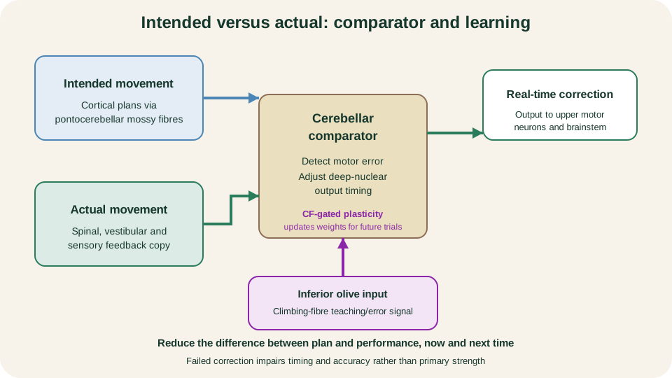
Comparator loop: intended plan and actual performance are compared; deep-nuclear output corrects in real time, while inferior olivary climbing-fibre input helps gate circuit-weight updates for future movements.
SimpleMed original educational diagram
Predecessor motor system lessons describe how upper motor neurons order force and direction. This lesson adds the error-reducing module that keeps those orders accurate as the body and environment change.
Motor Learning
Motor learning here means durable improvement of a movement map when conditions change, not mere one-trial correction. Classic illustrations include long-term adjustment of vestibulo-ocular reflex gain when visual magnification changes, and adaptation of reaching when forces or visual feedback are altered. An intact cerebellum is required for many such adaptations, even when strength remains normal.
Climbing fibres are widely modelled as teaching or error signals. A complex spike can mark an unexpected outcome and alter the future effectiveness of concurrent parallel-fibre synapses onto the same Purkinje cell. Parallel-fibre long-term depression is one well-studied candidate mechanism for that change, but it is not the sole settled account of cerebellar learning. Plasticity is distributed: synapses in cortex, deep nuclei and related brainstem sites can all shift, and multiple induction rules operate on different timescales. For first-year teaching, retain three claims with restraint: climbing-fibre activity carries high-salience error or teaching information; mossy-parallel pathways carry the rich context of what was intended and sensed; learning emerges from coordinated weight changes rather than from a single mandatory molecular switch.
Failure of learning does not equal paralysis. Patients may still generate force, yet they struggle to recalibrate timing when a tool, lens or surface changes. That distinction returns in the clinical signs section: the deficit is quality control, not power generation.
Why Limb Signs Are Ipsilateral
Lateralised cerebellar lesions usually disturb the ipsilateral limbs. The explanation is circuit geometry, not a single crossing. Consider a right-arm skilled movement. Left motor cortex issues the descending command. Corticopontine fibres reach the left basilar pons; pontocerebellar axons then cross through the middle peduncle into the right cerebellar hemisphere. The right cerebrocerebellum therefore receives the cortical plan for the right arm.
Right deep-nuclear output leaves largely through the right superior peduncle and crosses at its midbrain decussation towards left thalamus and cortex. Left cortex then drives the right arm through corticospinal fibres that cross at the pyramidal decussation. Cerebellar influence on the limb therefore crosses twice on its outward route, once at the midbrain decussation of the superior cerebellar peduncle and once at the pyramidal decussation, returning control to the same side as the cerebellar hemisphere. The incoming cortical plan has its own crossing in the pontocerebellar projection. A right cerebellar lesion therefore disrupts correction of the right arm even though primary corticospinal power pathways are intact.
Spinal mossy input reinforces the same laterality. Many proprioceptive spinocerebellar routes reach the cerebellum with ipsilateral emphasis for trunk and limb segments. Do not claim that the corticospinal decussation alone explains cerebellar laterality; it is one step in a multi-crossing architecture that maps each hemisphere onto the same-side body.
Signs from Failed Correction
Cerebellar clinical features are failed corrections of rate, range, rhythm and force, not primary paresis. Strength can be preserved while accuracy collapses. Disease catalogues and the DANISH mnemonic belong with broader Movement Disorders teaching; examination technique is outside this lesson. The anatomy already predicts the pattern.
Dysmetria is mis-scaled reach: the limb overshoots or undershoots a target because range is not updated against sensory feedback. Intention tremor grows as the limb approaches the target, when error correction is most demanded. Dysdiadochokinesia is irregular, poorly timed alternating movement, reflecting loss of rapid reciprocal control. Ataxia is disordered multi-joint coordination; gait ataxia is wide-based and staggering when axial and lower-limb spinocerebellar control fails. Nystagmus and related gaze instability follow vestibulocerebellar disruption of eye-head coordination. Scanning or ataxic speech is irregular, broken articulation when oro-laryngeal timing circuits are involved.
Map the sign to the division when possible: flocculonodular and vestibular connections for gaze and balance; vermis and intermediate zones for posture and limb execution; lateral hemisphere and dentate for skilled sequencing and speech motor timing. In every case the mechanism is the same teaching spine. Intended and actual trajectories are no longer brought into register, so output no longer repairs the difference. The patient moves, but the movement is late, mistimed or poorly aimed.
References and Further Reading
- Organisation of the Cerebellum, Purves et al., Neuroscience.
- The Cerebellum, Purves et al., Neuroscience.
- Principles of operation of a cerebellar learning circuit, Herzfeld et al.
- Revisiting the symptoms and signs of cerebellar syndrome, Manto et al.
- Neuroanatomy, Cerebellum, Jimsheleishvili and Dididze.
Reviewed by: Dr. Marcus Judge
- 178
