

Next Lesson - The Hypothalamus and Limbic System
Core
Shared Hair-Cell Rule
Hearing and balance begin with the same cellular rule. Each hair cell carries a staircase of stereocilia. Tip links couple neighbouring stereocilia so that deflection of the bundle applies force to mechanically gated cation channels at the tips. Deflection toward the taller stereocilia increases channel open probability. Potassium-rich endolymph then drives K+ into the cell, depolarising the receptor potential. Voltage-gated calcium entry at the basolateral pole triggers glutamate release onto primary afferents of the vestibulocochlear nerve (cranial nerve VIII). Deflection toward the shorter stereocilia reduces open probability, hyperpolarises the cell and lowers transmitter release.
That bidirectional coding is especially clear in vestibular organs, where hair cells maintain a tonic baseline firing rate in their afferents. Motion can raise or lower that rate relative to rest, so both directions of head movement remain informative. Vestibular hair cells also keep a true kinocilium that defines morphological polarity of the bundle. Mature mammalian cochlear hair cells lose the kinocilium after development; their staircase still has a tall-to-short axis that sets excitatory direction, but teaching language should reserve kinocilium talk for vestibular receptors.
Stimulus origin differs while the transducer does not. Sound pressure ultimately shears the cochlear bundle. Head rotation and linear acceleration shear vestibular bundles through endolymph inertia or otoconial mass. In both systems the pathway is stimulus, mechanical deflection, receptor potential, VIII afferent spike train, then central computation and useful output.
Cochlear Mechanics and Organ of Corti
Gross external and middle-ear anatomy is taught in Anatomy of the Ear. This lesson starts where stapedial motion becomes fluid pressure. Stapes footplate pressure at the oval window displaces cochlear fluids; the round window membrane bulges in the opposite direction and provides compliant relief so the almost incompressible fluid column can move. Perilymph fills scala vestibuli and scala tympani. Endolymph fills scala media (cochlear duct) and carries the high K+ concentration that powers hair-cell transduction. The organ of Corti sits on the basilar membrane within scala media. When the basilar membrane rises and falls, the overlying tectorial membrane creates a shearing motion across the stereocilia.
Inner hair cells form a single row and are the principal afferent transducers: most spiral-ganglion axons contact them and carry the frequency- and intensity-coded message toward the brainstem. Outer hair cells form three rows and act as a cochlear amplifier. Their electromotility changes cell length in time with the receptor potential, feeding mechanical energy back into the basilar membrane. That active boost raises sensitivity near threshold and sharpens frequency selectivity beyond what a passive travelling wave alone would achieve. Loss of outer hair-cell function therefore reduces gain and frequency resolution even when some inner hair cells remain.
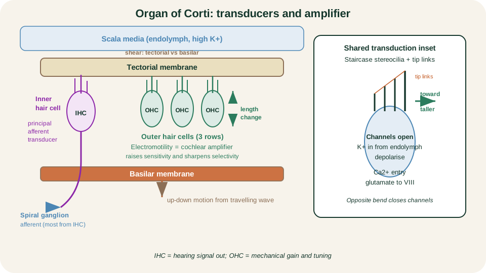
Organ of Corti: basilar motion and tectorial shear bend stereocilia; inner hair cells drive most afferents, outer hair cells amplify and sharpen the travelling wave.
SimpleMed original educational diagram
Travelling Wave and Tonotopy
Sound does not light the whole cochlea at once. A travelling wave propagates from the base toward the apex and peaks where basilar-membrane properties match the driving frequency. Near the oval-window (basal) end the membrane is relatively narrow and stiff, so high frequencies peak there. Toward the apex the membrane is wider and more compliant, so low frequencies peak there. Intensity mainly changes how strongly a place is driven and how broadly neighbouring places are recruited; the primary frequency map remains a place code along the coil.
That place code is tonotopy. Spiral-ganglion neurons inherit the characteristic frequency of their cochlear place and carry it into the cochlear nuclei. Successive stations of the central pathway preserve orderly frequency maps, so cortex still receives a systematic high-to-low layout rather than a scrambled catalogue of pitches. For first-year teaching, remember the mechanical rule first: base high and stiff, apex low and compliant, then note that the map continues centrally.
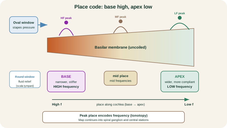
Tonotopy: travelling-wave peak place encodes frequency; high at base near the oval window, low at the compliant apex.
SimpleMed original educational diagram
Central Auditory Pathway
Spiral-ganglion bipolar neurons form the cochlear division of VIII and terminate first in the ipsilateral cochlear nuclei. From there the pathway is no longer a single compulsory chain. Axons diverge into multiple brainstem routes that include the superior olivary complex and the lateral lemniscus. Many fibres cross, so both ears are represented bilaterally above the cochlear nuclei. A main teaching route then continues to the inferior colliculus in the midbrain, the medial geniculate body of the thalamus, and primary auditory cortex on the superior temporal plane in the region of Heschl's gyri. Parallel streams for timing, intensity and spectral features travel together; the serial list is a scaffold, not a claim that every neuron visits every station in lockstep.
The superior olivary complex is the first major site specialised for binaural comparison. Interaural time and intensity differences help localise sound in space. Because representation is bilateral after the cochlear nuclei, a unilateral lesion of midbrain, thalamus or auditory cortex usually impairs localisation, discrimination or complex processing rather than silencing one ear completely. By contrast, damage confined to the cochlea or cochlear nerve removes or distorts input from that ear and produces ipsilateral sensorineural failure.
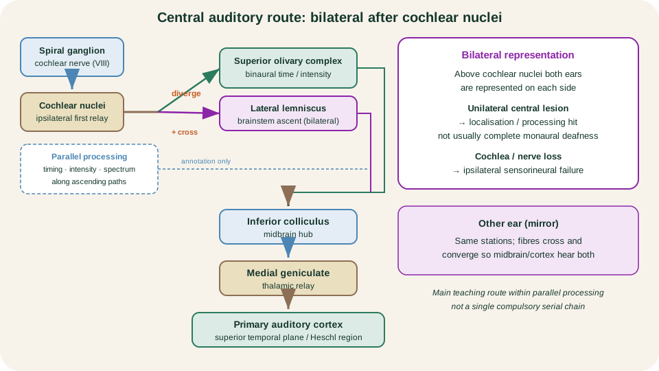
Main auditory teaching route with bilateral divergence after cochlear nuclei; superior olive compares binaural timing and intensity cues within parallel processing.
SimpleMed original educational diagram
Vestibular Sensors
Three semicircular ducts lie in approximately orthogonal planes and detect angular acceleration of the head. Each duct ends in an ampulla that houses a crista of hair cells capped by a gelatinous cupula. When the head begins to rotate, endolymph lags by inertia, deflects the cupula and bends the stereocilia. Afferent firing rises on one side of a push-pull canal pair and falls on the opposite side, giving a signed rotation signal. During a constant-velocity turn, endolymph and duct eventually move together, cupular deflection decays, and the canal no longer reports a maintained fixed head position. Canals are sensors of change in angular velocity, not of static orientation alone.
The utricle and saccule are otolith organs. Their macular hair cells are embedded beneath an otolithic membrane loaded with dense calcium carbonate otoconia. Linear acceleration or a change in head orientation relative to gravity shears that heavy membrane across the macula. At first-year depth, teach the utricle as mainly sensitive to horizontal-plane linear forces and head tilt about horizontal axes, and the saccule as mainly sensitive to vertical-plane linear forces. Both organs use the same hair-cell rule, but their mechanical load is mass under gravity rather than cupular fluid drag.
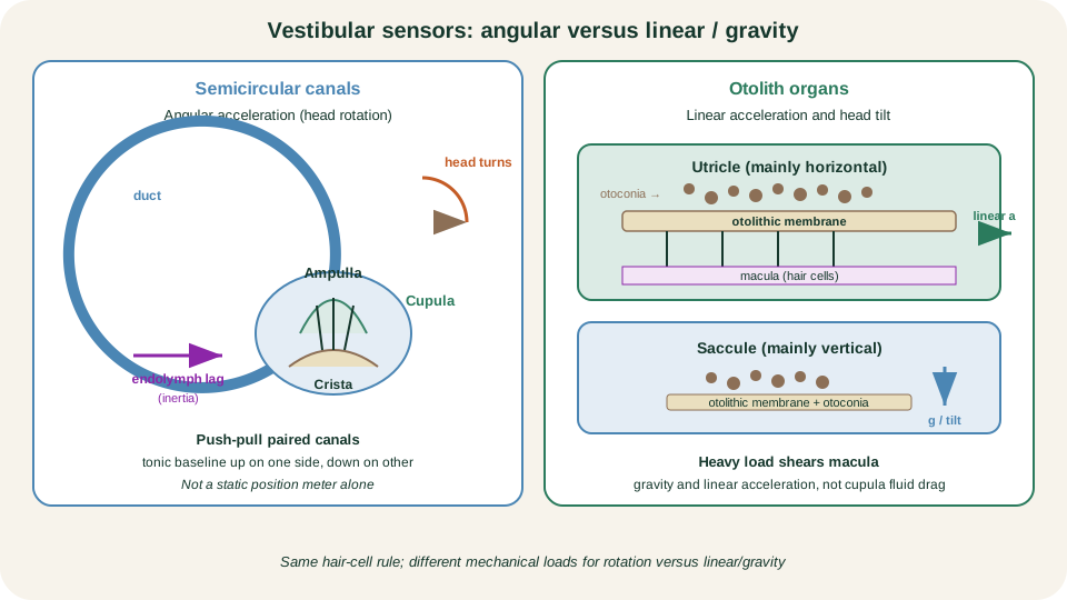
Vestibular sensors: canals and cupula for angular acceleration; macular otolithic membrane and otoconia for linear acceleration and gravity-related tilt.
SimpleMed original educational diagram
Central Vestibular Pathways and VOR
Vestibular bipolar neurons in the vestibular ganglion form the vestibular division of VIII and project to vestibular nuclei in the brainstem and to the cerebellum. From the nuclei, outputs reach ocular motor nuclei, spinal motor circuits that adjust posture, and thalamic-cortical routes that contribute to motion perception and spatial orientation. The same primary signal therefore stabilises gaze, supports balance and informs conscious sense of movement.
The vestibulo-ocular reflex (VOR) is the clearest short-latency example. Its purpose is gaze stability: when the head turns, the eyes counter-rotate so that the image stays nearly still on the retina. Horizontal VOR can be taught as a three-neuron arc at usable depth. Primary canal afferents synapse in vestibular nuclei; secondary vestibular neurons cross or relay to abducens and, via the medial longitudinal fasciculus (MLF), to medial rectus motoneurons in the oculomotor nucleus; the third neuron is the extraocular motoneuron itself.
Worked direction, fixed for examination: head turns right. Right horizontal-canal afferents increase firing; left horizontal-canal afferents decrease. The nuclear and MLF circuit activates left lateral rectus and right medial rectus so both eyes move left, opposite the head. That is the compensatory slow phase of the VOR. Without it, each head movement would smear the visual scene.
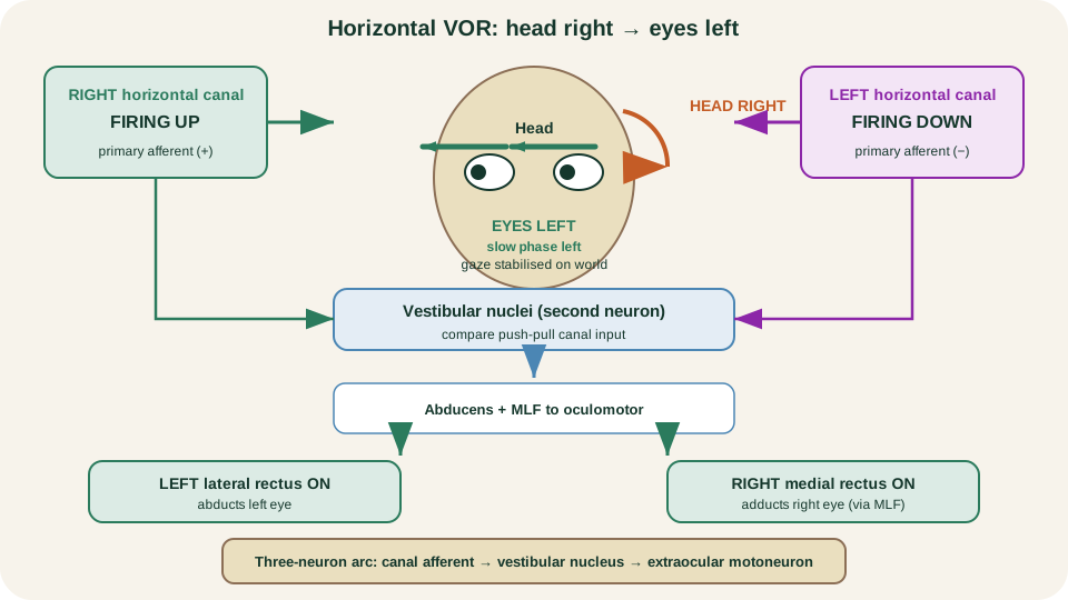
Horizontal VOR: head right raises right-canal and lowers left-canal firing; eyes drive left via left lateral rectus and right medial rectus to stabilise gaze.
SimpleMed original educational diagram
Physiological Nystagmus
During sustained head rotation in the dark or when the VOR is free to express itself without visual suppression, eye movement often becomes a sawtooth. The slow phase is the vestibular compensatory drift opposite the direction of head rotation. The fast phase is a central reset saccade that snaps the eyes back in the direction of rotation so that another slow phase can begin. Clinical naming follows the fast phase: right-beating nystagmus means the quick component beats right.
Physiological nystagmus is therefore not a disease label. It is the combination of a vestibular slow eye movement and a brainstem-generated quick reset. Disease catalogues, bedside testing batteries and caloric mnemonics such as COWS sit outside this lesson. Keep the mechanism: slow phase equals VOR compensation; fast phase equals central reset; name the beat by the fast phase.
Mechanism-Led Failure
Failure patterns follow the stations already taught. Conductive failure attenuates mechanical transfer in the outer or middle ear. Sound energy reaches the oval window poorly, yet the cochlear hair cells and nerve may still be intact, so bone-conducted vibration can still drive the inner ear. Sensorineural failure damages hair-cell transduction, the cochlear amplifier or the auditory nerve itself; place coding and intensity coding degrade at source. Named ear diseases are catalogued in Ear Pathology; here the rule is mechanical versus transducer-nerve failure.
Post-cochlear central lesions usually spare monaural detection more than they spare localisation and fine discrimination, because each ear is already represented bilaterally above the cochlear nuclei. A unilateral midbrain or cortical injury therefore tends to blur spatial and complex processing rather than produce complete deafness of one ear. Cochlear or nerve loss remains ipsilateral.
On the vestibular side, asymmetric firing between the two labyrinths creates a false-motion signal. The brain interprets the imbalance as continuous rotation or tilt, yielding vertigo, nystagmus and postural imbalance even when the head is still. Bilateral loss of VOR gain fails to stabilise gaze during head movement, so the visual world appears to bounce (oscillopsia) whenever the head moves. None of these statements is a diagnostic checklist or treatment guide; each is the map read backwards from interrupted mechanics, transduction or central comparison.
References and Further Reading
- Hair Cells and the Mechanoelectrical Transduction of Sound Waves, Purves et al., Neuroscience.
- The Auditory System, Purves et al., Neuroscience.
- Sound, Gottfried, Neurobiology of Sensation and Reward.
- Neuroanatomy, Auditory Pathway, Peterson et al.
- The Vestibular System, Purves et al., Neuroscience.
- Vestibular Hair Cells, Purves et al., Neuroscience.
- A brief review of the clinical anatomy of the vestibular-ocular connections, Bronstein et al.
Reviewed by: Dr. Marcus Judge
- 130
