

Next Lesson - Anxiety and Anxiety Disorders
Core
A subarachnoid haemorrhage (SAH) occurs when there is bleeding into the space surrounding the brain and spinal cord beneath the arachnoid mater (the middle of the three membranes, called the meninges, that cover them). It is a clinical emergency and must be recognised and treated rapidly to prevent irreparable complications and death.
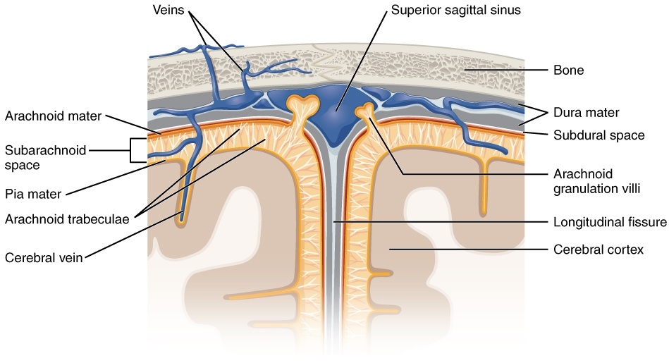
Diagram - The meningeal layers surrounding the brain. The bleed takes place in the subarachnoid space
Source illustration: OpenStax, source, CC BY 4.0.
Overall, the most common cause of a subarachnoid haemorrhage is head trauma; however, in spontaneous (non-traumatic) cases, the usual cause is rupture of a cerebral aneurysm. Trauma includes road traffic accidents and falls.
SAH can also occur spontaneously, commonly following rupture of a cerebral aneurysm. Aneurysm formation involves blood-flow forces and multifactorial arterial wall remodelling (many factors change the structure of the artery wall), including degeneration of the internal elastic lamina (the elastic layer beneath the inner lining) and the tunica media (the muscular middle layer). It cannot be explained simply by erosion of the intima (the inner lining of the artery).

Diagram - A cerebral aneurysm in the brain
Public Domain Source by National Institutes of Health [Public domain]
Non-traumatic causes of an SAH are related to the modifiable and non-modifiable risk factors a patient can present with. Risk factors are not direct causes of a haemorrhage but can increase the risk of developing a bleed.
Cerebral aneurysms often arise at arterial branch points, where local flow patterns act on susceptible vessel walls. Common sites include the anterior communicating artery complex (where the two anterior cerebral arteries are joined), the internal carotid–posterior communicating artery junction and middle cerebral artery bifurcations (points where an artery divides in two).
Modifiable risk factors are factors that can be reduced through changes to the patient’s lifestyle or medications, and include cigarette smoking, heavy alcohol use and hypertension.
Non-modifiable risk factors are those that cannot be changed through any intervention and include connective tissue disorders (such as Ehlers-Danlos syndrome, an inherited condition that weakens connective tissue), autosomal dominant polycystic kidney disease (an inherited condition with many fluid-filled cysts in the kidneys), a positive family history of subarachnoid haemorrhage and female sex (aneurysmal SAH is slightly more common in women than men). These risk factors can all contribute to the formation of aneurysms in the brain. Some raise the pressure of the blood in the vessels (for example hypertension) and others weaken the arterial wall (for example connective tissue disorders). An arteriovenous malformation (AVM, an abnormal tangle of arteries and veins) is a separate cause of SAH.
When the arterial wall gives way a large volume of arterial blood is released very quickly into the subarachnoid space, found between the pia mater (the thin innermost membrane, which sits directly on the brain) and the arachnoid mater. This event triggers a cascade of complex mechanisms in the brain that can be split into Early Brain Injury and Delayed Cerebral Ischaemia.
This occurs immediately after vessel rupture and encompasses the direct effect of the haemorrhage on the brain tissue and the compensatory mechanisms of the brain. The direct effects of the bleeding are the most common cause of death and disability after SAH.
The leakage (extravasation) of a high volume of blood initially causes a mechanical compression of the brain, resulting in direct damage to the cerebral tissue. Secondary to this, the mixing of the blood with cerebrospinal fluid (CSF) can impede normal CSF clearance, resulting in an acute phase hydrocephalus (a build-up of CSF in the brain) in approximately 20-30% of patients.
The increased volume in the subarachnoid space causes a rapid rise in the intracranial pressure (ICP, the pressure inside the skull) which correspondingly decreases the cerebral perfusion pressure (CPP, the pressure that drives blood through the brain), resulting in a reduced flow of blood to the brain tissue. (For more information on cerebral autoregulation, the brain’s way of keeping its own blood flow steady, see our article on Raised Intracranial Pressure). Arteriolar dilation (widening of the small arteries) reduces vascular resistance and attempts to support cerebral blood flow. This dilation does not restore a critically low CPP; severe pressure elevation and impaired autoregulation can overwhelm this compensation, producing acute global ischaemia.
Following this transient period of ischaemia, there is gradual reperfusion of the brain tissue as the blood flow returns which causes the added problem of cerebral reperfusion injury (damage caused when blood flow returns to tissue that has been short of oxygen). Oxidative damage occurs in the brain due to the increased production of reactive oxygen species (unstable, damaging oxygen molecules, also called free radicals; see our article on Oxidative Stress). These radicals act by directly damaging the neurovascular structures in the brain, increasing inflammation, breaking down the blood-brain barrier and releasing more vasoconstrictors.
Alongside these mechanisms, the extravasated blood itself is an irritant to the brain tissue, resulting in spasming of the arteries, inflammation and cerebral oedema. There is also the inappropriate production of microthrombi (tiny blood clots), secondary to the activation of platelets which attempt to clot the original haemorrhage. The microthrombi block the blood supply further.
Summary of early brain injury: a big haemorrhage compresses the brain tissue and impairs drainage of CSF, both of which raise intracranial pressure. This decreases the cerebral perfusion pressure, cutting off blood supply to the brain and encouraging the formation of reactive oxygen species. As reperfusion occurs, oxidative stress further damages the brain, causing oedema and further swelling.
In the days following SAH, some patients develop delayed cerebral ischaemia (DCI), with a new neurological deficit or reduced consciousness. DCI is multifactorial (it has many causes); vasospasm (narrowing) of the large arteries is only one contributor. Urgent specialist assessment is needed to distinguish it from other causes of deterioration, including rebleeding and hydrocephalus. The first investigation for unexplained neurological deterioration after SAH is a non-contrast CT head scan.
A subarachnoid haemorrhage presents with a sudden onset, ‘thunderclap’ headache (a severe headache that typically peaks in intensity within 1 to 5 minutes), with patients commonly describing it as the worst headache they have ever experienced. The headache then persists.
Other associated signs may include vomiting, seizures, neck stiffness and a decreased or absent level of consciousness. Raised intracranial pressure explains some of these, such as a reduced level of consciousness, but it is not the only cause. In severe SAH, global ischaemia (reduced blood flow to the whole brain) can also cause unconsciousness. The patient’s level of consciousness must be monitored carefully, as it directly correlates to the extent of their neurological dysfunction and can be predictive of their outcome. However, a person with SAH may have none of these signs.
It is very important to differentiate an SAH from meningitis. Both can present similarly, but the rapid identification and treatment of SAHs can help to minimise the risk of mortality and any further complications.
Patients presenting with a suspected SAH should always have an urgent CT scan of the head, without contrast (no injected dye). This helps to identify the presence of subarachnoid blood, which will guide any following management. A positive result is seen when there is an area of increased density around the Circle of Willis (the ring of arteries at the base of the brain), depicting blood in the basal cisterns (the CSF-filled spaces at the base of the brain).

Image - A CT slice of the human brain. In the centre, there are hyper-dense (brighter) areas corresponding to blood in the subarachnoid basal cisterns (area highlighted by red arrow)
Source illustration: Lipothymia, source, CC BY-SA 3.0.
After SAH is confirmed, offer CT angiography (a CT scan with injected dye that shows the blood vessels) promptly to identify the cause and guide specialist treatment.

Image - Angiogram of an aneurysm in a cerebral artery
Source illustration: Lucien Monfils, source, CC BY-SA 3.0.
If a CT done within six hours of onset is reported and documented by a radiologist as showing no subarachnoid haemorrhage, do not routinely offer lumbar puncture (a needle in the lower back to collect CSF); discuss alternative diagnoses with a senior clinician and seek specialist advice. After a negative CT performed more than six hours after onset, consider lumbar puncture, allowing at least 12 hours from onset and checking for contraindications.
When lumbar puncture is indicated, diagnose SAH if CSF shows elevated bilirubin (xanthochromia, a yellow discolouration of the CSF) on spectrophotometry (a laboratory test that measures how the sample absorbs light); visual inspection alone is insufficient.
All patients with a confirmed SAH must be placed under continuous observation, especially monitoring for any further neurological deterioration or systemic side effects of the event, such as a myocardial infarction or pulmonary oedema. Urgently discuss with a specialist neurosurgical centre whether the patient needs to be transferred there. Provide effective analgesia, including opioids (strong painkillers such as morphine) when needed, and account for their sedating effects and effects on the pupils during neurological assessment. Decisions about antiplatelets (drugs that stop platelets clumping together) or anticoagulants require specialist risk assessment. Once the culprit aneurysm (the aneurysm that bled) has been secured by coiling or clipping (described below), do not withhold these drugs solely because of the history of aneurysmal SAH.
There are two main components of managing a patient with an SAH:
- Reducing delayed ischaemic complications. Consider enteral nimodipine (a calcium-channel blocker given by mouth or by feeding tube) after confirmed SAH. Its clinical benefit is not adequately explained by prevention of angiographic vasospasm (narrowing of the arteries, seen on an angiogram) alone; DCI has multiple mechanisms.
- Prevention of a re-bleed. This is done by repairing the affected blood vessel, so the aneurysm does not burst again. This can be done in one of two ways: endovascular coiling (done from inside the blood vessels) or neurosurgical clipping. NICE recommends offering coiling, or clipping if coiling is not suitable. An interventional neuroradiologist (a doctor who treats blood vessel problems using catheters) and a neurosurgeon decide together, and treatment should happen at the earliest opportunity because the risk of re-bleeding is highest within 24 hours of the onset of symptoms.
- Coiling involves a catheter being inserted through an artery in the groin and up to the brain. Small platinum coils are then passed through the tube and into the aneurysm, preventing any further blood from entering it.
- Clipping on the other hand involves the surgeon accessing the aneurysm via a flap of bone in the skull (craniotomy) and securing a small metal clip across the base of the aneurysm. This cuts off the blood supply to the aneurysm and lowers the risk of re-bleeding. Aneurysms can come back after treatment, so follow-up brain imaging may be offered.
Edited by: Dr. Maddie Swannack
Reviewed by: Dr. Thomas Burnell and Dr. Marcus Judge
- 806



