

Next Lesson - Subarachnoid Haemorrhage
Core
Intracranial pressure (ICP) is the measure of the pressure that the contents of the skull exert in a closed environment. Things inside the skull that contribute to this pressure are blood (both venous and arterial supplies), cerebrospinal fluid (CSF), and brain tissue.
In adults, this pressure is usually between 7-15 mmHg when lying flat. Reference values for children and infants are not well established, but ICP is generally thought to be lower than in adults. Raised ICP is usually defined as a sustained intracranial pressure above around 20 mmHg in adults, particularly in the context of acute brain injury. This can be caused by an increase in any of the constituents of the cranium: blood in the vasculature, CSF in the ventricles or the brain tissue itself. Along with pathological causes, physiological causes of an increased ICP include performing the Valsalva manoeuvre (breathing out hard against a closed airway, as when straining).
The diagram below shows what elements are found inside the skull, and maintain its pressure.
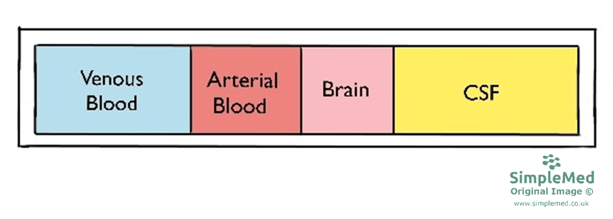
Diagram - The elements found inside the skull
SimpleMed original by Dr. Keertana Anne
The adult skull is effectively rigid. An increase in the volume of one intracranial component can initially be offset by displacement of CSF and reduction of intracranial venous blood volume, limiting the pressure rise. This is why an increase in one element must be compensated by a decrease in the volume of another, in order to maintain equilibrium and preserve a normal ICP. This is illustrated by the Monro-Kellie doctrine.
The main compensatory reserves are displacement of CSF and reduction of intracranial venous blood volume. The doctrine does not require either the displacement of CSF or the reduction in venous blood volume to happen before the other. Once this reserve is exhausted, small further volume increases can cause a steep rise in ICP.
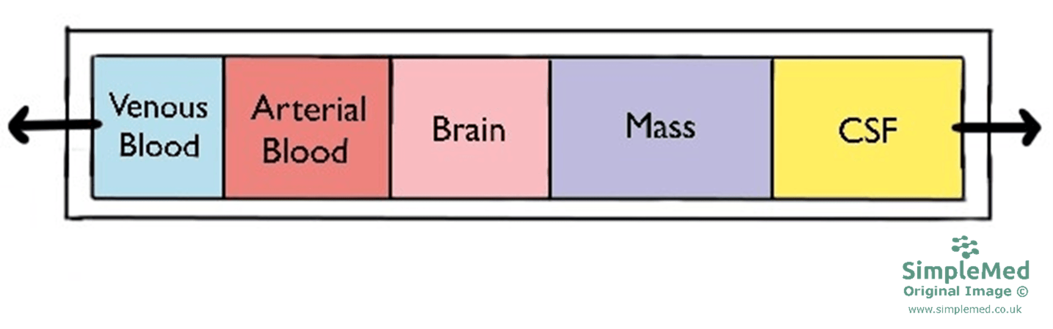
Diagram - An increase in any one of the components of the cranium or an introduction of a new compartment causes a decrease in the others
SimpleMed original by Dr. Keertana Anne
Once compensatory displacement of CSF and venous blood is exhausted, further volume increases raise ICP; arterial blood does not simply become the next compartment to drain. If mean arterial pressure does not rise sufficiently, the increased ICP reduces cerebral perfusion pressure (CPP), threatening blood flow and risking ischaemia. CPP is the pressure gradient driving cerebral perfusion; actual blood flow also depends on vascular resistance and autoregulation (the ability of the brain’s blood vessels to keep blood flow steady when blood pressure changes).
CPP is calculated using the equation:
Cerebral perfusion pressure (CPP) = Mean arterial pressure (MAP) - Intracranial pressure (ICP)
Reference values and a clinical treatment target are:
- CPP: a treatment target of 60-70 mmHg is recommended in severe traumatic brain injury, with the optimal threshold depending on autoregulation
- MAP: 70 - 110 mmHg
- ICP: 7 - 15 mmHg
At a given MAP, a rise in ICP reduces CPP. Clinical treatment targets depend on the underlying condition and the patient's autoregulatory status.
The Cushing’s Triad is a presentation consisting of systolic hypertension, bradycardia (slow heart rate) and irregular breathing. These occur as part of the Cushing Reflex, the body’s emergency response to very high ICP, which can indicate imminent brain herniation and death. The reflex is caused by the decrease in cerebral perfusion pressure (as mentioned above), which causes cerebral ischaemia (too little blood supply to the brain) as lactate and other waste products of metabolism build up in the brain.
This cerebral ischaemia triggers a sympathetic nervous response in the body which acts on α1-receptors (receptors on blood vessel walls that respond to noradrenaline) and causes peripheral arterial constriction. This increases systemic arterial pressure and leads to systemic hypertension. The systolic blood pressure increases much more than the diastolic, which results in increased pulse pressure (a wider gap between the systolic and diastolic pressures).
The increase in blood pressure is detected by baroreceptors (pressure sensors) in the aortic arch and the carotid sinuses (a widening at the point where each common carotid artery divides in the neck), and these activate the parasympathetic nervous system. This acts on muscarinic (M2) acetylcholine receptors in the heart and slows the heart rate, leading to bradycardia. The increased parasympathetic response also causes an increase in the production of stomach acid, leading to a higher rate of ulcers being formed - called Cushing’s Ulcers. Remember, though, that Cushing’s Triad is a very serious sign that brain herniation may be about to happen. Many of these patients do not survive long enough to develop stomach ulcers.
Raised ICP also has a compressive effect on the brainstem and reduces its blood supply. This is where the respiratory centres are found and damage to these causes irregular breathing, therefore completing the Cushing’s triad. Together, these three signs show that the patient can deteriorate rapidly, and this can quickly lead to death.
As mentioned earlier in the article, a rise in ICP will initially cause venous blood and CSF to drain from the cranium, as these are at the lowest pressure. Once these compensatory mechanisms fail, the next step is the compensatory movement of brain tissue in any way that it can. This is called herniation, as it is the movement of an organ out of the region it is normally contained in. This is similar to an abdominal hernia, where bowel tissue pushes out through the wall of the abdomen.
If the skull is intact, the brain tissue must herniate across structures such as the falx cerebri (the fold of tough membrane between the two cerebral hemispheres), the tentorium cerebelli (the fold of membrane between the cerebrum above and the cerebellum below), and through the foramen magnum (the large hole at the base of the skull). If a skull fracture is present, the brain tissue may herniate out of the fracture site. The herniation of brain tissue can put extreme amounts of pressure on the brain and cut off its blood supply, which can often be fatal.
There are multiple types of brain herniation.
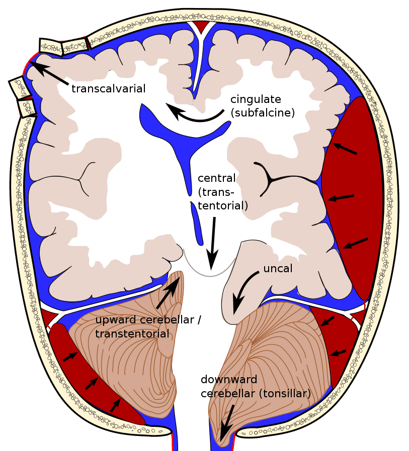
Diagram - The different types of brain herniation
Creative commons source by RupertMillard, modified from Delldot [CC BY-SA 4.0 (https://creativecommons.org/licenses/by-sa/4.0)]
Cerebellar (or tonsillar) herniation, also known as ‘coning’, occurs downwards through the foramen magnum. This can cause compression of the lower brainstem and part of the cervical spinal cord. This type of herniation can result in impaired respiratory and cardiac function, as seen in Cushing’s Triad.
Subfalcine herniation occurs across the midline. The cingulate gyrus (a fold of brain tissue on the inner surface of each hemisphere, next to the midline) moves across the falx cerebri, which separates the two hemispheres of the brain. This is most likely to damage the blood vessels in that area of the brain (including the anterior cerebral artery).
Uncal herniation occurs when an area of the temporal lobe, the uncus, herniates downwards towards the brainstem. This primarily affects the midbrain (the top part of the brainstem) and can also compress the oculomotor nerve (CN III, which moves the eye), resulting in an oculomotor nerve palsy (weakness of the nerve, giving a ‘down-and-out’ eye position). This can progress to central herniation.
Central herniation occurs when the diencephalon and areas of the temporal lobes herniate underneath the free edge of the tentorium cerebelli. The diencephalon (the deep central part of the brain, which includes the thalamus and hypothalamus) normally processes sensory information and emotions, and also controls hormone production. This type of herniation can cause paralysis of upward eye movements, resulting in the patient having ‘sun-setting’ eyes.
External (or transcalvarial, meaning through the skull vault) herniation occurs through a breach in the cranium, following a traumatic head injury or surgical intervention (such as a craniectomy, the removal of part of the skull).
Causes
There are four main causes of increased ICP. Three of these are due to an increase in the original components of the cranium:
Hypercapnia (a high level of carbon dioxide in the blood, also called hypercarbia) causes cerebral arteriolar vasodilatation, increasing cerebral blood flow and blood volume. This can raise ICP, particularly when compensatory CSF and venous-volume reserve is limited. See OpenAnesthesia: Hypercarbia.
An increased volume of blood inside the cerebral vessels can be due to conditions such as malignant hypertension (severe elevation in blood pressure causing loss of cerebral autoregulation and increased cerebral blood volume). It can also be due to superior vena cava obstruction. This is caused by external compression of the superior vena cava, the large vein that drains venous blood from the head, and it increases the venous blood volume in the cranium.
This is caused by a condition called hydrocephalus, which causes an accumulation of CSF inside the ventricles (the fluid-filled spaces deep in the brain). Hydrocephalus can either be congenital (present from birth) or acquired (developing after birth, for example after a head injury, bleeding, infection or tumour).
Congenital causes can include a deformity to the structure of the brain: such as neural tube defects (the spine or skull not closing properly in the embryo) or stenosis (narrowing) of the cerebral aqueduct, the channel between the third and fourth ventricles. Children present with bulging fontanelles (the ‘soft spot’ of the head) and sun-setting eyes. Acute management involves neurosurgical diversion of CSF to reduce intracranial pressure, and long-term management includes the placement of a ventricular shunt. These shunts continuously drain CSF from the ventricular system in the brain to either the peritoneal cavity (the space around the abdominal organs; V-P shunt) or the atrium (an upper chamber of the heart; V-A shunt). An external ventricular drain (EVD) is not a shunt. It is a temporary tube that drains CSF directly out of the body (to the external environment).
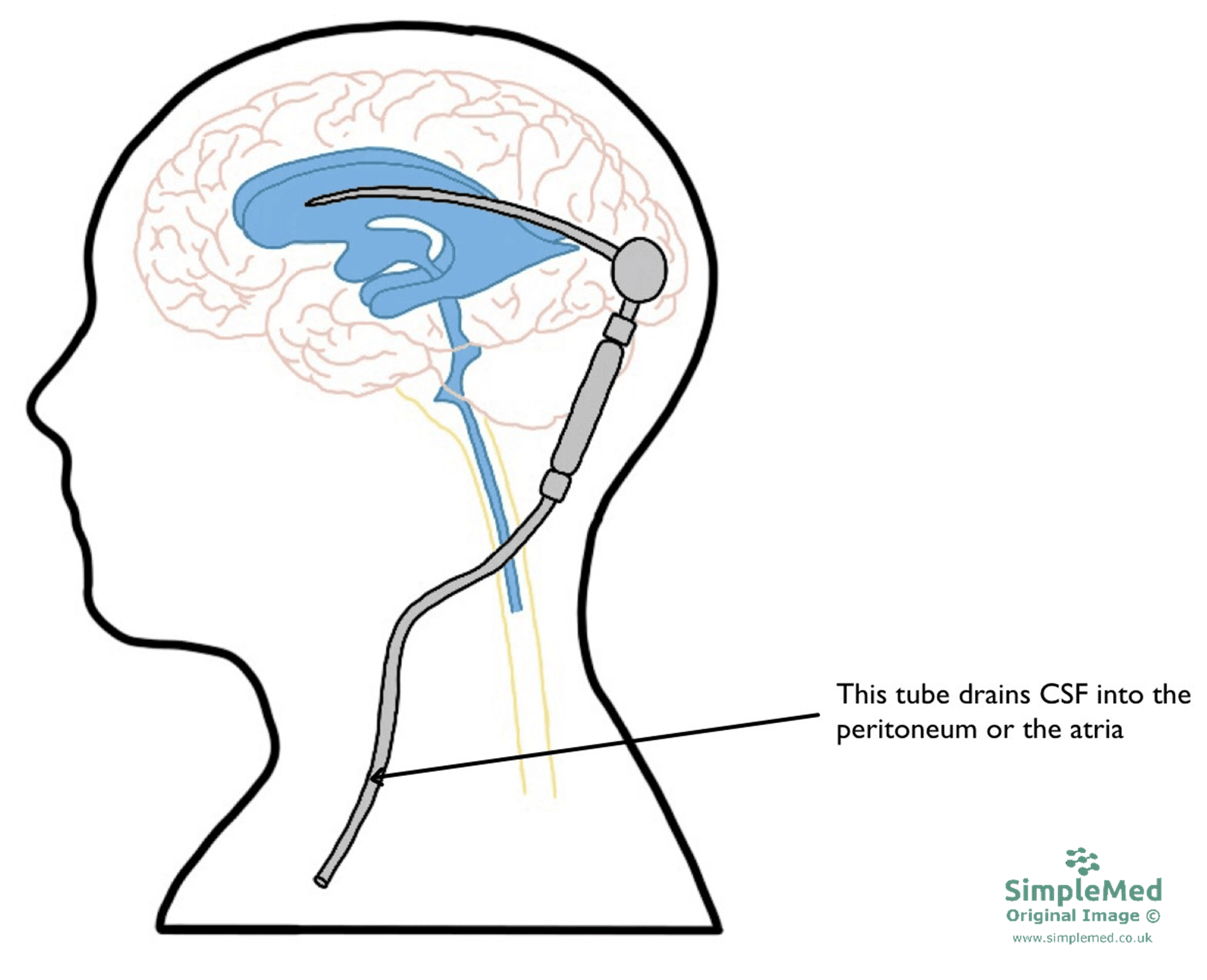
Diagram - Where ventricular shunts are inserted to drain CSF from the brain
SimpleMed original by Dr. Keertana Anne
An increase in the volume of brain tissue inside the cranium is caused primarily by the swelling of the tissue. This is known as cerebral oedema and is the build-up of fluid in the intracellular and extracellular compartments of the brain tissue. Oedema can be caused by a wide range of injuries to the brain tissue, including but not limited to: ischaemic stroke, traumatic brain injury and infection (such as meningitis or encephalitis, which is inflammation of the brain tissue itself).
The final cause of raised intracranial pressure comes from abnormal masses in the brain which can be tumours, abscesses or haemorrhages. These cause a ‘mass effect’ (the mass takes up space and squeezes the surrounding brain) and can cause damage to the components in the cranium.
Idiopathic Intracranial Hypertension
When there isn’t a definable cause for the increase in ICP, the condition is called Idiopathic Intracranial Hypertension (IIH). The pathophysiology (how the disease develops) is not fully understood, but it primarily affects obese women of childbearing age. Diagnosis needs brain imaging that shows no mass or hydrocephalus (a build-up of CSF), followed by a lumbar puncture (a needle in the lower back to sample CSF). This must show a high opening pressure (the CSF pressure at the start of the procedure, above 25 cm of CSF) and normal CSF composition. A high opening pressure on its own is not enough to make the diagnosis. Treatment includes weight loss and measures to reduce CSF production, such as acetazolamide.
In the early stages of raised ICP, the patient may present with the following signs and symptoms:
- Headache - this is usually worse in the morning (as lying flat reduces venous drainage from the brain, increasing intracranial pressure) and when bending or coughing (this increases the pressure further). As the condition progresses, the headache may become continuous.
- Difficulty concentrating or focusing
- Confusion or drowsiness - confusion may be very subtle. A falling level of consciousness, shown by a falling Glasgow Coma Scale score (a score of how awake and responsive the patient is), is an important warning sign.
- Nausea and vomiting
- Double vision - the patient has a poor accommodation reflex, may report worsening eyesight or specific visual field defects, and can develop papilloedema.
- Seizures
- Focal neurological signs - including changes in behaviour and difficulty in moving or talking.
In the later stages when the ICP is too high and compensatory mechanisms fail, brain herniation occurs, along with developing Cushing’s reflex.
In patients that present with the above symptoms, the following investigations would take place:
- A full history - if the patient is also experiencing seizures, a collateral history (an account from someone who saw the seizures) is also important.
- Neurological exam - full cranial and peripheral nerve exam, to look for focal signs such as a cranial nerve palsy or weakness.
- Examination of the eyes - fundoscopy (looking at the back of the eye, to check for papilloedema), pupillary reflexes, and visual fields.
- Lumbar puncture (a needle in the lower back to sample CSF) - this measures the pressure of the CSF. It is used mainly when IIH is suspected, and only after imaging has excluded a mass lesion or hydrocephalus. It must not be done if there are new focal neurological signs (including seizures), abnormal pupil reactions, or a severely reduced or rapidly falling level of consciousness, because of the risk of brain herniation.
- CT scan - this is used to look at cross-sectional images of the brain.
Once the patient has been diagnosed with having a raised ICP, the next steps would be to try and decrease the pressure on the brain tissue.
The initial goal would be to protect the brain tissue. This would be achieved by ensuring the patient is being oxygenated well and has a blood pressure high enough to maintain the cerebral perfusion pressure, using fluids and, if needed, drugs that raise blood pressure (vasopressors). The patient’s head is also usually raised to about 30 degrees to improve the venous drainage from the brain.
Direct treatment of the raised ICP includes giving the patient Mannitol, which acts by increasing the osmotic pressure outside the blood-brain barrier (the tight lining of the brain’s capillaries) and causes the fluid from brain cells to flow into the plasma. 3% hypertonic saline (a concentrated salt solution) is an alternative that works in a similar way.
As a temporary measure, an external ventricular drain could be inserted, connecting the ventricular system to the external environment and draining off excess CSF. This would need to be performed as an inpatient procedure and would temporarily reduce the pressure on the brain.
In some extreme cases where all the above fail, a decompressive craniectomy can be considered. This is a surgical procedure where part of the skull is temporarily removed to allow a swollen brain room to expand without being compressed. In the RESCUEicp trial of patients with traumatic brain injury and a raised ICP that did not respond to other treatment, craniectomy lowered the death rate at 6 months (26.9% versus 48.9%), but a higher proportion of patients were left in a vegetative state or with severe disability. To reduce the risk of brain injury, the patient may require protective measures such as a helmet while the bone defect remains. Then, once the raised ICP has resolved the opening in the skull is closed again, by a cranioplasty (an operation to repair the skull).
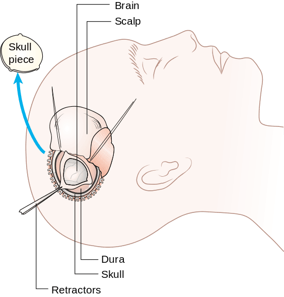
Diagram - A decompressive craniectomy
Creative commons source by Cancer Research UK [CC BY-SA 4.0 (https://creativecommons.org/licenses/by-sa/4.0)]
Edited by: Dr. Maddie Swannack
Reviewed by: Dr. Thomas Burnell and Dr. Marcus Judge
- 1005



