

Next Lesson - Somatosensory System
Core
The cerebrospinal fluid (CSF) is a fluid that surrounds the brain and spinal cord. It is colourless and clear, and there is approximately 150 ml of CSF within the central nervous system at any one time. The CSF has a number of functions, which fall into two main groups: mechanical support and protection, and metabolic support. The CSF acts as a cushion for the brain and protects it from knocks against the hard skull. It also helps to support the brain inside the cranial cavity (the space within the skull), so that the brain does not sag into the spinal canal. The CSF also has metabolic functions: it helps to deliver nutrients, such as glucose, to the central nervous system and to clear away waste products.
The brain develops as a tube, meaning that in a fully developed brain, there is a hollow space in the centre. These cavities within the brain are called ventricles, and four of them together form the ventricular system: the left lateral ventricle, the right lateral ventricle, the third ventricle and the fourth ventricle. For more information about the development of the brain, please see the lesson Development of the Nervous System. For the blood supply to the brain, see Arterial Supply to the Brain.
The CSF is produced from structures contained within the ventricular system called the choroid plexus (a network of tiny blood vessels lined by cells that make CSF). Portions of the choroid plexus are found in all of the ventricles, so CSF is made throughout the ventricular system. The lateral ventricles make more CSF than any other ventricle.
The lateral ventricles are the largest ventricles in the brain. Each is a C-shaped cavity made up of a body and three horns (curved extensions): the frontal, temporal and occipital horns.
Each lateral ventricle connects to the third ventricle through its own interventricular foramen (an opening, also called the foramen of Monro). The third ventricle lies in the midline between the two thalami.
The third ventricle connects to a long tube called the cerebral aqueduct, which drains into the diamond-shaped fourth ventricle. CSF passes from each lateral ventricle through its interventricular foramen into the third ventricle, then through the cerebral aqueduct into the fourth ventricle.
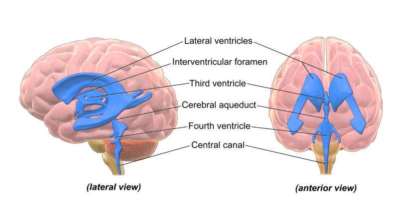
Diagram - The Ventricular System
Licensed under Creative Commons, Source.
The CSF drains from the fourth ventricle in a number of ways.
The first route is through the central canal of the spinal cord, which runs down the middle of the cord. Only a small amount of CSF goes this way. The canal is very narrow, and in adults it is mostly closed.
The second route carries the majority of the CSF. It leaves through three apertures (openings) in the fourth ventricle, shown in the image above. They let CSF pass from the ventricles of the brain into the subarachnoid space (the fluid-filled space around the brain and spinal cord). There is a pair of lateral apertures, which are the two arm-like structures beside the fourth ventricle on the anterior view above. There is also a single median aperture on the dorsal (back) side of the fourth ventricle, seen on the lateral view above.
These apertures are named after people (they are eponyms): the lateral apertures are known as the foramen of Luschka, and the median aperture is known as the foramen of Magendie. A memory aid is that Lateral and Luschka both start with ‘L’ and Median and Magendie both start with ‘M’.
From the subarachnoid space, most CSF is absorbed through arachnoid granulations (small projections of the arachnoid mater that push into the venous sinuses) into the superior sagittal sinus (a large channel for venous blood along the top of the brain), and so into the venous system. Newer evidence suggests that some CSF also drains by other routes, such as lymphatic vessels in the dura mater (the tough outer layer of the meninges) and along the sheaths around cranial and spinal nerves. The image below shows the circulation of CSF through the ventricular system and around the brain in more detail.
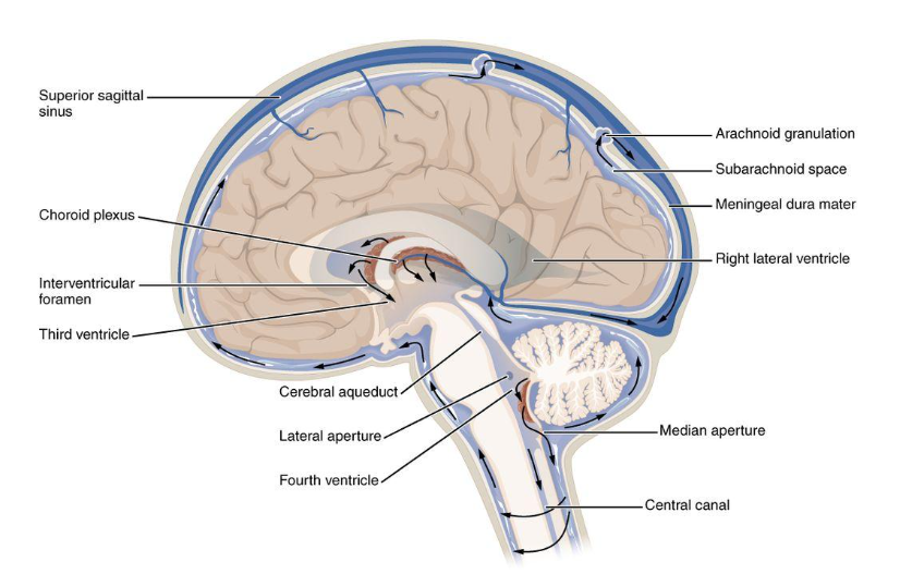
Diagram - The Circulation of the Cerebrospinal Fluid through the Ventricular System and Subarachnoid Space
Licensed under Creative Commons, Source.
Edited by: Dr. Marcus Judge
Reviewed by: Adrian Judge
- 793


