

Next Lesson - Reticular Formation
Core
The cerebral cortex can be compared to a massive computer, continuously firing neurons and working via a sequence of complex inputs and outputs. The cortex is made up of grey matter. Most of it (the neocortex) is arranged into 6 layers containing cell bodies and dendrites; the hippocampus and olfactory cortex (the smell area) have only three layers.
- Inputs to the cortex include the thalamus and directly from other areas in the cortex (these can perform actions without any sensory input).
- Outputs are to nerve tracts, which are bundles of axons in the CNS. There are three types of these:
- Association fibres connect nearby regions of the same hemisphere.
- Commissural fibres connect the two hemispheres.
- Projection fibres to the brainstem and cord.
- The inputs connect to the outputs and are processed by interneurons (small neurones that link one neurone to another within the brain), and the complexity of these connections gives rise to different types of behaviour and emotions.
Localisation of Functions in the Lobes
Each of the cerebral hemispheres is split into four lobes: frontal, temporal, parietal and occipital. Each of these carries a different purpose related to the everyday functioning of the human body.
The frontal lobe is found in the most anterior part of the brain and extends back as far as the central sulcus (the groove that separates the frontal and parietal lobes).
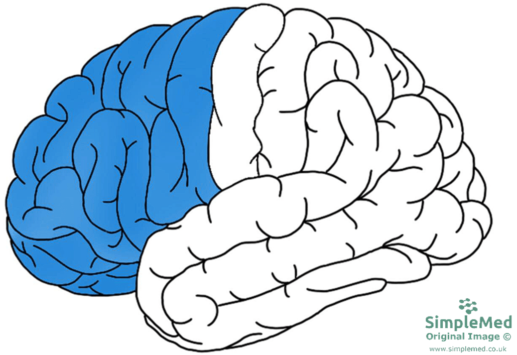
Image - Frontal lobe highlighted
SimpleMed original by Dr. Keertana Anne
It carries functions such as:
- Motor - the primary motor cortex (the area that sends commands for voluntary movement) is found in the precentral gyrus (a ridge of cortex), which sits just anterior to the central sulcus.
- Expressing Speech - Broca’s area is found here, in the left hemisphere in about 95% of people (less often in left-handers).
- Cognition - the prefrontal cortex (the front part of the frontal lobe) is involved in functions such as memory, planning and problem-solving.
- Behavioural Regulation - drinking excess alcohol can lead to frontal lobe dysfunction, which causes inappropriate behaviour.
- Continence - areas on the inner surface of the frontal lobe, including the paracentral lobules (one on the inner surface of each hemisphere), are part of a wider brain network that controls urination. Control of defecation also depends on the brain.
The prefrontal cortex is one of the last parts of the brain to mature, continuing to develop until the end of adolescence. Control of the bladder and bowel also depends on physical, thinking and emotional development, which most children have reached by 18 to 30 months of age, so babies take a long time to gain these functions.
The parietal lobes are paired lobes (one in each hemisphere) that sit posterior to the frontal lobe, just behind the central sulcus.
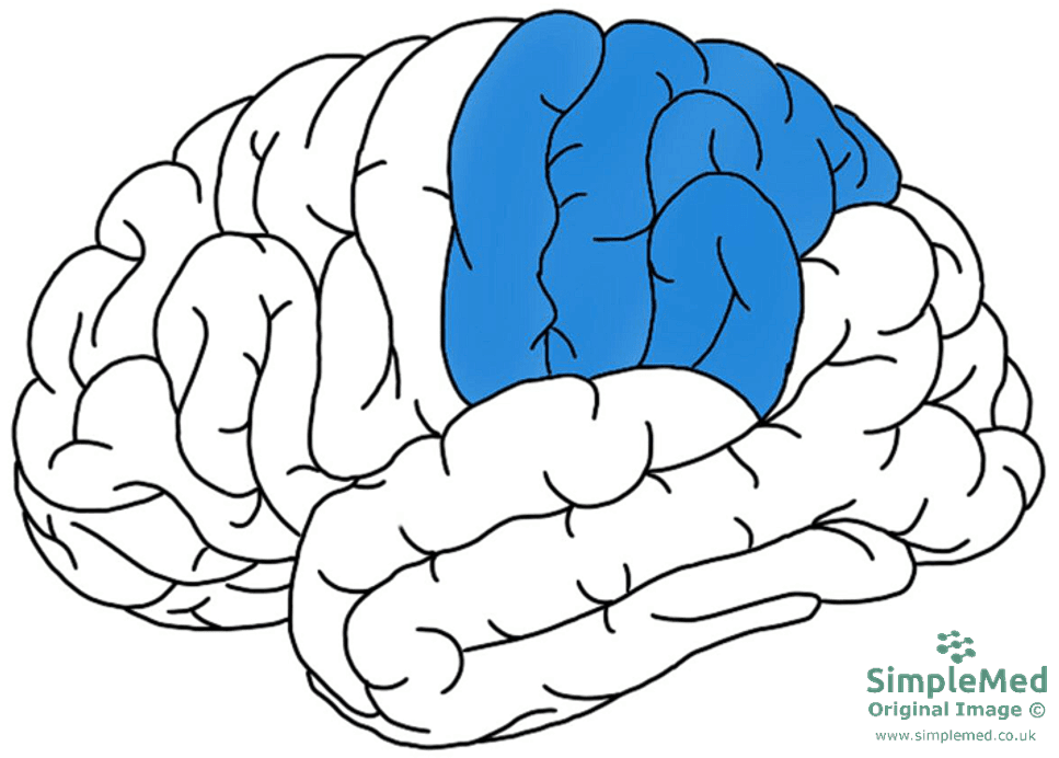
Image - Parietal lobe highlighted
SimpleMed original by Dr. Keertana Anne
It carries functions such as:
- Sensation - the postcentral gyrus (the ridge of cortex just behind the central sulcus) contains the primary sensory cortex. Sensory inputs from the skin relay through the thalamus to the parietal lobe.
- Understanding Speech - Wernicke’s area is located in the posterior part of the temporal lobe, close to the junction with the parietal lobe. It is listed here because of this position next to the parietal lobe. Similar to Broca’s area, it is usually found in the left hemisphere.
- Awareness of external environment and body. This relies on the lobe interpreting sensory information; it lets you know the location of parts of your body in space.
- Working with the frontal lobe to perform calculation and writing tasks.
The temporal lobes are paired lobes (one in each hemisphere) that sit below the parietal lobes on either side. Their main jobs include making sense of sounds, including speech, and helping to form and store memories.
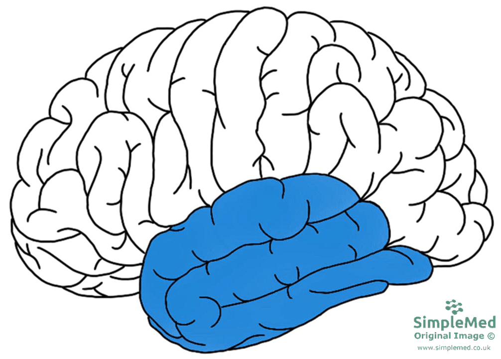
Image - Temporal lobe highlighted
SimpleMed original by Dr. Keertana Anne
It carries functions such as:
- Hearing and Olfaction (Smell) - hearing is mediated by the primary auditory cortex in the temporal lobe, while olfaction involves cortical areas within the temporal lobe as part of a wider network. Damage can disturb the recognition of sounds, including speech, and of smells. Seizures that start in the temporal lobe can cause auditory or olfactory hallucinations (hearing or smelling things that are not there).
- Memory - the hippocampus (a curved structure on the inner surface of the temporal lobe) helps to form and consolidate factual (declarative) information into long-term memory. Over time, these memories are stored in the cerebral cortex.
- Emotion - the hippocampus and amygdala (an almond-shaped structure involved in emotions such as fear) are part of the limbic system, a set of structures involved in emotion and memory. They have a complex effect on emotion and are thought to be related to psychiatric disorders.
The occipital lobe is located at the most posterior region of the brain, and sits just behind the parietal and temporal lobes. The primary function involves the processing of visual information in the brain.
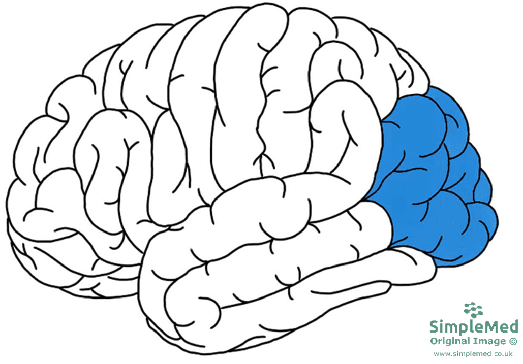
Image - Occipital lobe highlighted
SimpleMed original by Dr. Keertana Anne
It carries functions such as:
- Vision - the primary visual cortex sits here and receives visual information from the eyes via the optic nerves and tracts, with relay through the thalamus. See the article on the Visual System for more information. The primary visual cortex receives the visual information and begins to process it. Neighbouring visual areas then analyse particular features, such as colour (area V4) and movement (area V5). The processed information is sent to other areas of the brain, so they are able to respond appropriately.
Certain functions of the brain are better represented in one hemisphere than the other. Knowing where these functions are mostly handled helps doctors predict the causes of neurological deficits (loss of a normal function, such as weakness or loss of speech) and work out which area of the brain is affected.
The left hemisphere is typically dominant for language, while the right hemisphere has a prominent role in spatial attention. Most complex activities depend on networks spanning both hemispheres. Lateralisation (how far one side of the brain handles a function) varies between individuals and between functions. For example, in one study of healthy adults, language was mainly handled by the right hemisphere in 4% of strong right-handers and 27% of strong left-handers.
A lesion (an area of damaged tissue) affecting the right parietal lobe or its attention networks can cause left hemispatial neglect: reduced awareness of the left side of the body or surrounding space. This reflects disrupted attention rather than a simple division of every function between hemispheres. Its severity and recovery vary, and in some patients other networks can compensate. On a clock-drawing task, a patient may leave the left side of the clock empty and crowd the numbers onto the right.
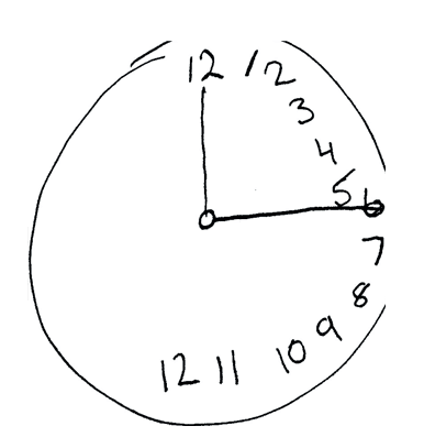
Image - Hemispatial neglect. A clock-drawing task may reveal reduced attention to the left side of space
Public Domain Source by Dhru4you [Public domain]
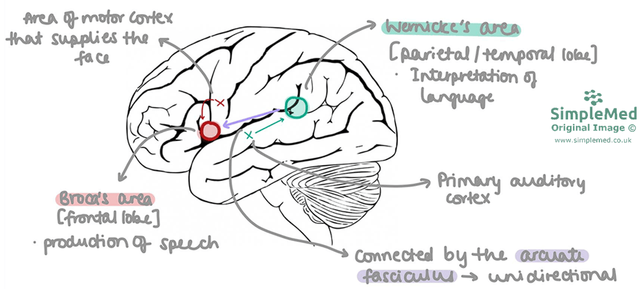
Diagram - This is the pathway for repeating the heard word
SimpleMed original by Dr. Keertana Anne
The process of language comprehension and the production of speech takes place in the left hemisphere of the majority of the population. The image above shows the connective pathways between different areas in the brain and how these all work together to produce the spoken word.
The primary auditory cortex (PAC) in the temporal lobe receives the sound signals sent from the ear. Wernicke’s area is found at the parieto-temporal junction (where the temporal and parietal lobes meet) in close proximity to the PAC, and it is responsible for decoding and interpreting these sounds. This information is then transmitted along the arcuate fasciculus (a bundle of white matter fibres that links the two areas of grey matter, Wernicke’s and Broca’s, and appears to carry signals in both directions) to Broca’s area in the frontal lobe. Broca’s area is responsible for producing speech, and it coordinates with an area of the primary motor cortex that supplies the muscles of the face, pharynx and larynx.
Damage to the individual areas can cause different types of speech deficits. The left hemisphere is dominant for language processing in about 95% of the population (less often in left-handers). So it is usually damage to the left half of the brain that causes these aphasias (language impairments).
- Wernicke’s Aphasia - arising from damage to Wernicke’s area (posterior temporal lobe, near the parietal lobe). Patients suffer from receptive aphasia, resulting in fluent speech (speech that flows easily) that is nonsensical, with wrong or substituted words (paraphasias). The patient can still speak fluently because Broca’s area is intact, but has a marked impairment in understanding spoken language and is often unaware of it.
- Broca’s Aphasia - arising from damage to Broca’s area (frontal lobe). Patients suffer from expressive aphasia, causing non-fluent, staccato speech (halting and effortful, in short phrases that leave out small words). The patient understands what is being asked, but struggles to get the words out.
- Conduction Aphasia - classically arising from damage to the arcuate fasciculus (more recent work also points to the cortex where the temporal and parietal lobes meet). The patient is able to comprehend auditory inputs and produce fluent speech, but make sound errors (phonemic paraphasias, where sounds within words are swapped or added). They are specifically unable to repeat a heard word.
The following pathways use the same basic areas of the brain (Broca’s and Wernicke’s) in order to produce speech from a variety of inputs.
- Pathway for speaking a word that you are thinking of (a thought word)
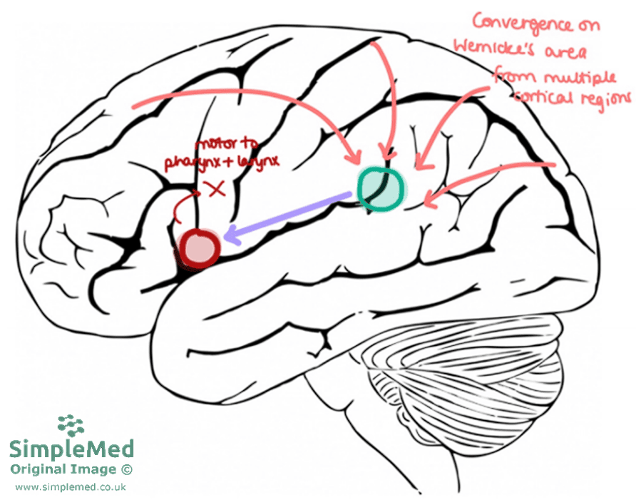
Diagram - Pathway for speaking a word that you are thinking of (a thought word)
SimpleMed original by Dr. Keertana Anne
- Pathway for speaking a written word
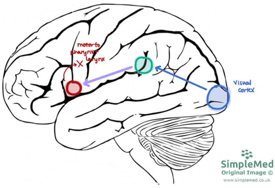
Diagram - Pathway for speaking a written word
SimpleMed original by Dr. Keertana Anne
As mentioned above, the temporal lobe and its associated structures are primarily responsible for the creation and consolidation of memories.
Memories are thought to be stored across large areas of the brain and are classified into two different groups: declarative and non-declarative. Declarative (or explicit) memories are factual information and events, and tend to be stored in the cerebral cortex. Non-declarative (or implicit) memories include learnt skills and habits (such as motor skills), which depend on subcortical structures (structures deep to the cortex) such as the basal ganglia (deep grey matter involved in movement and habit) and cerebellum. They also include priming (an unconscious effect of earlier exposure) and simple conditioned responses, and implicit emotional learning depends on the amygdala. Declarative memory is the conscious storage and recollection of data, while non-declarative is unconscious.
Memories can also be divided by how long they last into short-term (seconds to about half a minute) and long-term memories. The declarative and non-declarative groups above are the two main types of long-term memory. Short-term memories can be converted to long-term through the process of consolidation, allowing for declarative or non-declarative memories to be stored in the locations mentioned above. Factors influencing the conversion of these memories include rehearsal (repeating the information), association (linking it to things already known) and emotional context. Emotionally arousing events tend to be remembered better, because the amygdala strengthens their consolidation. In post-traumatic stress disorder (PTSD), distressing memories of a traumatic event keep returning as flashbacks, intrusive images or nightmares.
The hippocampus (part of the temporal lobe) is essential to consolidate declarative memories into the cerebral cortex. It receives inputs from multiple areas of the brain (visual, auditory and sensory areas, and the limbic system, a set of structures involved in emotion and memory), which makes it very good at linking stimuli to a memory.
Learning and memory are thought to depend on lasting changes in the strength of neuronal synapses. One important example is long-term potentiation (a lasting strengthening of a synapse after repeated activity). It involves changes in glutamate receptors (receptors for glutamate, an excitatory neurotransmitter), which strengthen the synapse and are important in learning and memory (See article on Cells and Components of the Nervous System for more information on long-term potentiation).
Edited by: Dr. Maddie Swannack
Reviewed by: Dr. Thomas Burnell and Dr. Marcus Judge
- 999

