

Next Lesson - Production and Circulation of CSF and Related Disorders
Core
The central nervous system (CNS) is one of the first body systems to begin development and the last one to complete it (continuing after the birth of the baby). Because its development takes so long, there is a long period in which it can be damaged, from early pregnancy through to adolescence.
The nervous system begins to develop in the 3rd week following fertilisation. It is formed from the ectoderm layer (the outer germ layer) of the trilaminar disc (the flat, three-layered disc of the early embryo). A process called neurulation forms the basis of the central nervous system. It begins when the ectodermal layer of the embryo specialises to form neuroectoderm (the part of the ectoderm that becomes nervous tissue). This is induced by a rod of tissue in the midline called the notochord, which is formed from mesoderm (the middle germ layer). The neuroectoderm forms the neural plate (a thickened plate of tissue) along the back (the dorsal surface) of the trilaminar disc. The border of this neural plate goes on to form neural crest cells, which will be covered later in this article. From about day 18 (in week 3), a groove forms within the neural plate and causes it to fold in on itself. The folds then fuse and separate off as the neural tube by about week four of development (see Diagram 1).

Diagram 1 - Neurulation. This process involves the formation of the neural tube and neural crest cells from neuroectoderm
Public Domain Source by NikNaks [Public domain]
The closure of the neural tube takes approximately 10 days to complete, from about day 18 to about day 28 after fertilisation. It runs from the middle of the embryo (near the junction of the future brain and spinal cord) to either end. Signals from the notochord help to control how the neural plate bends. In chick and mouse embryos, the cells in the midline of the neural plate, directly above the notochord, form a hinge (the median hinge point), which helps the plate to bend. Neuropores (cranial and caudal) are the openings in the neural tube that remain at either end until the tube closes. The cranial neuropore closes at about day 24 and the caudal neuropore at about day 26.
Neural tube defects arise from the failure of either neuropore to completely close, although the most extreme form, craniorachischisis (described below), arises when closure fails to start. In the UK, the 20-week scan (an ultrasound scan offered between 18 and 21 weeks of pregnancy) checks for anencephaly (a condition in which large parts of the brain and skull are missing) and open spina bifida (a gap in the spine, with the spinal cord or its coverings pushing out through it). A raised maternal serum α-fetoprotein (a protein made by the fetus, which would normally be contained within the fetal body) can also point to a neural tube defect before birth. However, this test is not specific, because the level can also be raised by defects in other systems. There are many causes of neural tube defects, but taking folic acid (a B vitamin) can prevent many cases. In the UK, the advice is to take 400 micrograms of folic acid every day. Ideally, start 3 months before conception (or as soon as the pregnancy is known) and continue until the end of week 12 of pregnancy. Women at higher risk are advised to take a higher dose of 5 mg every day. Examples are women who have had a previous pregnancy affected by a neural tube defect, women who have diabetes and women who take certain epilepsy medicines. From 13 December 2026, folic acid must also be added to non-wholemeal wheat flour in the UK. The advice to take a folic acid supplement continues.
Anencephaly results from the failure of the cranial neuropore to close. Large parts of the brain and the skull do not form, and the face and neck may also be affected. There is no treatment. If the pregnancy continues, most babies (up to 3 out of 4) are stillborn. Those born alive die shortly after birth, and only rarely live for a few days.
Spina bifida is due to the failure of the caudal neuropore to close. It is most common in the lumbosacral region (the lower back and sacrum), but can occur anywhere. In spina bifida, there is an incomplete closure of the vertebrae, which can lead to the protrusion of the spinal cord out of the spinal column. Babies born with open spina bifida, such as myelomeningocele, often have hydrocephalus (a build-up of fluid in the brain, which puts pressure on it). Hydrocephalus can damage the brain and cause learning difficulties (cognitive delay). There are multiple types of spina bifida and the severity ranges from mild to life-threatening.
- Spina bifida occulta is the mildest form. There is an opening in the vertebrae but no protrusion through it, so the spinal cord itself is not at high risk. It does not usually cause symptoms, so most people do not know they have it, and it is often found by chance on a scan done for another reason. A few people have problems such as pain or numbness and need treatment.
- Meningocele occurs when only meninges and cerebrospinal fluid protrude through the vertebral column but the spinal cord remains in place. This means that the meninges could be damaged, but the spinal cord should generally be unaffected.
- Myelomeningocele is the most severe form and is the protrusion of the spinal cord and meninges through the opening in the vertebrae. This is the most dangerous form of spina bifida, because the spinal cord is at risk.
Rachischisis is the failure of the neural tube to close during neurulation, resulting in exposed neural tissue along the affected region. The most extreme form is craniorachischisis, in which closure never starts, so the hindbrain and the whole spinal neural tube stay open. This is lethal before or at birth. If only the spine is affected, as in open spina bifida, survival after birth is generally possible, but there is usually neurological impairment.
Formation of the Nervous System
The central nervous system is made up of several components, all of which develop from the neural tube (see Diagram 2).
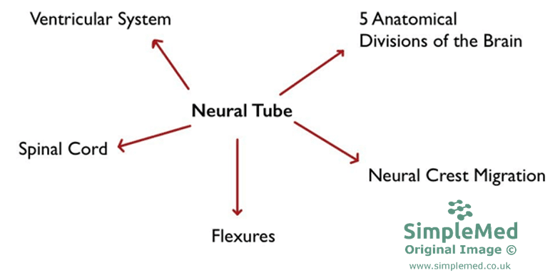
Diagram 2 - The different components that make up the CNS
SimpleMed original by Dr. Keertana Anne
Patterning and Organisation of the Neural Tube
The wall of the neural tube is made up of neuroepithelial cells (the first stem-like cells lining the neural tube). These differentiate into neuroblast cells (immature nerve cells) to form the mantle layer (grey matter). Nerve fibres emerge from the grey matter to form the marginal layer (white matter). The ventral part of the mantle layer is called the basal plate while the dorsal part is called the alar plate. The basal plate forms the motor areas and the alar plate forms the sensory areas.
The roof and floor plates (the thin strips of tissue along the dorsal and ventral midline of the neural tube) regulate how the dorsal and ventral areas take on different roles (known as patterning). This patterning differs along the length of the neural tube (see Diagram 3 for the patterning in the spinal cord).
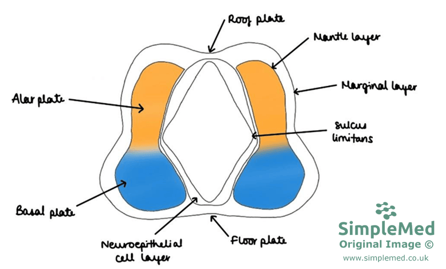
Diagram 3 - Cross-section of the neural tube in the spinal cord during development
SimpleMed original by Dr. Keertana Anne
The ventricles of the brain are a set of 4 connected spaces. Cerebrospinal fluid (CSF) is produced within them by the choroid plexuses (the vascular tissue lining the ventricles that makes CSF). Refer to our article on the production and circulation of CSF for more information. The ventricles are formed from the lumen of the neural tube (see Diagrams 4 and 5).
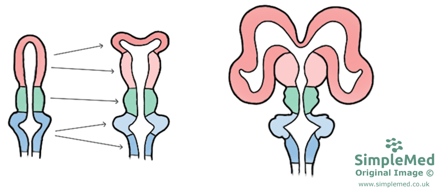
Diagram 4 - The formation of the ventricles from the lumen of the neural tube. The diagram also shows the development of the anatomical divisions of the brain
SimpleMed original by Dr. Keertana Anne
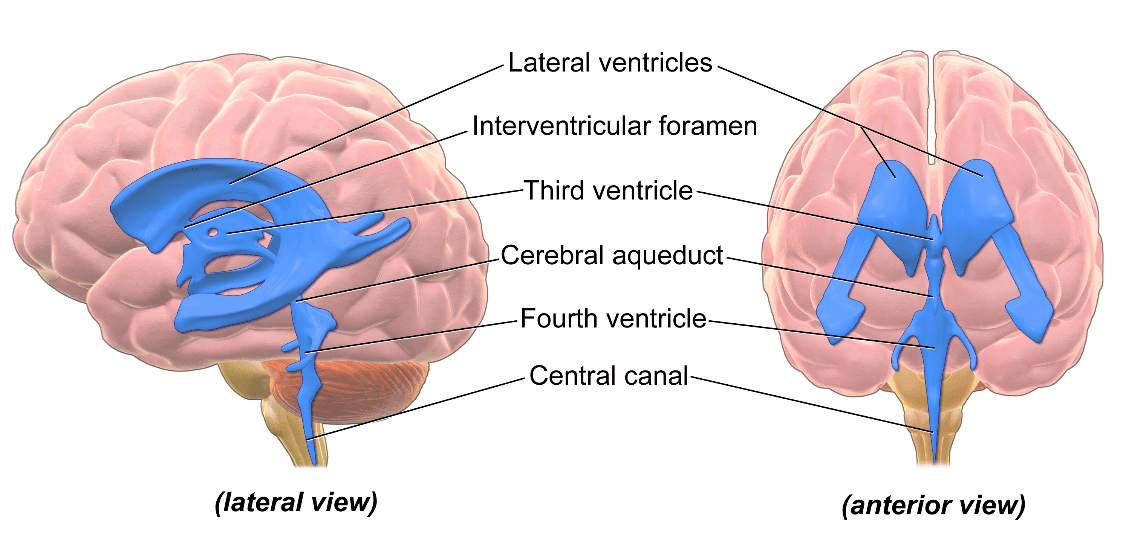
Diagram 5 - The position of the ventricular system in the brain
Creative commons source by BruceBlaus [CC BY-SA 4.0 (https://creativecommons.org/licenses/by-sa/4.0)]
Most of the neural tube becomes the spinal cord. The elongation of the spinal cord takes place in tandem with the growth of the vertebral column until the third month (at which point they are the same length). However, after the third month the vertebral column grows faster than the spinal cord. As a result, the conus medullaris (the tapered lower end of the spinal cord) appears to ascend, and in the adult it lies at around the level of the first or second lumbar vertebra (L1 or L2). Each spinal nerve must still leave the vertebral canal through the intervertebral foramen (the gap between two vertebrae) at the same level as before, so only the roots of the spinal nerves must elongate. The elongated roots form the cauda equina (Latin for horse's tail), the bundle of lumbar and sacral nerve roots below the end of the spinal cord. (See Diagram 6).
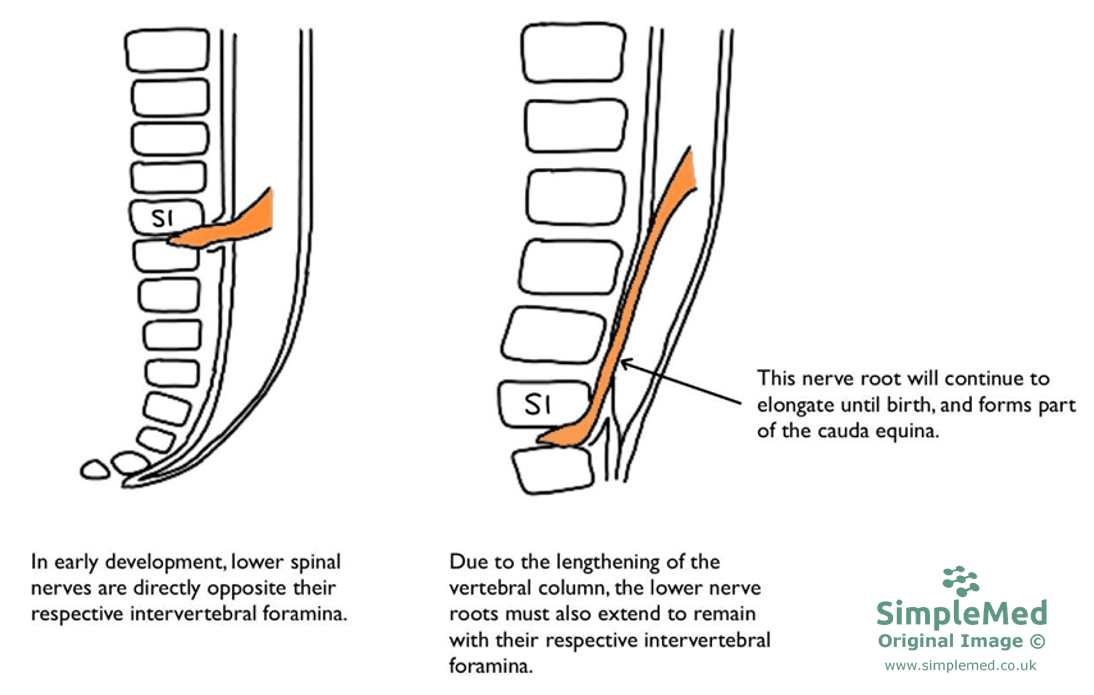
Diagram 6 - Elongation of nerve roots in the spinal cord
SimpleMed original by Dr. Keertana Anne
The formation of the brain happens in two stages. The first stage begins during neural fold formation (when the raised edges of the neural plate form), where three primary areas can be distinguished:
- The forebrain - the prosencephalon
- The midbrain - the mesencephalon
- The hindbrain - the rhombencephalon
After the closure of the neural tube, these three areas become the primary brain vesicles (swellings at the cranial end of the neural tube). In the second stage, the primary brain vesicles form secondary brain vesicles at 5 weeks of development (see Diagram 7).
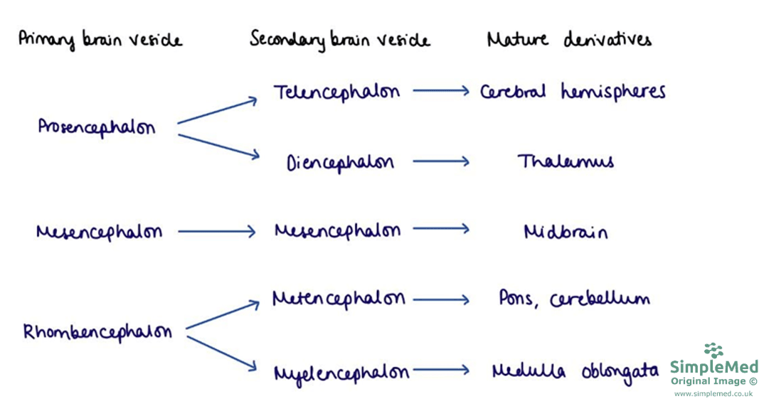
Diagram 7 - The stages of the anatomical divisions of the brain and their mature derivatives
SimpleMed original by Dr. Keertana Anne
The growth of the cranial neural tube exceeds the space available, and this limitation causes the brain to fold up forming flexures (see Diagram 8). The flexures formed include the cephalic flexure (in the midbrain region), the cervical flexure (between the hindbrain and spinal cord), and the pontine flexure (named after the pons, part of the brainstem).
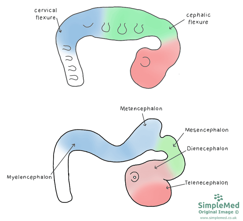
Diagram 8 - Flexures of the brain in relation to the primary and secondary brain vesicles
SimpleMed original by Dr. Keertana Anne
Neural crest cells are formed from the lateral border of the neural plate (see Diagram 1). They go on to migrate (move) to the rest of the body and form a diverse population of cells (see Table 1).

Table 1 - Derivatives of neural crest cells
SimpleMed original by Dr. Keertana Anne
This migration and specialisation can be vulnerable to both genetic and environmental insult (damage from faulty genes or from outside factors). This damage may affect one type of cell derived from the neural crest or several types. They result in neurocristopathies (disorders of neural crest development), such as DiGeorge syndrome (also known as 22q11.2 deletion syndrome) or Treacher Collins syndrome (a genetic condition that affects the development of the face).
Reviewed by: Dr. Thomas Burnell and Dr. Marcus Judge
Edited by: Dr. Maddie Swannack
- 3854


