

Next Lesson - The Spine
Core
A joint is defined as the point in the body where two bones meet. As a result, a joint doesn’t just include obvious, freely movable joints such as the knee and shoulder but also features such as the joins between bones in the skull.
Joints balance the properties of movement and stability. This means that joints with a high range of movement are often more unstable, such as the shoulder joint, which dislocates relatively easily. It also means that joints with lower range of movement are often more stable such as the sutures in the skull.
There are three main types of joint in the body:
Fibrous joints are strong, stable joints, with tough fibrous connective tissue binding the bones together. As a result, fibrous joints have very limited movement. An example of this would be the joints between skull bones, known as sutures. This stability helps to protect the brain. Skull sutures allow some movement until about 20 years of age, after which they become fixed. In a baby the skull bones are not yet fused, so the skull can change shape as it passes through the birth canal. Another group, the syndesmoses (bones held together by a sheet of fibrous membrane), allows slight movement. An example is the middle radioulnar joint between the shafts of the two forearm bones.
Cartilaginous joints unite bones through cartilage and provide stability. Primary cartilaginous joints, or synchondroses, use hyaline cartilage (smooth, glassy cartilage) and are functionally immobile; growth plates in growing bones are examples. Secondary cartilaginous joints, or symphyses, use fibrocartilage (tougher cartilage containing strong fibres) and allow slight movement. Examples include the pubic symphysis and the intervertebral discs between adjacent vertebral bodies.
Synovial joints are the most mobile of all the types of joints. The bone ends are capped with articular (joint-surface) cartilage, which is usually hyaline (smooth and glassy) and reduces friction and absorbs shock. A fibrous capsule surrounds the joint. Its inner lining, the synovial membrane, makes the synovial fluid that fills the joint cavity. These joints often have associated ligaments or menisci (crescent-shaped pads of cartilage that cushion the joint). There are six main types of synovial joints in the human body:
This type of joint is relatively simple. It involves one bone with a rounded end inserting into a corresponding cup. Common examples of this in the body are the hip joint and the shoulder joint. This type of joint allows multidirectional movement, including rotation, flexion, extension, abduction and adduction. Muscles move the joint, while the ligaments and muscles that support it limit how far it can move.

Diagram of a ball and socket joint.
SimpleMed original by Dr. Maddie Swannack.
This type of joint is very similar to a ball and socket joint except that the ball is elliptical in shape. Also known as a condyloid joint, this joint allows flexion, extension, abduction, adduction and circumduction (moving in a circle). It does not allow true axial rotation (spinning around the bone’s own long axis), as the oval shape of the bone cannot spin in the oval shaped socket. An example of this is the radio-carpal joint (the wrist joint).
This type of joint involves one bone ending in a cylindrical shape, which fits into a trough in another bone. An example of this would be the elbow. Due to the cylindrical shape of the articular surface (where the bones contact each other) this joint only allows movement in one plane, bending and straightening (flexion and extension), like a hinge in a door that can only move along one axis.

Diagram of a hinge joint.
SimpleMed original by Dr. Maddie Swannack.
This type of joint is made up of a peg in a hole. This joint only allows rotational movement. This joint occurs between the atlas (C1) and the dens (the peg-shaped projection of the axis, C2), the first two neck vertebrae (the median atlantoaxial joint). Ligaments limit its movement and help to keep it stable. One of them, the transverse ligament of the atlas, holds the dens in place. The joint can still become unstable, for example after injury.
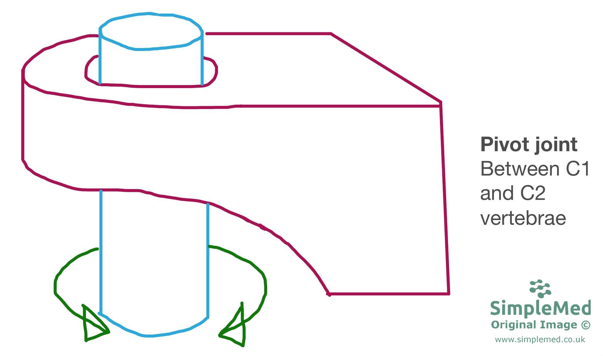
Diagram of a pivot joint.
SimpleMed original by Dr. Maddie Swannack.
Plane joints have relatively flat articular surfaces that allow small gliding movements, limited by the joint capsule and ligaments. Examples include the intercuneiform joints (between the wedge-shaped cuneiform bones of the midfoot) and the second to seventh sternocostal joints (between the breastbone and the rib cartilages). The first sternocostal joint is a primary cartilaginous synchondrosis, rather than a plane synovial joint.
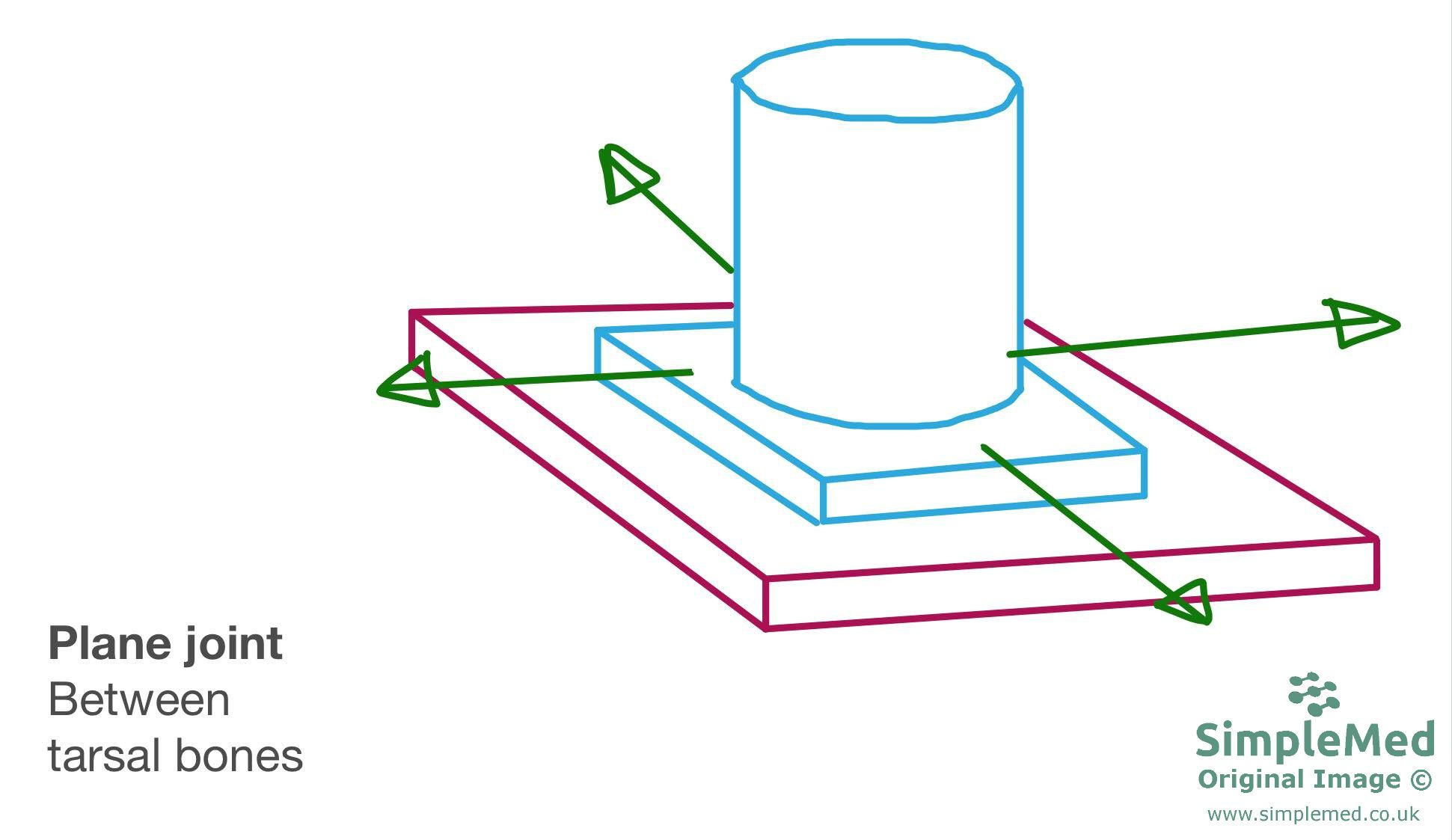
Diagram of a plane joint.
SimpleMed original by Dr. Maddie Swannack
Saddle joints are named after their resemblance to a horse’s saddle. The articular surfaces are shaped like Pringles: each surface curves inwards (concave) in one direction and outwards (convex) in the other. This allows movement in two arcs that are perpendicular to each other. An example of this joint in the body is between the trapezium (one of the carpal bones) and the 1st metacarpal bone (the hand bone at the base of the thumb).
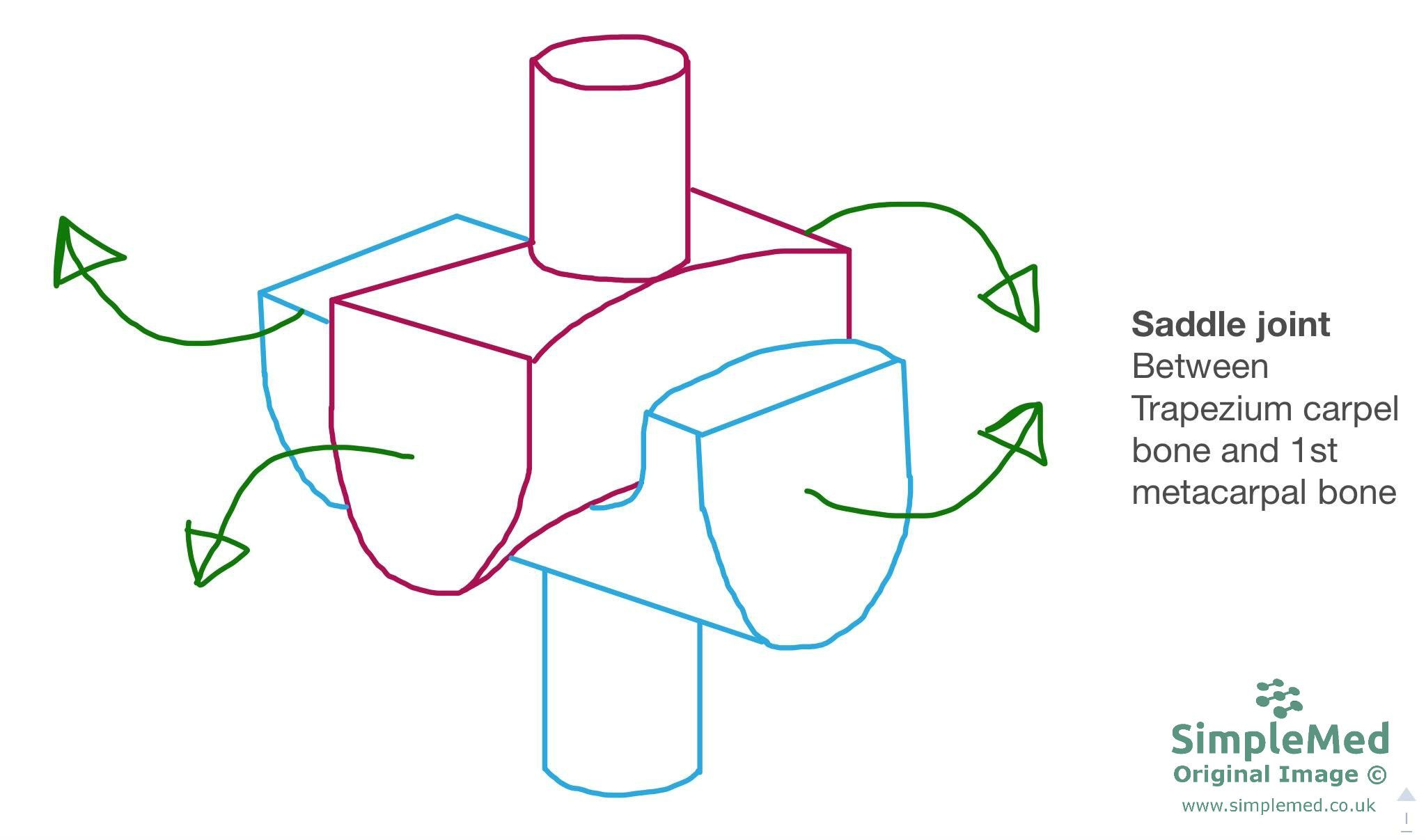
Diagram of a saddle joint.
SimpleMed original by Dr. Maddie Swannack
For information about the pathology of each type of joint, check out the Clinical Conditions section of the Musculoskeletal unit.
Edited by: Dr. Ben Appleby
Reviewed by: Dr. Marcus Judge
- 15699
