

Next Lesson - Conditions of the Foot
Core
This article is closely associated with the article on the Lumbar Plexus, as both the lumbar and sacral plexuses supply peripheral nerve innervation to the lower limb.
The sacral plexus is a convergence of spinal nerves that occurs lateral to the sacrum. It includes input from six spinal nerves (L4 [lumbar nerve 4], L5, S1 [sacral nerve 1], S2, S3 and S4) and gives rise to several branches. The five major nerves discussed here are superior gluteal, inferior gluteal, sciatic, posterior femoral cutaneous and pudendal.
Each of these peripheral nerves will be discussed individually throughout the article.
The mnemonic “Sassy Irish Sailors Pester Parrots” recalls the order of the five nerves in this teaching list: superior gluteal, inferior gluteal, sciatic, posterior femoral cutaneous and pudendal. These list positions do not describe an invariant anatomical sequence of branch origins, and smaller branches are not included in the mnemonic.
The five major nerves discussed here leave the pelvis through the greater sciatic foramen. This is not the route of every sacral plexus branch: the nerve to levator ani passes directly to the pelvic floor within the pelvis.
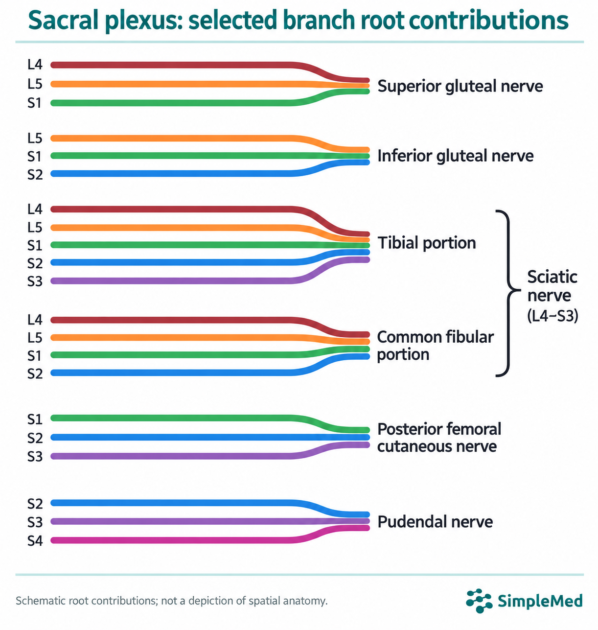
Schematic - Root contributions to selected branches of the sacral plexus. Superior gluteal: L4, L5 and S1. Inferior gluteal: L5, S1 and S2. Tibial portion: L4, L5, S1, S2 and S3. Common fibular portion: L4, L5, S1 and S2. Posterior femoral cutaneous: S1, S2 and S3. Pudendal: S2, S3 and S4. The tibial and common fibular portions form the sciatic nerve (L4–S3). This schematic shows root contributions, not spatial anatomy or the order in which branches arise.
SimpleMed root-contribution schematic, 2026. Previous diagram by Dr. Maddie Swannack.
Peripheral Nerves of the Sacral Plexus
The superior gluteal nerve is a major branch of the sacral plexus.
It leaves the pelvis through the greater sciatic foramen, superior to the piriformis muscle.
It has three spinal nerve contributions: L4, L5 and S1.
The motor functions of the superior gluteal nerve include the gluteus minimus, gluteus medius and the tensor fascia lata.
It has no sensory functions.
The inferior gluteal nerve is listed second in the five-name mnemonic used here.
It leaves the pelvis through the greater sciatic foramen, inferior to the piriformis muscle.
It has three spinal nerve contributions: L5, S1 and S2.
The motor function of the inferior gluteal nerve is the innervation of gluteus maximus.
It has no sensory functions.
The sciatic nerve is the largest nerve in the body, approximately 2cm wide, and is listed third in the five-name mnemonic used here. It can be considered to be two separate nerves within the same fibrous sheath: the tibial nerve and the common peroneal nerve (also known as the common fibular nerve). In most people, the sciatic nerve separates into the two constituents just superiorly to the popliteal fossa, but they may separate at any point superior to this.
It leaves the pelvis through the greater sciatic foramen, emerging inferiorly to the piriformis muscle (in most people). It passes over the muscles of the deep gluteal region and enters the posterior thigh by passing deep to the long head of biceps femoris. Within the posterior thigh, it gives off the branches to the hamstring muscles, and the adductor magnus (which is half innervated by the sciatic nerve and half innervated by the obturator nerve). When the sciatic nerve reaches the superior border of the popliteal fossa, it divides into the tibial and common peroneal nerves.
Spinal Nerve Contributions
- Sciatic - to the sciatic nerve as a whole, the following nerves contribute: L4, L5, S1, S2, and S3.
- Tibial - to the tibial portion of the sciatic nerve, the following nerves contribute: L4, L5, S1, S2, and S3 (all of the spinal nerves that contribute to the sciatic nerve branch into the tibial nerve).
- Common Peroneal (Common Fibular) - the common fibular portion receives L4, L5, S1 and S2 (L4–S2). The sciatic nerve as a whole remains L4–S3.
- The sciatic, tibial and common peroneal nerves all have individual motor functions:
- Sciatic - posterior compartment of the thigh (biceps femoris, semitendinosus, semimembranosus) and the hamstring portion of adductor magnus.
- Tibial - posterior compartment of the leg (gastrocnemius, soleus, plantaris, popliteus, tibialis posterior, flexor digitorum longus and flexor hallucis longus) and some of the intrinsic muscles of the foot.
- Common Peroneal - anterior compartment of the leg (tibialis anterior, extensor digitorum longus, extensor hallucis longus), the lateral compartment of the leg (fibularis longus and fibularis brevis), and the rest of the intrinsic muscles of the foot.
- In summary the sciatic nerve and its derivatives supplies the posterior thigh and the entire lower leg.
- The sciatic nerve itself does not provide any sensory innervation, but the tibial and common peroneal branches do.
- Tibial - sole of the foot.
- Common Fibular - its branches supply the anterolateral lower leg and the dorsum of the foot, including the first interdigital cleft via the deep fibular nerve.
The posterior femoral cutaneous nerve is listed fourth in the five-name mnemonic used here.
It has three spinal nerve contributions: S1, S2, and S3.
It leaves the pelvis via the greater sciatic foramen, inferior to the piriformis. It then travels inferiorly, deep to gluteus maximus, and down the posterior thigh to the knee.
The posterior femoral nerve has no motor functions.
Its sensory innervation includes the posterior surface of the thigh, as well as the perineum.
The pudendal nerve is listed fifth and last in this five-name mnemonic; it is not the final anatomical branch of the entire plexus.
It leaves the pelvis via the greater sciatic foramen, then enters the perineum via the lesser sciatic foramen. It moves between the ischium and the rectum.
It has contributions from three spinal nerves: S2, S3, and S4.
The motor functions of the pudendal nerve include the skeletal muscles of the perineum, the external urethral sphincter and the external anal sphincter. This gives rise to the saying ‘S2, S3 and S4 keep sh*t off the floor’ referencing the roots of the pudendal nerve.
The sensory functions of the pudendal nerve include the penis, the clitoris and the skin of the perineum.
Other Branches of the Sacral Plexus
Along with the five major peripheral nerves produced by the sacral plexus, there are a number of smaller branches that are not shown on the diagram. They are generally nerves that are directly supplying muscles:
- Nerve to piriformis
- Nerve to obturator internus
- Nerve to quadratus femoris
The Pelvic Splanchnic Nerves are separate from these muscular branches and provide parasympathetic innervation to the hindgut.
Pathologies of the Sacral Plexus
Damage to the Lumbosacral Plexuses
Damage to the lumbar or sacral plexuses (or both) can be very serious and are suspected when the symptoms shown cannot be localised to a single peripheral nerve region.
It can present with neuropathic pain, numbness, weakness or wasting of muscles.
One of the main causes is diabetic amyotrophy, also known as lumbosacral radiculoplexus neuropathy. It is thought to occur due to an immune mediated inflammatory process affecting the nerves, often in people with diabetes. Immunosuppressive treatments such as corticosteroids have been used in some cases, but evidence for their benefit is mixed.
Another cause is local invasions, such as tumours. Management depends on the underlying cause and may include surgery, radiotherapy or other oncological treatments, with a risk of further nerve damage.
Piriformis Syndrome
Piriformis syndrome is a clinical syndrome caused by compression of the sciatic nerve by the piriformis muscle, resulting in pain, numbness and muscle weakness. Treatment options can range from analgesia and physiotherapy, to corticosteroid injections, to intraoperative relief of pressure.
Edited by: Dr. Ben Appleby
Reviewed by: Dr. Thomas Burnell
- 17007
