

Next Lesson - Structure of Bones
Core
Cells and Matrix
Cartilage is specialised connective tissue built for compression, smooth articulation and flexible support. Its bulk is extracellular matrix rather than cells. General fibre chemistry and the connective-tissue family map belong in Connective Tissue; this lesson asks how those components are arranged so cartilage can load-bear, grow and often fail to heal.
Chondroblasts secrete matrix in growing or remodelling cartilage. Once enclosed by their own product they occupy small matrix compartments called lacunae and are termed chondrocytes. The apparent gap around a cell can enlarge during tissue preparation and should not be mistaken for a large fluid reservoir in life. Isogenous groups mark recent interstitial division: daughters stay close until new matrix separates them.
In hyaline cartilage the dominant structural collagen is type II. Fibrils form a tensile network that limits expansion of the hydrated gel. Aggrecan, the large aggregating proteoglycan, carries densely sulphated glycosaminoglycan chains whose fixed negative charges retain mobile ions and water. Under compression, pressurised interstitial fluid within the restrained matrix carries much of the early load. The first-pass functional pair is type II collagen restraining a water-rich aggrecan gel.
Cell density is low and metabolism modest once growth slows. Chondrocytes maintain matrix by balanced synthesis and limited degradation. Vessels do not penetrate mature matrix, so nutrients, gases and wastes must diffuse across hydrated tissue. That geometry is the structural root of slow turnover and weak repair.
Mechanical Behaviour
Cartilage does not behave like a dry spring. Under sudden load, interstitial fluid pressurises and shares force across a wide area. Fluid then flows slowly through the porous proteoglycan mesh, so deformation is time-dependent: rapid loading feels stiff, prolonged loading allows creep, and unloading permits fluid return and shape recovery. Collagen restrains the gel laterally and in tension, preventing sideways extrusion.
Fluid pressurisation and solid-matrix stress act together. Early in a load cycle, fluid pressure carries much of the compressive force and shields the collagen-proteoglycan solid. As fluid redistributes, a larger fraction of load is borne by the solid network. Ignoring fluid flow underestimates shock absorption; ignoring collagen underestimates tensile restraint and recovery.
The result is biphasic, viscoelastic behaviour suited to joints and flexible frameworks. Cyclic loading with recovery intervals allows fluid to re-imbibe; continuous overload keeps fluid displaced and concentrates stress on the solid matrix. That mechanical story, not hardness alone, explains why cartilage can look intact yet fail after repeated abnormal loading.
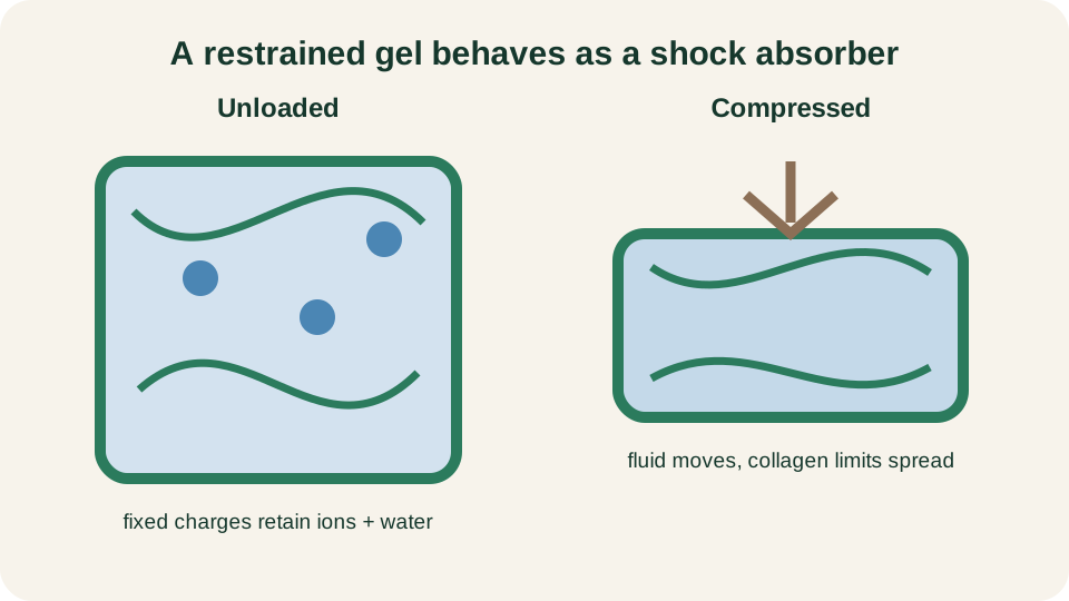
Aggrecan's fixed charges retain ions and water; compression redistributes fluid while collagen restrains matrix expansion.
SimpleMed original educational diagram
Three Types of Cartilage
Three histological types share the chondrocyte-in-lacuna plan but differ in matrix fibre content and sites.
Hyaline cartilage is the commonest. Its glassy matrix is rich in type II collagen and aggrecan. It forms the temporary fetal skeleton, growth-related cartilages of the young, costal cartilages, respiratory tract rings and most articular surfaces. Articular hyaline cartilage is a special case, modified by zone organisation and the absence of a free perichondrium.
Elastic cartilage adds a dense elastic-fibre network to a type II collagen and proteoglycan base. It is flexible and springy, as in the external ear, auditory tube and epiglottis. Elastic fibres restore shape after bending; the matrix remains avascular and diffusion-dependent.
Fibrocartilage meets tensile as well as compressive demand. It contains abundant type I collagen in ordered bundles, together with type II collagen and proteoglycans in cartilaginous regions. Type I is prominent in the fibrous component; fibrocartilage is neither pure type II tissue nor pure dense fibrous tissue. Sites include intervertebral discs, pubic symphysis, menisci and some tendon and ligament attachments to bone.
Type wording must stay careful. Hyaline and elastic cartilage are type II dominant. Fibrocartilage is dual: type I and type II both present, type I conspicuous in dense fibrous areas. Collagen type is a matrix signature, not a synonym for the tissue name.
Perichondrium and Nutrition
Many cartilages are wrapped by perichondrium: an outer fibrous layer continuous with surrounding connective tissue and an inner cellular layer that can supply chondroblasts for appositional growth. The perichondrium is vascular and a major nutrient source for the cartilage it invests. It is not universal. Fibrocartilage generally lacks a true perichondrium. Articular cartilage has no perichondrium on its free surface; its deep aspect meets calcified cartilage and subchondral bone rather than a vascular sheath.
Mature cartilage matrix contains no blood vessels, lymphatics or nerves, so chondrocytes have no direct blood supply. Nutrition is by diffusion from the nearest vascular bed or from synovial fluid. In perichondrium-covered hyaline and elastic cartilage, perichondrial vessels feed the outer matrix. In synovial joints, articular chondrocytes exchange metabolites mainly with synovial fluid produced by the vascular synovial membrane. Joint compression and release assist surface fluid mixing, but deep zones still depend on long diffusion paths.
Avascularity therefore links three facts: matrix turnover is slow once growth ends; inflammatory cells cannot recruit quickly within intact matrix; and repair is limited unless injury reaches vascular tissue.
Growth
Cartilage grows by two complementary mechanisms. Interstitial growth expands from within: chondrocytes divide inside lacunae and deposit new matrix between daughters, increasing volume and separating isogenous groups. It is important in young, soft matrix, including cartilaginous models that later contribute to bone length through endochondral pathways described in Structure of Bones.
Appositional growth adds matrix at a surface. Chondroblasts in the inner perichondrium lay down new layers from the outside. Where perichondrium is absent, that route cannot operate in the same way. Articular cartilage therefore has very limited adult capacity to thicken by either mechanism.
With maturation, matrix stiffens and division falls. Residual chondrocytes maintain composition more than they enlarge the tissue. Growth-plate cartilage remains a temporary engine of interstitial expansion until it closes; that endochondral sequence is reserved for the bone lesson.
Articular Cartilage Zones
Articular cartilage is hyaline cartilage organised into depth-dependent zones. From the joint surface downward the teaching sequence is superficial, middle, deep and calcified, with a tidemark at the mineralisation front toward subchondral bone.
In the superficial (tangential) zone, collagen runs largely parallel to the surface and chondrocytes are flattened. The zone resists shear and helps retain fluid in deeper matrix. The middle (transitional) zone has more rounded chondrocytes and obliquely organised collagen; proteoglycan content rises and compressive load-sharing begins. In the deep (radial) zone, collagen runs more perpendicular to the surface, chondrocytes often form columns, and high proteoglycan content maximises compressive stiffness. The calcified zone anchors cartilage to subchondral bone; the tidemark separates uncalcified from calcified cartilage.
Zone structure is functional architecture. Surface fibrillation begins where tangential collagen fails. Loss of deep anchoring or tidemark advance alters load transfer into bone. Zone language also clarifies partial- versus full-thickness lesion depth.
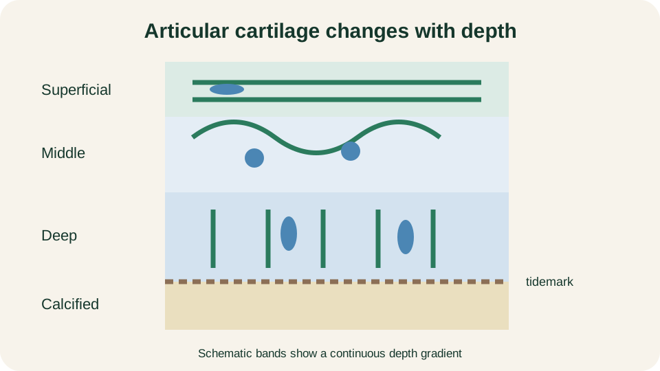
Schematic bands summarise a continuous depth gradient in collagen orientation and chondrocyte shape; the tidemark marks the calcified front.
SimpleMed original educational diagram
Limited Repair
Adult articular cartilage repairs poorly because the features that make it a durable bearing surface work against healing. There is no free-surface perichondrium to supply chondroblasts. Chondrocytes are sparse, enclosed and diffusion-dependent. Dense matrix blocks easy entry of progenitors into injuries that do not breach the tissue.
Partial-thickness lesions confined to uncalcified cartilage typically show little intrinsic healing. Adjacent chondrocytes may increase synthesis, but they rarely restore continuous, zonally organised type II matrix. The defect often remains as a stress-concentrating surface irregularity.
Full-thickness lesions that cross the tidemark, traverse calcified cartilage and reach vascularised subchondral bone allow blood and marrow elements into the defect, and repair tissue can form. That repair is commonly fibrocartilage-like rather than true hyaline articular cartilage: type I collagen is prominent, zonal architecture is incomplete and proteoglycan organisation is inferior. The fill may reduce the geometric hole, yet it is mechanically second-best under repetitive shear and compression and can later fibrillate and fail.
Outside joints, cartilages with perichondrium retain some appositional potential, but deep matrix injury still heals slowly and often incompletely. Across sites the rule holds: avascular matrix plus low cell mobility equals limited regeneration of native structure.
Clinical Consequences
When the restrained-gel design fails, joint surface integrity fails with it. Proteoglycan loss reduces fluid pressurisation and compressive stiffness. Collagen network damage allows abnormal swelling, then fissuring. Surface fibrillation can advance toward deeper delamination. Without restored architecture, local synthetic attempts leave the tissue vulnerable. Osteoarthritis can develop through this process, but it is a whole-joint disorder: synovium, subchondral bone, ligaments and periarticular tissues also contribute alongside cartilage damage.
Meniscal and disc fibrocartilage show the same principle under mixed tension and compression: fibre disruption impairs load distribution and can accelerate adjacent hyaline wear. Elastic cartilage heals imperfectly for the same avascular reason, though it is less often a major joint problem.
The educational endpoint is mechanistic, not therapeutic. Cartilage works because collagen restrains a water-rich proteoglycan gel and fluid movement spreads compression. It injures badly because chondrocytes have no direct blood supply, nutrition is diffusion-limited and adult repair often substitutes mechanically inferior fibrocartilage for organised hyaline architecture. Those facts prepare bone structure and later orthopaedics without prescribing management.
Reviewed by: Dr. Marcus Judge
- 314
