

By Dr. Jennifer Hubball and Dr. Marcus Judge
Next Lesson - The Brachial Plexus
Core
Dermatomes and myotomes are areas of skin and muscle supplied by a single spinal nerve. A sound understanding of dermatomes and myotomes is key for localising spinal cord pathologies, understanding and locating referred pain (pain felt in a different place from where it starts) and understanding the impacts of damage to particular nerves. Examining myotomes and dermatomes, and knowing where they are on the body, is a key part of a neurological examination. This matters most when a patient has a suspected spinal cord injury.
Basic Development of the Spinal Cord
By the end of the fourth week of development, the two edges of the neural folds (ridges of tissue) have joined together over the neural groove (a channel along the back of the embryo), forming the neural tube. This process of forming the neural tube is known as neurulation. The neural tube will develop to form the central nervous system. Some of the ectodermal cells (cells from the outer layer of the early embryo) migrate away as neural crest cells. These give rise to some structures of the peripheral nervous system.
As the neural tube is forming, the paraxial mesoderm (sections of mesoderm, the middle layer of the early embryo, that align next to the neural tube) separates into blocks called somites. These somites further differentiate into sclerotomes and dermatomyotomes. The sclerotomes give rise to vertebrae and ribs. Dermatomyotomes give rise to the dermis (the deeper layer of the skin) via the dermatomes, and the skeletal muscle via the myotomes.
Because the somites are associated with the neural tube, dermatomyotomes are associated with specific levels of the neural tube. This means the structures formed from each dermatomyotome will take their nerve supply from an individual spinal cord segment of the neural tube. As a result, the skin and muscle derived from a single dermatomyotome have a common spinal nerve supply, creating dermatomes and myotomes.
During fetal life, the vertebral column grows faster than the spinal cord, so the lower end of the cord ends up at a higher level relative to the vertebrae. By around the time of birth it is close to its adult level. In adults, the spinal cord runs from the medulla oblongata (the lowest part of the brainstem) to the conus medullaris (the tapered lower end of the cord), usually at the L1 to L2 level. The spinal nerve roots below this level run down the vertebral canal (the central channel of the spine) as a bundle known as the cauda equina, Latin for horse's tail. This lets them still leave the vertebral column below the correct vertebrae.
The spinal cord gives off 31 pairs of spinal nerves: 8 cervical (C), 12 thoracic (T), 5 lumbar (L), 5 sacral (S) and 1 coccygeal (at the coccyx, the tailbone). Each pair leaves the vertebral column at its own level. Spinal nerves C1 to C7 originate above their associated vertebrae. C8 originates between C7 and T1 as there is no C8 vertebra. From T1 downwards, spinal nerves exit the spine below their corresponding vertebrae.
A dermatome is an area of skin supplied by a single spinal nerve. Two dermatome maps are widely used: the Keegan and Garrett map (1948) and the Foerster map (1933). In this article we will use the Foerster map, as it correlates better with clinical practice. Any dermatome map is only an estimate, because many areas of skin are supplied by at least two spinal nerves.
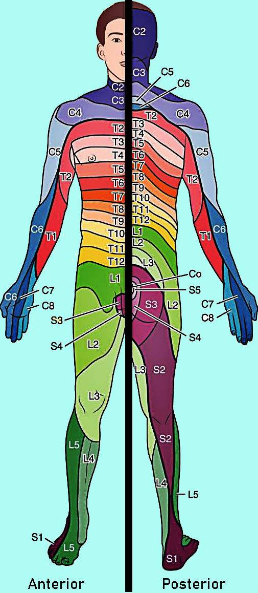
Image showing the pattern of dermatomes on the body.
Source: Creative Commons 4.0, Wikimedia.
A myotome is a group of muscles supplied by a single spinal nerve. A myotome is different from a motor unit, which consists of a motor neurone and the skeletal muscle fibres that it supplies.
The movement-related myotome functions for the upper and lower limbs are listed below:
C5: shoulder abduction and external rotation
C6: elbow flexion, wrist extension, and supination (turning the palm up)
C7: elbow extension, wrist flexion, and pronation (turning the palm down)
C8: finger flexion and extension (shared with C7), and thumb extension
T1: finger abduction and adduction
L2: hip flexion
L3: knee extension, and hip adduction
L4: ankle dorsiflexion (pulling the foot up towards the shin)
L5: great toe extension, and hip abduction
S1: ankle plantar flexion (pointing the foot down)
S2: knee flexion, and great toe flexion
Some other key functions of spinal nerve myotomes, alongside some helpful mnemonics for remembering them are:
- S2, S3 and S4 supply the muscles that control the bowel and bladder (continence), namely the external anal and external urethral sphincters, and some muscles of the sex organs
- Remembered as “S2,3,4 keep the s*** off the floor”
- C3, C4 and C5 supply the diaphragm
- Remembered as “C3,4,5 keep the diaphragm alive”
Edited by: Dr. Ben Appleby and Dr. Maddie Swannack
- 29915
