

Next Lesson - Alcohol Metabolism
Core
The Blocked-pathway Model
An inborn error of metabolism is usually an inherited defect in an enzyme, transporter or cofactor-handling step. The most useful starting diagram is a pathway with substrate A converted through intermediate B into product C. If the B-to-C step slows, material before the block accumulates and product after the block becomes deficient. Flux may also escape through a normally minor side-pathway, creating a metabolite that is newly abundant or toxic.
These four consequences are not mutually exclusive. Accumulated substrate can enter the brain or draw water into cells. Product deficiency can impair membrane synthesis, signalling or growth. A side-product can inhibit other enzymes. Reduced flux can also limit ATP production during fasting or illness. The affected tissue is often the one with the highest demand, weakest alternative pathway or greatest exposure to the accumulating material.
Laboratory patterns follow the same arrows. Measure before the block and a substrate or related metabolite may rise. Measure after it and a product may fall. If a volatile, organic-acid or amino-acid side-pathway opens, a new marker can appear. A normal concentration at one calm moment does not always exclude a flux disorder, because feeding, fasting, catabolism and residual activity change how hard the pathway is being driven.
Not every block is the catalytic centre of an enzyme. A membrane transporter can prevent substrate from reaching the correct organelle, or prevent product leaving it. A missing activator can leave an intact hydrolase unable to meet its substrate. Defective cofactor synthesis or recycling can reduce several enzymes that share the helper. The same accumulation-deficiency logic still works, but only after the compartment and required partner are drawn correctly.
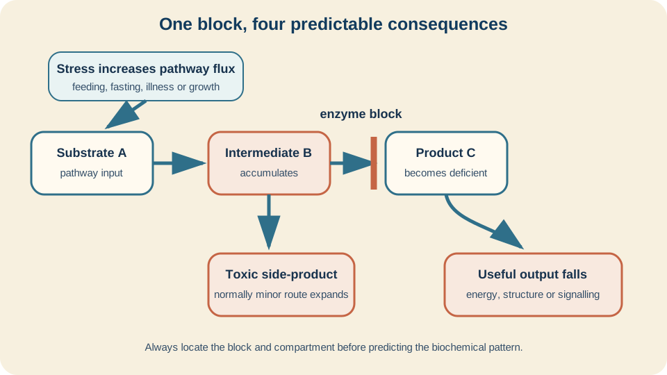
Start every inborn error by asking what accumulates, what becomes deficient, where flux is diverted and which tissue needs the missing output.
SimpleMed original educational diagram
Inheritance and Variable Severity
Many enzyme deficiencies are autosomal recessive. Two pathogenic alleles reduce effective activity, while parents with one altered allele often retain enough reserve to remain well. Important exceptions exist, including X-linked and mitochondrial disorders, so inheritance should be read from the specific pathway rather than assumed from the phrase "metabolic disease".
Genotype does not translate into one fixed percentage of symptoms. Some variants leave residual enzyme activity; others disrupt folding, targeting or stability. Cofactor availability, competing enzymes and transport between compartments modify effective flux. Fever, fasting or infection increases catabolism and can expose a defect that was previously compensated. The phenotype therefore reflects reserve under demand, not just whether an enzyme is labelled present or absent.
This explains threshold behaviour. A pathway may cope while flux stays below a critical level, then decompensate when substrate supply rises or alternative fuel disappears. It also explains why early intervention can matter even when a newborn appears well: preventing the first major accumulation may protect tissue that cannot recover after injury.
Residual activity also changes the diagnostic signal. A severe block may produce a striking marker at baseline, while a milder variant generates an abnormal pattern only under metabolic stress. Two variants in one person can contribute different amounts of activity. Modifier genes and organ maturity change downstream handling. This is why a pathway model predicts a spectrum rather than a one-gene, one-appearance rule.
Small-molecule Examples
In phenylalanine hydroxylase deficiency, including classical phenylketonuria, conversion of phenylalanine towards tyrosine is impaired. Phenylalanine accumulates and enters the brain through a shared large-neutral-amino-acid transporter, disturbing the availability of other amino acids and brain development. Tyrosine becomes relatively conditionally essential. PKU is therefore mainly a substrate-toxicity model with a downstream product consequence, not simply a missing pigment pathway.
In classic galactosaemia, deficiency of galactose-1-phosphate uridylyltransferase impairs normal handling of galactose-1-phosphate. After milk feeding, galactose-related metabolites accumulate and can damage liver, kidney, lens and brain. The mechanism shows why timing and exposure matter: an affected newborn can be well before the relevant substrate load, then deteriorate as pathway flux rises.
Maple syrup urine disease results from deficient branched-chain alpha-ketoacid dehydrogenase activity. Leucine and related branched-chain amino acids or ketoacids accumulate. The shared complex creates a three-substrate pattern, while leucine has particular neurotoxic importance. Catabolic illness releases amino acids from body protein, increasing pathway input even if external intake has not risen.
These examples should remain maps rather than catalogues. Ask which substrate enters, which step fails, which metabolite best reflects the block and why a particular tissue is vulnerable. Existing amino-acid and carbohydrate lessons retain the detailed chemistry.
Ammonia and the Urea Cycle
Amino-acid breakdown transfers nitrogen towards ammonia. The hepatic urea cycle converts that nitrogen into urea for excretion. If a cycle enzyme or transporter is deficient, ammonia can rise because nitrogen disposal cannot match production. Ammonia crosses into the brain, where astrocytes incorporate it into glutamine. Osmotic stress, altered neurotransmitter handling and energy disturbance contribute to cerebral dysfunction.
The position of a urea-cycle block changes associated metabolites, but hyperammonaemia is the shared emergency mechanism. Catabolism increases nitrogen delivery, so infection or fasting can precipitate deterioration. This differs from a primary organic acidaemia, although both can present with encephalopathy. Acid-base pattern, glucose, ketones and pathway-specific metabolites help separate mechanisms; ammonia should not be treated as merely a liver-function label.
Glycogen and Lysosomal Storage Patterns
Storage disorders require a compartment. In a glycogen-storage disease, excess or structurally abnormal glycogen accumulates in cytosol because synthesis, breakdown or glucose release is impaired. Liver-dominant defects tend to disturb fasting glucose handling and cause hepatomegaly. Muscle-dominant defects tend to limit ATP supply during contraction. The stored polymer and missing fuel output both matter.
A lysosomal-storage disease has different topology. Macromolecules enter lysosomes for stepwise degradation. Loss of one hydrolase, activator or trafficking step leaves partially degraded material trapped inside the organelle. Lysosomes enlarge and disturb cell function, particularly in long-lived cells. Substrate distribution helps predict whether nervous system, bone, reticuloendothelial tissue or other organs dominate.
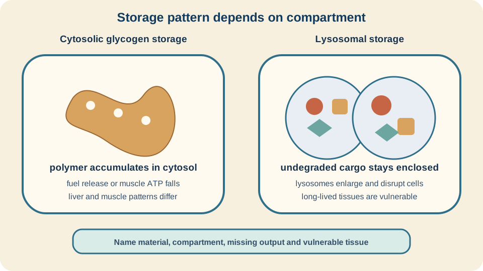
Storage is not one mechanism: cytosolic fuel polymer and lysosomal undegraded cargo produce different patterns.
SimpleMed original educational diagram
Newborn Screening
Newborn blood-spot screening samples capillary blood onto filter paper during the first days of life. A laboratory can measure metabolites, proteins or enzyme-linked markers from a small standardised specimen. The purpose is not to test for every rare disorder. A screening programme selects conditions for which a reliable early marker and an effective pathway after detection can improve outcome before symptoms would normally prompt testing.
A screening threshold balances missed cases against false-positive recalls. High sensitivity is valuable when delay risks irreversible injury, but no threshold perfectly separates disease from health. Prematurity, transfusion, parenteral nutrition, timing and illness can change markers. An out-of-range result therefore triggers urgent assessment and confirmatory biochemical or genetic testing; it is not a final diagnosis by itself.
Marker choice should sit close enough to the defect to be sensitive while remaining stable enough for population sampling. Tandem mass spectrometry can measure patterns of amino acids and acylcarnitines in one run. Immunoassays or enzyme-activity measurements suit other conditions. Ratios can be more informative than one concentration because they compare material before and after a step. A second-tier assay can add specificity before recall, but confirmation still uses a separate diagnostic pathway.
The pathway must close safely. The sample is linked to the correct baby, collected in the right window, transported promptly, analysed under quality control and followed by a recorded result. Positive or uncertain results require rapid specialist communication. Screening value comes from the whole journey, not from the analytical instrument alone.
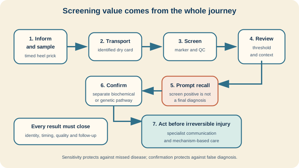
A blood-spot result is one governed step in a journey from early sampling to confirmation and action before irreversible injury.
SimpleMed original educational diagram
Mechanism-based Principles
Mechanism suggests several general responses without turning this lesson into a treatment protocol. Reducing substrate input can lower flux into a blocked pathway. Supplying a deficient product can bypass a missing output. Some variant enzymes retain activity that increases when enough vitamin-derived cofactor is available. Removing a toxic metabolite or diverting it into an excretable form can protect vulnerable tissue.
Enzyme replacement supplies functional activity to accessible compartments and is particularly useful as a principle in selected lysosomal diseases. It does not automatically cross every biological barrier or reverse established injury. Cell or gene-based approaches aim further upstream, but delivery, immune response, tissue access and durable expression remain part of the mechanism problem.
The direction of a response can also confirm the map. If lowering substrate reduces the accumulating marker, input flux mattered. If a cofactor increases residual activity only for selected variants, the protein was impaired rather than completely absent. If circulating enzyme improves visceral storage but not a protected neural compartment, tissue delivery is the limiting step. Mechanism predicts both benefit and boundary.
The reusable sequence is simple: draw the pathway and compartment, mark the block, predict accumulation and deficiency, add the stress that increases flux, then ask why screening changes timing. That model connects apparently different disorders while leaving detailed PKU, galactose, glycogen and amino-acid chemistry in their existing lessons. It also prepares the metabolic context for Alcohol Metabolism.
Reviewed by: Dr. Marcus Judge
- 126
