

Next Lesson - Sepsis
Core
When Gram Staining Is Unhelpful
A Gram stain asks a specific structural question: does a bacterial envelope retain the crystal-violet-iodine complex during decolourisation? Thick peptidoglycan usually retains it and appears purple; a thin peptidoglycan layer behind an outer membrane usually loses it and takes the pink counterstain. The process is explained fully in Gram Staining and Infection Investigations. Its result is useful, but it is not a universal bacterial detector.
A blank or faint film has two broad explanations. First, the process may have failed: too few organisms were present, the specimen was poorly chosen, the film was over-decolourised, or prior growth conditions altered staining. Second, the organism may sit outside the stain's useful design. A lipid-rich coat can obstruct ordinary dye behaviour, a wall-less cell cannot show a peptidoglycan reaction, an extremely thin cell may lie below routine bright-field resolution, and an intracellular organism may be scarce outside host cells. These are predictable biological exceptions, not a miscellaneous list.
This distinction prevents a common reasoning error. Absence of stainable forms is not proof that bacteria are absent, and it is equally weak proof that an unusual organism must be present. The specimen, expected burden and suspected biology decide the next test. Ask which microbial feature the method could measure here rather than treating Gram staining as a universal detector.
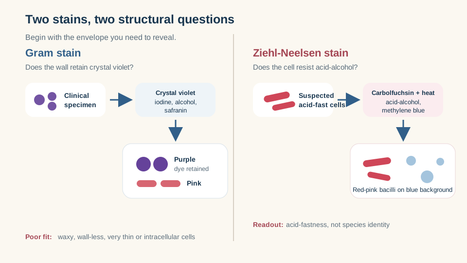
Choose the stain for the envelope: Gram staining reads peptidoglycan behaviour, while Ziehl-Neelsen staining reveals acid-fast cells.
SimpleMed original educational diagram
Mycolic Acids and Acid-fast Staining
Mycobacteria possess peptidoglycan, but it is linked through arabinogalactan to a dense outer region rich in very long-chain mycolic acids. This wax-like layer has low permeability and gives the cell a different staining problem from an ordinary Gram-positive bacterium. Mycobacteria may stain weakly or irregularly with the Gram method, so their envelope is tested with an acid-fast stain instead.
In the Ziehl-Neelsen sequence, carbolfuchsin is driven into the envelope with heat. Acid-alcohol then removes dye from non-acid-fast material, but mycolic acid-rich cells retain the red-pink carbolfuchsin. A blue counterstain provides contrast. The name therefore describes the decisive property: resistance to decolourisation by acid-alcohol. Acid-fast microscopy can show that acid-fast bacilli are present, but species identification and susceptibility questions require culture or molecular methods. The existing Tuberculosis lesson retains disease presentation and management.
The envelope also slows exchange with the environment, while many medically important mycobacteria grow slowly. Culture therefore remains valuable but takes longer than the rapid stain. A nucleic-acid test can identify target sequence sooner, whereas culture demonstrates viable organisms and enables further testing. Faster sequence detection and richer growth-based information answer different needs.
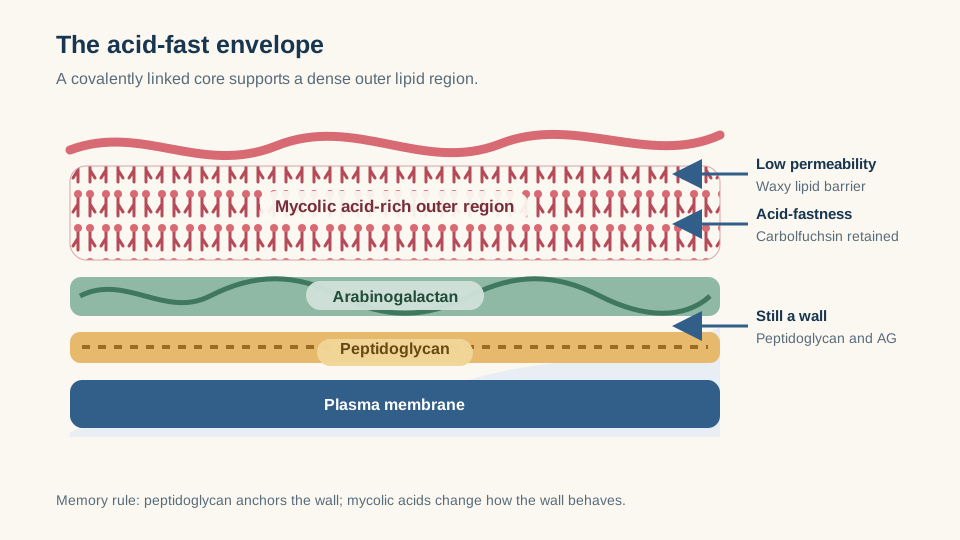
Mycolic acids sit beyond peptidoglycan and arabinogalactan, creating a lipid-rich permeability barrier and the acid-fast reaction.
SimpleMed original educational diagram
Intracellular Survival and Granulomas
Phagocytosis normally places a bacterium inside a phagosome, which matures, acidifies and fuses with lysosomal compartments. Virulent mycobacteria can disrupt this sequence, including phagosomal acidification and phagosome-lysosome fusion, so viable organisms may persist within macrophages. Their intracellular address shelters them from mechanisms that work best in extracellular fluid, while infected macrophages present microbial antigens to T cells.
Activated T cells and macrophages then organise a granuloma: a structured collection of macrophage-lineage cells, lymphocytes and surrounding tissue responses. Containment is the usual aim, while eradication is not guaranteed. The same structure can limit spread yet preserve a niche in which organisms persist. Tissue injury arises substantially from the host cell-mediated response as well as from the microbe. Microscopy, culture and nucleic-acid tests therefore still answer different questions, and treatment-target reasoning must weigh both the lipid-rich envelope and intracellular access.
Granuloma is therefore a tissue-level answer to persistence, not a synonym for any intracellular infection. Chlamydia and Rickettsia require host cells too, but their teaching mechanisms and diagnostic patterns are considered separately below.
Chlamydia and Rickettsia
Obligate intracellular bacteria require viable host cells to complete replication. They are genuine bacteria with DNA, RNA and ribosomes, but routine cell-free agar cannot reproduce the host environment they need. Their small size and intracellular position also make an ordinary Gram film insensitive.
Chlamydia solves transmission and replication with two forms. The elementary body is compact, infectious and non-dividing. It enters a host cell within a membrane-bound inclusion and reorganises into the larger reticulate body, which is non-infectious but metabolically active and replicates. Reticulate bodies then condense into elementary bodies for release and infection of further cells. Separating the jobs is the memory rule: elementary enters; reticulate replicates. Nucleic-acid amplification is useful because it can detect organism-specific sequence directly without growing this cycle on ordinary media.
Rickettsia species are also small obligate intracellular bacteria. Many replicate within vascular endothelial cells, so culture requires living cells and specialist containment rather than a standard plate. Direct molecular detection can be useful when microbial material is present, while paired serology can demonstrate a developing host antibody response later. Serology tracks the host over time instead of substituting for early direct detection.
The contrast with ordinary culture is decisive. Supplying nutrients in agar is sufficient only when the bacterium can run its own replication programme outside a host cell. Obligate intracellular organisms require the biochemical setting of viable cells, so routine culture conditions answer the wrong biological question.
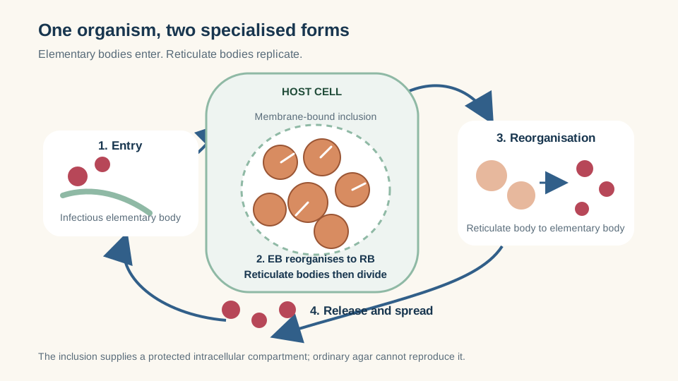
Chlamydia separates entry from replication: elementary bodies infect, reticulate bodies multiply inside an inclusion.
SimpleMed original educational diagram
Mycoplasma without a Wall
Mycoplasma removes the Gram stain's central target: it has a plasma membrane but no peptidoglycan cell wall. Sterols incorporated into the membrane add stability, while the absence of a rigid wall permits variable, pleomorphic shapes. Crystal violet retention cannot classify a structure that is not present, and ordinary light microscopy is further limited by the cells' very small size.
The missing wall also predicts a treatment constraint at the level of mechanism. Drugs whose selective target is peptidoglycan synthesis have no target in Mycoplasma. This is not the same as acquired resistance in a walled bacterium; it is intrinsic non-susceptibility created by anatomy. Specialised culture is demanding and slow, so nucleic-acid tests are often more practical for direct detection.
Place the two envelopes side by side. Mycobacteria keep a highly modified wall that changes permeability and staining; Mycoplasma simply has no wall for the stain to read. The Gram film can look unhelpful in either case for opposite reasons.
Spirochaetes: Thin Form, Different View
Spirochaetes are elongated helical bacteria. Their flagella lie in the periplasm and wind around the cell cylinder rather than projecting freely into the environment. Rotation of these endoflagella twists the whole flexible body, producing corkscrew motility that helps movement through viscous material. The cell is so slender that a routine bright-field Gram film may not resolve it reliably, even though it possesses an envelope.
Dark-field microscopy increases contrast by showing the thin moving organism bright against a dark background; fluorescence-based methods can also make slender cells visible. Direct viewing needs suitable fresh material and expertise. When width defeats routine microscopy and culture is difficult, work shifts toward motility imaging or toward host and molecular signatures rather than another ordinary Gram film.
Shape alone is not the limitation. A thick spiral cell would collect enough stain to be seen. It is the combination of a very small diameter and limited contrast that makes spirochaetes unreliable targets for routine bright-field Gram examination.
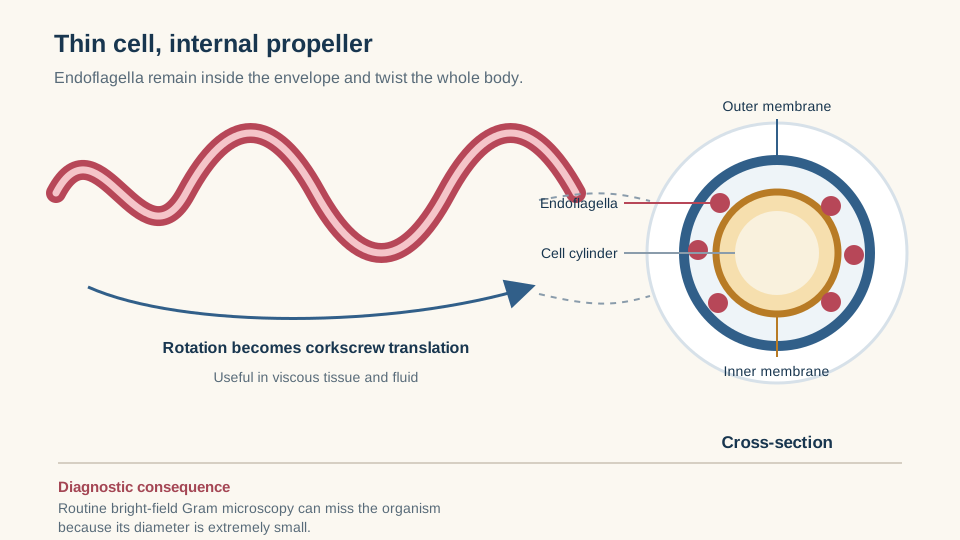
Periplasmic endoflagella twist the slender cell body, producing corkscrew movement while remaining difficult to resolve on a routine Gram film.
SimpleMed original educational diagram
Choosing the Diagnostic Method
Start with the biological obstacle, then choose what can still be measured:
- Microscopy gives rapid structural information when enough organisms are visible and an appropriate stain or optical method exists. Gram and acid-fast films answer different envelope questions; dark-field microscopy solves a contrast problem.
- Culture proves viable growth and supplies organisms for further characterisation, but it slows when growth is intrinsically slow and fails on routine media when living host cells or specialised nutrients are required.
- Serology detects the host response. It is valuable when the organism is elusive, but antibodies take time to develop and may persist, so timing and paired results can matter.
- Nucleic-acid amplification detects organism-specific DNA or RNA without ordinary culture. It is powerful for small or intracellular organisms, although sequence detection does not by itself prove viability.
The practical map is therefore mechanistic. A waxy envelope points to acid-fast staining; intracellular dependence points away from routine agar; no wall explains both Gram invisibility and the absence of a peptidoglycan drug target; extreme thinness points to contrast-enhancing microscopy or indirect detection. Rather than discarding the Gram method, use the organism's biology to decide which laboratory question comes next.
Always match the method to an appropriate specimen and time point. A powerful assay cannot recover material that was never sampled, and an antibody test taken too early may precede the response it is meant to detect.
Reviewed by: Dr. Marcus Judge
- 135
