

Next Lesson - Streptococci
Core
Practical Identification Map
Gram-positive bacteria are not identified by a single microscopic feature. Shape supplies the first practical split: cocci are roughly spherical, while bacilli are rods. Arrangement then records how daughter cells remain attached after division. Irregular clusters suggest staphylococci, chains point toward streptococci, and pairs or short chains are common for enterococci. These patterns reflect division planes and residual wall links rather than decorative naming.
Morphology never identifies a species alone. Similar cocci can belong to different genera, films can be mixed, and important organisms may stain unevenly. The laboratory therefore uses shape and arrangement only to narrow the differential, then applies envelope clues, oxygen preference and simple enzyme tests. Envelope chemistry and stain technique belong in Gram Staining and Infection Investigations; generic cell structure and growth physiology remain in Bacterial Structure, Classification and Growth.
For rods, oxygen habit and spore formation reorder the map. Oxygen-tolerant spore-formers sit with Bacillus; anaerobic spore-formers sit with the clostridia. Listeria is a Gram-positive rod that does not use endospores as its signature survival strategy, so it must not be forced into the spore-forming box by default. The rest of this lesson follows that bench order: cocci first with catalase and coagulase, then compact rod contrasts, then clostridial spore and toxin logic.
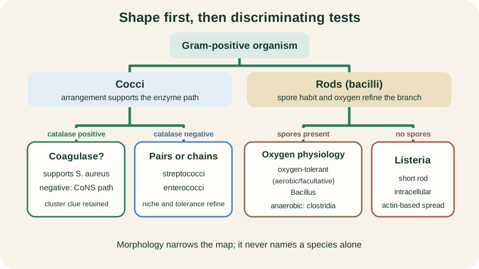
Practical map from Gram-positive morphology to catalase, coagulase and oxygen-spore branches.
SimpleMed original educational diagram
Catalase and Coagulase Reasoning
Catalase decomposes hydrogen peroxide to water and oxygen. On the bench, bubbling after peroxide is added is a rapid screening result. Among common Gram-positive cocci, staphylococci are catalase positive, whereas streptococci and enterococci are catalase negative. The test therefore separates the cluster-forming staphylococcus pathway from the chain- and pair-forming paths taught more fully in Streptococci.
Catalase is a screening split, not a species name. A positive result supports a staphylococcus-like organism but does not prove S. aureus. Coagulase then refines staphylococci. Free coagulase binds host prothrombin to form staphylothrombin, whose protease activity converts fibrinogen into fibrin; this produces a clot in the tube coagulase test. A slide test instead detects cell-bound clumping factor through visible agglutination. Traditionally, these coagulase reactions help distinguish S. aureus from coagulase-negative staphylococci (CoNS). Modern systems may confirm identity with additional phenotypic or genotypic markers, but the teaching logic remains: catalase first among Gram-positive cocci, then coagulase within the catalase-positive group.
Always keep the hierarchy honest. Arrangement suggests a genus family; catalase sorts major coccal groups; coagulase sorts staphylococci; definitive species identity still requires culture context and confirmatory methods. No single enzyme replaces that full path.
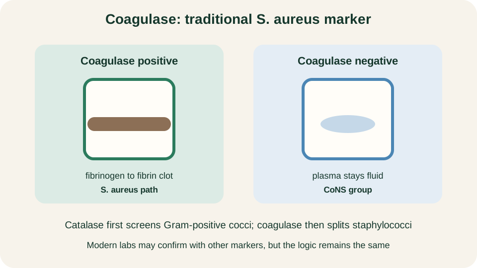
Coagulase as the traditional split between Staphylococcus aureus and coagulase-negative staphylococci.
SimpleMed original educational diagram
Staphylococcus aureus
Staphylococcus aureus is the coagulase-positive staphylococcus of greatest medical importance. Cells are Gram-positive cocci that typically form irregular clusters. The organism is catalase positive and often produces golden-pigmented colonies on ordinary media, although pigment is neither constant nor diagnostic. Nasal and skin carriage explain why the same species can appear as a coloniser in one setting and a pathogen in another.
Virulence is modular rather than a single weapon. Surface proteins support adhesion and immune interference; protein A binds the Fc region of IgG and can blunt opsonisation. Exotoxins extend injury beyond local growth: pore-forming cytotoxins damage host cells, staphylococcal enterotoxins are superantigens and can cause food-borne intoxication, toxic shock syndrome toxin-1 dysregulates T-cell activation, and exfoliative toxins cleave desmoglein in the epidermis. Capsule and biofilm-related polymers further help persistence on tissues and devices. Community surface behaviour after attachment is developed in Biofilms.
Mechanistically, therefore, S. aureus couples a classic laboratory identity (catalase positive, coagulase positive, clustered cocci) to a flexible toxin and adhesion toolkit. Clinical syndromes are many, but the student task here is to remember why the organism can both colonise quietly and injure remotely through secreted proteins.
Coagulase-negative Staphylococci and Enterococci
Coagulase-negative staphylococci share the catalase-positive coccus plan with S. aureus but lack the traditional coagulase marker. Staphylococcus epidermidis is the teaching prototype. These organisms are common skin flora and become clinically significant mainly when foreign material or breached barriers allow them to persist. Their virulence story is quieter than that of S. aureus: adhesion to plastics, extracellular polysaccharide matrix and biofilm maturation matter more than dramatic superantigen shock. That is why a CoNS isolate from a blood culture bottle should prompt a colonisation-versus-infection question rather than an automatic diagnosis.
Enterococci are Gram-positive cocci that are catalase negative and often appear as pairs or short chains. They are natural gut inhabitants. They tolerate harsh conditions that many streptococci do not, including growth in bile and elevated salt, which helps laboratories separate them from ordinary streptococci. Enterococcus faecalis and Enterococcus faecium are the two species most often discussed in human medicine. Their importance lies in opportunistic invasion from the gut or urinary tract and in intrinsic or acquired antimicrobial tolerance patterns taught separately in Antibiotics.
Do not confuse the two groups because both can appear on devices or in healthcare settings. CoNS sit on the catalase-positive staphylococcal branch; enterococci sit on the catalase-negative coccal branch beside streptococci. Arrangement, catalase and clinical niche keep the map clean when colony appearance alone is ambiguous.
Listeria and Bacillus Contrasts
Listeria monocytogenes is a short Gram-positive rod that is catalase positive and does not depend on endospore formation for its core teaching identity. It is a facultative intracellular pathogen: after uptake it escapes the phagosome, and its surface protein ActA recruits host actin polymerisation for cell-to-cell spread while limiting exposure to extracellular defences. Tumbling motility at room temperature is a classic laboratory clue. Food-borne transmission and the ability to grow at refrigeration temperatures follow from its environmental hardiness, but the mechanistic point is intracellular lifestyle rather than a long disease catalogue.
Bacillus species are aerobic or facultatively anaerobic spore-forming rods. Endospore formation, introduced in I01, lets vegetative cells convert into highly resistant dormant forms when conditions deteriorate. Bacillus anthracis and Bacillus cereus illustrate two toxin-centred stories within one genus family: anthrax toxins disrupt host signalling at a systemic level, while B. cereus food poisoning reflects preformed or intestinal toxins rather than invasive spore germination in every case. For this lesson, remember the shared design: oxygen-tolerant rods that can leave a spore state, then return to toxin-producing vegetative growth.
Contrast the three rod strategies. Listeria invests in intracellular survival without making spores the signature. Bacillus combines oxygen-tolerant growth with spores and toxins. Clostridia, next, combine anaerobic growth with spores and toxins. Oxygen physiology and spore habit therefore separate genera that all look like Gram-positive rods on a rushed first glance.
Anaerobic Spore-forming Clostridia
Clostridia are anaerobic Gram-positive rods capable of endospore formation. Spore resistance explains persistence in soil, dust, food and the gut lumen when vegetative cells would die. Sporulation is not reproduction: one vegetative cell yields one spore, and germination later restores one vegetative cell. Disease often begins only after germination in a suitably reduced niche, followed by growth and toxin production.
Many clinically important clostridia are toxin-driven. Local tissue necrosis, food-borne intoxication or neurological syndromes can appear out of proportion to the number of organisms seen, because soluble toxins carry the injury. Clostridioides difficile (still often discussed with the clostridial group) is the compact gut example: spores persist in the environment and support transmission; vegetative cells expand when the normal microbiota is disturbed; large clostridial toxins A and B glucosylate small GTPases, especially Rho-family proteins, disrupting enterocyte cytoskeleton and barrier function. The teaching sequence is therefore spore persistence, germination, vegetative expansion and toxin-mediated epithelial injury.
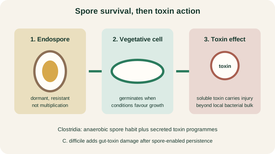
Clostridial logic: resistant spore, germination to a vegetative rod, then toxin-mediated host injury.
SimpleMed original educational diagram
Tetanus versus Botulinum Neurotoxins
Clostridium tetani and Clostridium botulinum produce related zinc metalloprotease neurotoxins that cleave SNARE proteins required for synaptic vesicle fusion. The clinical pictures are opposite because the toxins reach different synapses. Tetanus toxin is taken up at peripheral terminals and transported retrogradely into the central nervous system, where it blocks release of inhibitory neurotransmitters from interneurons that normally restrain motor neurons. Unopposed motor drive produces spastic paralysis and characteristic muscle rigidity.
Botulinum toxin acts mainly at peripheral cholinergic terminals, especially the neuromuscular junction. Cleavage of SNARE components prevents acetylcholine release onto skeletal muscle, so the motor command never arrives and paralysis is flaccid. The same molecular family therefore yields tetanic spasm or descending flaccid weakness according to which synapse is silenced. Food-borne botulism reflects preformed toxin; wound and infant forms reflect toxin production after germination in a permissive niche. The mechanism lesson is synaptic address, not a treatment algorithm.
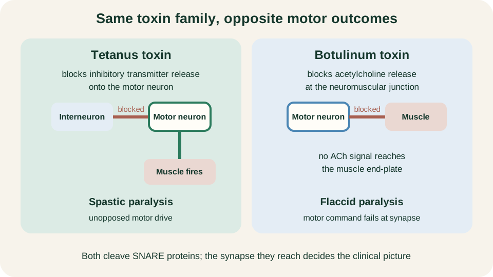
Tetanus toxin removes inhibition of motor neurons; botulinum toxin blocks acetylcholine at the neuromuscular junction.
SimpleMed original educational diagram
Identification Reasoning Flow
Bring the steps back into one bench-facing order. Start with the Gram film: Gram-positive cocci or Gram-positive rods? For cocci, note cluster, chain or pair arrangement, then ask catalase. Catalase-positive cocci enter the staphylococcus path; coagulase then separates S. aureus from CoNS. Catalase-negative cocci move toward streptococci and enterococci, with enterococci further suggested by gut origin and tolerance traits. For rods, ask whether spores and oxygen physiology point to Bacillus, clostridia or a non-spore specialist such as Listeria.
Finally, decide whether the clinical problem is driven by invasion, biofilm persistence or toxin action at a distance. S. aureus can do all three in different settings. CoNS emphasise device biofilm. Clostridial disease often elevates toxin logic above local bacterial bulk, with tetanus and botulinum as the purest synaptic examples. Antimicrobial choice and resistance tables stay in Antibiotics. Your task in this article is the reasoning map: shape and arrangement open the file, catalase and coagulase sort the cocci, and spore plus oxygen plus toxin target explain the major Gram-positive rods.
Reviewed by: Dr. Marcus Judge
- 146
