

Next Lesson - Hepatitis
Core
Virion Without a Factory
A virus is an obligate intracellular infectious agent: it carries genetic instructions for its own proteins but does not possess a complete independent manufacturing system. The extracellular particle is the virion. Inside it sits the viral genome (DNA or RNA), protected by a protein capsid. Genome plus capsid form the nucleocapsid. Some virions also carry a lipid envelope studded with viral attachment proteins; others present those proteins on a naked capsid surface. Attachment proteins determine which cells the particle can dock with, and therefore help set tissue tropism before any intracellular step begins.
This incomplete autonomy is the teaching spine. Viruses encode structural proteins, regulatory proteins and, in many families, dedicated polymerases. They still depend on host ribosomes for protein synthesis, and on host energy, membranes and metabolic precursors. Do not picture a particle that borrows every enzyme: the point is dependency on a living cell, not absolute enzyme poverty. Productive infection therefore needs a cell that is both susceptible (expresses a usable receptor or entry pathway) and permissive (supplies the intracellular conditions the viral programme requires). Receptor binding alone does not guarantee that the cycle can finish. That dual requirement explains why the same virus may attach widely yet replicate productively only in selected cell types, and why selective therapy is hard when many steps sit close to host machinery.
Enveloped versus Non-enveloped
Non-enveloped virions expose a protein capsid to the environment. Enveloped virions wrap the nucleocapsid in a host-derived lipid bilayer into which viral glycoproteins are inserted. Those surface proteins mediate attachment and, for many enveloped viruses, membrane fusion at the plasma membrane or after endocytosis. The envelope is therefore both a delivery tool and a vulnerability: lipid coats tend to be disrupted by drying, detergents and many surface disinfectants more readily than tough naked capsids.
Treat stability language as tendency, not law. Non-enveloped virions are often more environmentally robust and more likely to survive on surfaces or in faecal-oral routes, while enveloped viruses often favour close contact, droplets, aerosols or body fluids. Counter-examples and intermediate behaviours exist, so do not convert envelope status into an absolute transmission rule. Dose, moisture, temperature and host behaviour still dominate real transmission. The practical contrast is physical chemistry of the outer coat, not a fixed clinical algorithm.
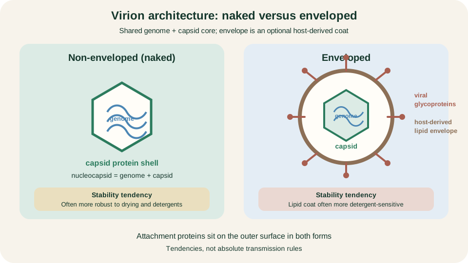
Naked and enveloped virions share genome plus capsid; the envelope adds host lipid and viral glycoproteins with different stability tendencies.
SimpleMed original educational diagram
Genome Strategy Map
A useful preclinical map groups viruses by how they reach translatable mRNA, not by memorising every family. Double-stranded DNA genomes are transcribed toward mRNA using viral and/or host transcriptional machinery. Positive-sense single-stranded RNA can often act directly as mRNA on host ribosomes, though the virus still needs to produce RNA-dependent RNA polymerase to complete its cycle. Negative-sense RNA cannot be translated until a complementary positive strand is made; the incoming virion therefore usually carries its own RNA-dependent RNA polymerase. Double-stranded RNA likewise depends on a virion-associated transcriptase because the host does not maintain a standard RNA-to-RNA copying pathway for foreign genomes.
Retroviral RNA follows a different constraint: reverse transcriptase copies RNA into DNA, and integrase helps establish a DNA provirus that host polymerase can then transcribe. Exceptions and refinements exist across real families; the map is a manufacturing logic chart. It explains why some particles must package polymerase proteins and why selective antiviral design often aims at virus-specific nucleic-acid enzymes rather than at the shared ribosome.
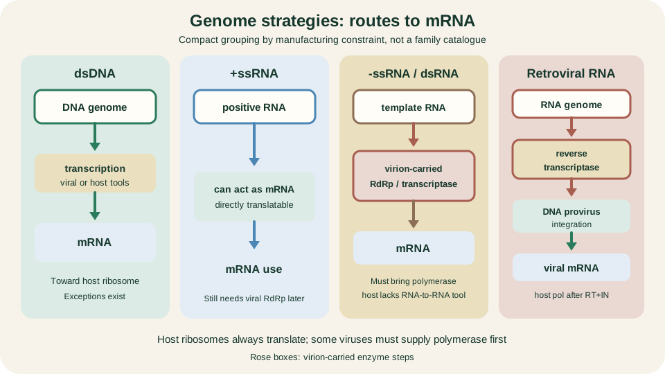
Routes to mRNA: dsDNA transcription, +ssRNA as message, -ssRNA/dsRNA with virion polymerase, retroviral DNA provirus.
SimpleMed original educational diagram
Replication Cycle
Despite huge diversity, a shared sequence of events organises thinking. Attachment links viral surface proteins to host receptors. Entry follows by fusion, endocytosis or related routes. Uncoating releases the genome into the right cellular compartment. Genome replication then proceeds by the strategy class above. Viral mRNA production and translation supply structural and non-structural proteins; host ribosomes do the protein synthesis. Assembly packages new genomes, often with maturation cleavage steps that convert immature particles into infectious ones. Release returns particles to the extracellular space so a new round of host cells can be reached.
Keep susceptibility separate from permissiveness throughout: a receptor may allow docking while missing intracellular factors abort the cycle. Release mode also varies. Many enveloped viruses bud through host membranes, acquiring envelope lipids as they exit. Many non-enveloped viruses are released when the cell ruptures. Neither pattern is universal, and some systems mix strategies or use specialised exit pathways. Timing of gene expression (early regulatory products before late structural products) is family-specific detail; the shared lesson is ordered dependency on host resources. The cycle is the scaffold for pathogenesis and for later antiviral target mapping, not a single cartoon that fits every family.
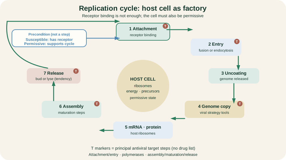
Attachment to release as a loop around host ribosomes, energy and precursors; target markers flag principal steps only.
SimpleMed original educational diagram
Cell Outcomes and Disease
Infection of a cell need not equal rapid death. Productive cytolytic infection yields new virions and destroys the cell. Productive non-cytolytic or persistent infection continues particle production while the cell survives for a time, sometimes with altered function. Abortive or restrictive infection begins but fails to complete the full programme because the cell is non-permissive or host defences interrupt key steps. The same virus can produce different outcomes in different cell types or at different stages of host immunity.
Disease mechanisms are correspondingly broader than lysis. Direct cell injury removes functional tissue. Altered secretion, signalling or antigen presentation can impair organ performance without frank necrosis. Some viruses drive syncytium formation or classic cytopathic effects visible in culture and sometimes in tissue. Host responses matter at least as much: innate sensing and adaptive immune effectors clear infection but also cause immunopathology through inflammation, cytotoxic killing of infected cells and immune-complex injury. Clinical syndromes therefore reflect both viral cytopathicity and the intensity of the host response. Do not reduce viral illness to "the virus exploded the cell." Link the general immune architecture in Innate Immunity and Adaptive Immunity without repeating those lessons here.
Latency and Reactivation
Latency is a distinct programme, not merely "quiet chronic infection." In latency the viral genome persists in the host cell with highly restricted gene expression and without continuous full production of infectious virions. The cell may look largely normal while still harbouring the genetic potential for later output. Reactivation resumes the productive gene cascade and can shed virus again, sometimes with clinical recurrence and sometimes as silent shedding. Chronic productive infection, by contrast, keeps generating particles over long periods; the host may control symptoms while replication continues at some level.
Herpesviruses are the brief standard illustration: neuronal or leukocyte reservoirs maintain latent genomes that can later reactivate under stress, immunosuppression or other triggers. The preclinical task is the logical contrast (restricted persistence versus ongoing production), not a catalogue of every latent pathogen. HIV and hepatitis viruses have their own dedicated articles for chronic or integrating lifestyles; use those neighbours rather than expanding them here.
Drift and Shift
Antigenic drift is gradual change in viral surface antigens driven by accumulating mutations, especially where polymerase error rates are high and immune selection favours escape variants. Over seasons, drift helps explain why population immunity and vaccine match can erode for mutable RNA viruses even without a new species appearing. Antigenic shift is different in kind and teaching scope: it denotes an abrupt major change in influenza A antigens, commonly when segmented genomes reassort inside a co-infected cell and package a novel combination of haemagglutinin and neuraminidase genes.
Never generalise shift to all viruses. Most agents lack influenza A's segmented reassortment pathway, and not every reassortment event becomes a pandemic. Animal reservoirs and human co-infection create opportunities for mixing, but ecological chance and further adaptation still decide impact. Drift is the everyday mutational walk; shift is an influenza-A special case of sudden genomic mixing. Keep those terms precise in clinical conversation.
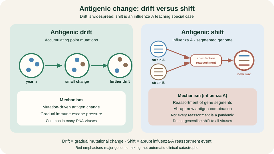
Drift: stepwise mutational antigen change. Shift: influenza A reassortment with a new antigen mix, not automatic pandemic status.
SimpleMed original educational diagram
Oncogenic Viruses
Some viruses contribute to cancer by disturbing the same control networks taught in cell biology. Viral oncoproteins may inactivate p53 or Rb, push growth-factor signalling, or otherwise unlock checkpoints that normally restrain proliferation or promote apoptosis. Integration or chronic regulatory interference can alter host gene expression near insertion sites or through viral transcriptional programmes. Long-standing inflammation and local immunosuppression add non-genetic routes to transformation by sustaining tissue damage and weakening immune surveillance. Infection is generally insufficient alone: host cofactors, time and additional genetic hits usually matter.
Brief illustrations only: high-risk human papillomaviruses and the E6/E7 logic against p53/Rb; Epstein-Barr virus and lymphoid or epithelial associations; hepatitis B and C through chronic hepatic injury and, for HBV, integration-related effects. Use The Cell Cycle, Checkpoints and Apoptosis for checkpoint detail, Hepatitis for hepatotropic viruses, and Human Immunodeficiency Virus for HIV-related immunodeficiency and secondary oncogenic risk. This section states principles; it does not replace those articles.
Diagnosis and Antiviral Targets
Start with specimen, anatomical site and timing. A perfect assay on the wrong sample or at the wrong phase of illness misleads. Nucleic acid amplification (PCR/NAAT) detects viral genome fragments with high analytical sensitivity; a positive result does not by itself prove that intact, infectious virions are present, especially after treatment or late in recovery. Antigen tests detect viral protein and depend on burden and assay design; they can be faster at the point of care but miss low-level infection. Serology measures host antibody (and sometimes antigen-antibody patterns) and mainly informs exposure, timing and immune status rather than immediate infectivity. Culture demonstrates replicating virus and supports characterisation, yet it is slow, specialist and unavailable for many agents. Method choice follows the biological question, not a single best test for all viruses.
Antiviral design follows the cycle. Attachment and entry steps, virus-specific polymerases, reverse transcriptase, integrase and protease, and assembly, maturation or release machinery are the classic target classes because they differ from essential host processes more than the shared ribosome does. Selective toxicity is easier when the drug hits a virus-encoded enzyme the host lacks. Drug names, regimens and resistance tables belong in Antivirals. Here the task is mechanistic mapping: know what you are measuring in the laboratory, and know which step a future drug might interrupt, without turning this lesson into a prescribing guide.
Reviewed by: Dr. Marcus Judge
- 181
