

Next Lesson - The Organisms Gram Staining Misses
Core
The Two-Membrane Envelope
Gram-negative bacteria are defined less by a single disease and more by a shared cell envelope. Outside the cytoplasm sits an inner (cytoplasmic) membrane, then a periplasmic space that contains a thin peptidoglycan layer, then an outer membrane. That architecture is the practical opposite of the thick outer wall emphasised for Gram-positive organisms in Staphylococci and Other Gram-positive Bacteria and in Streptococci. Generic wall chemistry and growth physiology remain in Bacterial Structure, Classification and Growth; stain technique belongs in Gram Staining and Infection Investigations.
The outer membrane is an asymmetric bilayer. Its outer leaflet is rich in lipopolysaccharide (LPS), while the inner leaflet is more phospholipid-like. Porin proteins form aqueous channels that admit small hydrophilic solutes but limit larger or hydrophobic molecules. The result is selective permeability: nutrients still enter, yet many environmental stresses and some antimicrobial classes meet a real barrier before they reach the periplasm or cytoplasm. Inside the periplasm, the thin peptidoglycan maintains shape and osmotic support without forming the thick external mesh of Gram-positive walls. Enzymes, binding proteins and intermediate metabolites share that aqueous compartment.
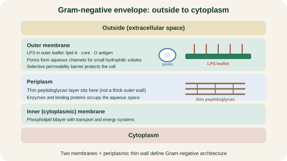
Outside to cytoplasm: LPS outer leaflet, porin, outer membrane, thin periplasmic peptidoglycan, then the inner membrane.
SimpleMed original educational diagram
LPS and Lipid A
LPS has three conceptual parts. Lipid A is the hydrophobic membrane anchor and the principal endotoxin motif recognised by innate immunity. A core oligosaccharide links lipid A to the O antigen, a repeating polysaccharide chain that projects outward and varies between strains. O-side-chain diversity underpins classical serotyping and antigenic labels, but the conserved danger signal for the host is lipid A.
Some mucosal organisms display a shorter related glycolipid called lipooligosaccharide (LOS). LOS retains membrane-anchored lipid A logic with a truncated outer carbohydrate domain; Neisseria is the later teaching example. Whether the molecule is long LPS or shorter LOS, the shared idea is an outer-leaflet glycolipid that both stabilises the envelope and can trigger host inflammation when released or exposed in large amounts. LPS is therefore structural armour for the bacterium and a potent innate immune ligand for the host, not merely a staining curiosity. Those dual roles explain why infection can remain local or escalate when large lipid A signals circulate.
Local Defence versus Systemic Endotoxin Injury
Host recognition of lipid A is a co-ordinated hand-off rather than a single receptor event. Lipopolysaccharide-binding protein (LBP) helps transfer LPS in body fluids. CD14 concentrates the ligand at the cell surface. MD-2 associates with Toll-like receptor 4 (TLR4) and completes the sensing complex. Engagement of TLR4 activates signalling that converges on transcription factors such as NF-κB and drives production of inflammatory cytokines, including tumour necrosis factor and interleukin programmes that recruit and activate immune cells.
At a contained focus of infection this response is useful. Local cytokine release promotes vascular permeability at the right scale, leukocyte recruitment and antimicrobial defences that help clear organisms. The same molecular pathway becomes harmful when activation is systemic and poorly controlled. Widespread cytokine excess promotes endothelial leak, inappropriate vasodilation, coagulation dysregulation with microvascular thrombosis, and progressive tissue hypoperfusion that clinically appears as shock. The student task here is mechanism, not a treatment algorithm: dose, distribution and host regulation decide whether TLR4 signalling defends or injures. Clinical recognition of sepsis is developed in Sepsis; antimicrobial selection stays in Antibiotics.
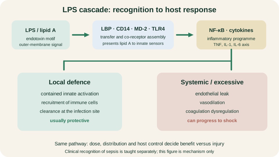
Lipid A sensing via LBP, CD14, MD-2 and TLR4: protective when local, injurious when systemic and excessive.
SimpleMed original educational diagram
Entry-Route Map
Once the shared envelope and endotoxin logic are clear, organism groups become easier if you organise by route and niche rather than by alphabetical catalogue. Ingestion selects for enteric organisms adapted to survive the gut lumen and interact with intestinal epithelium. Urinary ascent selects for adhesins that resist flushing and permit climb from the urethra toward the bladder and beyond. Respiratory mucosa and wet healthcare environments select for organisms that colonise airways or exploit damaged barriers and devices. Genital and nasopharyngeal mucosa favour diplococci specialised for mucosal attachment. The gastric mucus layer selects for acid-tolerant specialists that buffer their immediate microenvironment.
That map does not name every species, and it should not. It predicts which biological tools matter: toxin or invasion factors in the gut, adhesion in the urinary tract, nutritional fastidiousness or environmental hardiness in the airway and hospital water systems, LOS and mucosal colonisation for Neisseria, and urease for Helicobacter. Community surface persistence after attachment is developed further in Biofilms.
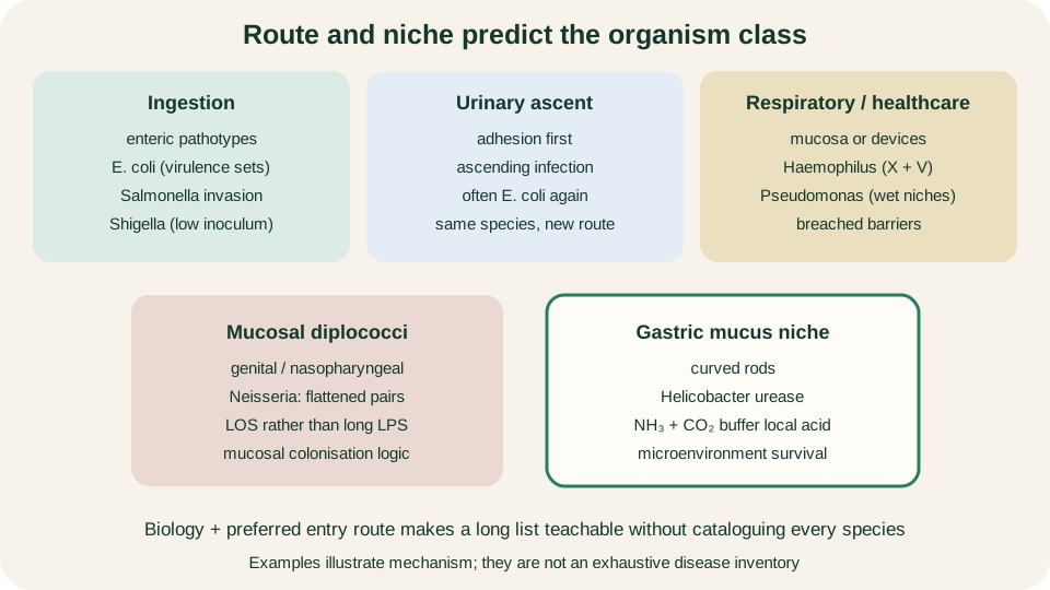
Route and niche organise Gram-negative teaching examples without turning the lesson into a species list.
SimpleMed original educational diagram
Ingested Enteric Organisms
Escherichia coli is a single species name that covers many behavioural pathotypes. Shared laboratory features can sit beside very different clinical mechanisms because virulence is modular and often plasmid- or island-encoded. Some pathotypes emphasise adhesion and enterotoxin production with watery diarrhoea; others emphasise intimate attachment and cytoskeletal rearrangement of enterocytes; still others emphasise Shiga-like toxins or extra-intestinal invasion. The teaching point is not a full syndrome catalogue. It is that one species can acquire different virulence packages and therefore different gut behaviours while remaining recognisably E. coli on the bench.
Salmonella illustrates epithelial invasion with the capacity for intracellular persistence. After ingestion, organisms interact with intestinal epithelium, enter host cells and can survive within a modified vacuolar niche. That intracellular lifestyle helps explain why some infections remain gastroenteritis while others seed deeper sites if dissemination occurs. Shigella is a colonic invasive contrast with a characteristically low infectious inoculum: relatively few organisms can establish disease because acid tolerance and efficient invasion of colonic epithelium reduce the usual dose barrier. Remember mechanism (invasion, low inoculum, colonic preference) rather than a long list of named syndromes. Across these enteric examples, shared LPS remains, but disease phenotype is driven by acquired tools matched to the intestinal route.
Ascending Urinary E. coli
Urinary tract infection commonly features E. coli again, but that is a route story, not a new species. Faecal flora near the perineum can reach the urethra. Adhesins, including fimbrial systems, allow attachment to uroepithelium so that micturition does not wash every organism away. From there, ascent to the bladder and potentially the upper tract follows anatomy and host factors rather than a change of bacterial identity.
Keep the conceptual link explicit. The same species that can cause enteric disease with particular virulence sets can also cause ascending urinary infection with adhesion-centred tools. You are not learning two unrelated organisms; you are learning how niche selects which traits matter. Imaging and antimicrobial tables sit outside this article. The preclinical task is adhesion plus ascent as the mechanistic core of a common Gram-negative route.
Respiratory and Healthcare Niches
Haemophilus influenzae is a small Gram-negative coccobacillus associated with respiratory mucosal disease more than with enteric or urinary ascent. It is nutritionally fastidious: classical teaching emphasises requirement for haemin (X factor) and nicotinamide adenine dinucleotide (V factor) for growth on ordinary media. Those factor needs explain specialised culture behaviour and keep the organism in a different laboratory branch from lactose-fermenting enteric rods, even though both groups are Gram-negative.
Pseudomonas aeruginosa is an aerobic, oxidase-positive, non-lactose-fermenting opportunist adapted to wet environments and damaged host barriers. Hospital water systems, moist devices and wounds or airways with impaired defences are classic teaching niches. The organism is not primarily a story of low-inoculum gut invasion; it is a story of environmental hardiness, oxidative metabolism clues on the bench and exploitation of breached barriers. Device-associated persistence connects naturally to biofilm biology without requiring a full resistance table here. Contrast the two: Haemophilus is a small, factor-dependent mucosal specialist; Pseudomonas is a tough, oxidase-positive environmental opportunist. Shared Gram-negative status and potential endotoxin exposure do not make their ecological strategies the same.
Neisseria and Helicobacter Contrasts
Neisseria species appear as flattened Gram-negative diplococci, often described as kidney-bean pairs. Their outer membrane displays LOS rather than long O-antigen LPS, which still presents lipid A logic to innate sensors but with a shorter carbohydrate domain. Mucosal niches (nasopharynx or genital mucosa depending on species) dominate the teaching picture: attachment, colonisation and local invasion of mucosal surfaces matter more than enteric fermentation maps. Morphology on Gram film therefore carries real weight, provided it is read with clinical route and then confirmed.
Helicobacter pylori is a curved Gram-negative organism specialised for the gastric mucus niche. Urease hydrolyses urea to ammonia and carbon dioxide, buffering the immediate microenvironment against acid and supporting survival where many bacteria fail. Motility through mucus and adhesion to the epithelium complete a local colonisation strategy. The lesson is ecological chemistry: urease is not an abstract enzyme badge, but a tool for living next to gastric acid. A positive urease clue in the right clinical context narrows probability; definitive identification still requires the broader laboratory pathway. On the route map, Neisseria is mucosal diplococci with LOS; Helicobacter is a curved gastric specialist whose urease creates a tolerable microclimate. Neither is taught well as a footnote to enteric MacConkey tables alone.
Compact Laboratory Reasoning
Bring the bench steps into one honest order. Start with specimen type and clinical route: stool, urine, respiratory sample, mucosal swab, gastric specimen or device-related material already reorder probability before any plate is inspected. Next, record morphology: rods, coccobacilli, diplococci or curved forms. Only then apply a short set of differential screens.
MacConkey agar is both selective and differential for many Gram-negative rods. Lactose fermentation acidifies colonies and helps separate fermenters such as many E. coli isolates from non-fermenters such as Pseudomonas. Oxidase testing supports the Pseudomonas path when positive and is commonly negative among Enterobacterales. Special growth requirements, especially X and V factors, pull Haemophilus into its own branch. Urease activity is a powerful clue for Helicobacter in a gastric context. None of these reactions names a species by itself. They narrow the map; confirmation follows with further phenotypic panels, antigen or molecular methods according to laboratory practice.
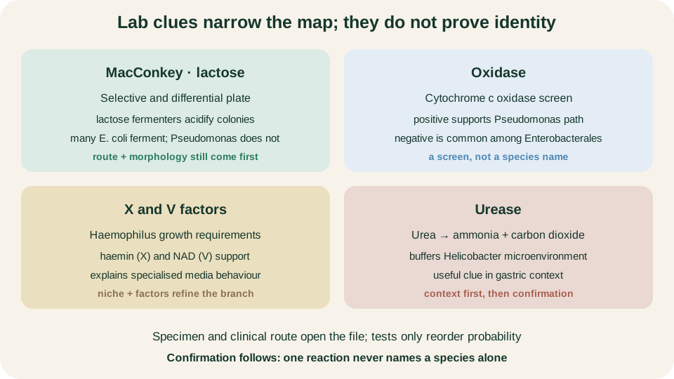
MacConkey lactose, oxidase, X/V factors and urease reorder probability; confirmation still required.
SimpleMed original educational diagram
Close the loop on the teaching spine. Two membranes, thin periplasmic peptidoglycan, LPS and porins define the shared biology. Lipid A sensing through LBP, CD14, MD-2 and TLR4 explains local defence versus systemic endotoxin injury. Route and niche then make enteric pathotypes, ascending urinary E. coli, respiratory and healthcare opportunists, mucosal Neisseria and gastric Helicobacter predictable without a catalogue. Specimen and route first, morphology next, then screens that narrow: never a single test that pretends to be a name.
Reviewed by: Dr. Marcus Judge
- 134
