

Next Lesson - Biofilms
Core
Prokaryotic Cell Plan
A bacterium is a prokaryotic cell. It has no membrane-bound nucleus or organelles such as mitochondria, and protein synthesis uses 70S rather than eukaryotic 80S ribosomes. Chromosomal DNA occupies a nucleoid without a nuclear envelope. Because transcription and translation share one compartment, messenger RNA can be translated while it is still being made, coupling gene expression tightly to environmental change.
The plasma membrane houses transport systems and, in many species, respiratory chains that generate proton motive force. Wall and any outer membrane form the cell envelope; capsules, flagella and pili may extend beyond it. These structures provide osmotic protection, energy generation, adhesion, motility or immune evasion. Clinically, envelope chemistry predicts Gram behaviour and drug permeability, while surface polymers can become antigens, vaccine targets or antiphagocytic shields.
Read every structure twice: as a survival tool, then as a diagnostic clue, virulence mechanism or drug target.
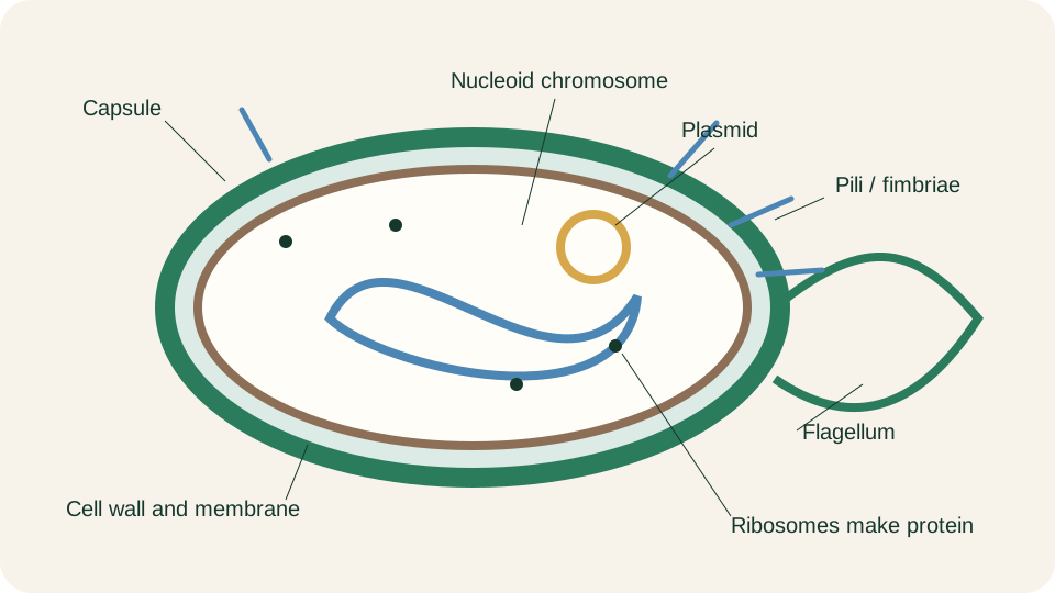
Bacterial cell plan with envelope, nucleoid, 70S ribosomes, capsule, flagellum and pili as survival tools and clinical landmarks.
SimpleMed original educational diagram
Cell Envelope Recap
The cell envelope is the layered shell outside the cytoplasm. Gram-positive bacteria have a thick peptidoglycan mesh, often containing teichoic and lipoteichoic acids. Gram-negative bacteria have thinner peptidoglycan, a periplasm and an outer membrane whose outer leaflet contains lipopolysaccharide (LPS). Thick peptidoglycan without an outer membrane produces one laboratory phenotype; thin peptidoglycan plus an LPS-bearing outer membrane produces another.
LPS is a potent driver of host inflammation when Gram-negative organisms release it, linking envelope chemistry to the systemic inflammatory response taught in later infection lessons. Peptidoglycan cross-links are absent from human cells, which is why agents that block wall synthesis can be selectively toxic to bacteria. The Gram-negative outer membrane also restricts entry of some large or hydrophobic molecules and therefore contributes to intrinsic antimicrobial susceptibility patterns. Envelope type is thus simultaneously a survival design, a virulence source and a pharmacological boundary.
Stain technique, film interpretation and the wider infection investigation workflow belong in Gram Staining and Infection Investigations. This section only records the structural contrast those methods exploit: peptidoglycan load, presence or absence of an outer membrane, and the clinical consequences that follow from each design.
External Structures
Many bacteria coat themselves with a glycocalyx that is usually polysaccharide and less commonly polypeptide. When the coat is discrete and tightly organised it is called a capsule; when it is loose and easily sheared away it is a slime layer. Capsules impair phagocytosis, help some pathogens avoid complement-mediated killing and may appear as clear haloes with appropriate preparations. Several medically important capsules are also vaccine antigens, so a coat built for bacterial survival can become both a virulence factor and a public-health target.
Flagella are rotary filaments that power swimming. Their number and placement, such as polar or peritrichous, affect movement and sometimes serological classification. Chemotaxis adjusts rotation so cells approach nutrients or avoid toxins, helping organisms find a niche before immune defences or competing flora clear them.
Pili and fimbriae are thinner fibres used mainly for adhesion to epithelium, abiotic surfaces and other cells. Adhesion can initiate colonisation of mucosa, catheters or prosthetic material. Specialised pili participate in conjugation, but horizontal-transfer genetics belong elsewhere. Surface community behaviour after attachment is developed in Biofilms.
Endospores
A limited set of genera, notably Bacillus and Clostridium, can convert a vegetative cell into an endospore when nutrients fail or environmental stress rises. The endospore is dehydrated, metabolically dormant and wrapped in protective layers that resist heat, desiccation, ultraviolet light and many chemical disinfectants far better than the parent vegetative form. Because of that resistance, spores persist in soil, dust and inadequately processed equipment and explain why some pathogens reappear long after apparent environmental cleaning.
Endosporulation is not reproduction. One vegetative cell yields one spore; germination under favourable moisture, temperature and nutrient conditions yields one vegetative cell again. There is no net increase in cell number during the spore cycle. The clinical meaning is persistence and later re-entry into growth, not multiplication by spores. When you meet a spore-forming pathogen, ask what allows the spore to survive outside the host and what conditions in tissue, food or culture medium permit germination back to a dividing vegetative state.
Genetic Organisation
Most bacteria organise their essential genome as a single circular chromosome compacted into the nucleoid. Genes required for a shared task are frequently arranged as operons: contiguous coding sequences transcribed as one messenger RNA under common control. Operons allow rapid, coordinated induction or repression when a nutrient appears or disappears, matching the short generation times of many free-living species. The chromosome therefore functions less like a static library and more like a compact operating system tuned for speed.
Plasmids are smaller DNA molecules that replicate separately from the chromosome. They often carry accessory genes useful in particular niches, including some virulence or resistance determinants, although neither property is exclusively plasmid-borne. The structural idea is modularity: core housekeeping usually resides on the chromosome, while optional survival functions may sit on extrachromosomal elements. Horizontal-transfer routes belong elsewhere.
Shape and Arrangement
Light-microscopic shape remains a rapid first classifier at the bench. Cocci are roughly spherical. Bacilli, or rods, are elongated. Curved rods include vibrio forms. Spiral organisms include relatively rigid spirilla and the more flexible spirochaetes. After division, cells may remain attached in characteristic arrangements: pairs (diplococci), chains (as in many streptococci), irregular clusters (as in staphylococci), or packets in a few genera. These patterns reflect the plane of division and the strength of residual wall links rather than arbitrary naming conventions.
Morphology never identifies a species alone. Genera can look similar, important pathogens may stain poorly, and mixed flora confuse arrangement. Shape narrows the differential and checks whether later culture is consistent with the original film. Combine it with envelope type, oxygen preference and clinical site.
Oxygen Requirements
Oxygen availability sorts bacteria into physiological classes that dictate both natural habitat and laboratory handling. Obligate aerobes require molecular oxygen as a terminal electron acceptor and grow only where it is present. Obligate anaerobes are inhibited or killed by oxygen and occupy niches that are deep, necrotic or otherwise chemically reduced. Facultative anaerobes grow with or without oxygen; many switch between respiration and fermentation as supply changes. Aerotolerant anaerobes do not use oxygen for energy yet survive its presence. Microaerophiles need oxygen, but only at tensions below atmospheric levels.
The chemical problem is reactive oxygen species (ROS). Superoxide, hydrogen peroxide and related oxidants damage DNA, proteins and lipids. Organisms that live in air usually deploy protective enzymes such as superoxide dismutase, catalase and peroxidase. Missing or limited defences help explain obligate anaerobiosis; abundant catalase activity underpins common laboratory identification tests. Oxygen class is therefore both an ecological statement and a practical instruction for transport and incubation.
Matching atmosphere to physiology is as important as choosing agar. An obligate anaerobe incubated in room air may be reported as no growth because of atmospheric error. A microaerophile forced into full aerobiosis or anaerobiosis may also grow poorly. Specimen transport and incubation must preserve the required oxygen tension.
These growth requirements also shape which organisms can occupy each body site. The resident community and its colonisation resistance are introduced in An Introduction to Infection; the wall, oxygen and nutrient mechanisms here explain why particular bacteria can persist within that community.
Growth Curve and Culture
In a closed batch system with finite nutrients, a bacterial population traces four phases. During lag phase, cells adjust to the new environment: they repair injury, transport substrates and synthesise the enzymes needed for growth, but viable number rises little. Log, or exponential, phase follows when division proceeds at a roughly constant rate under those conditions. The generation time is the interval required for the viable population to double. Generation times differ by species and by temperature, nutrients and oxygen, so labels such as fast or slow grower are always relative to the conditions used.
Stationary phase begins when nutrient exhaustion, toxic waste, acidity or crowding make the birth rate equal the death rate and net viable count plateaus. Death, or decline, phase is reached when losses exceed new growth and viable numbers fall. Total microscopic counts can remain high after viable counts drop, because dead, injured or temporarily non-culturable cells may still be visible. That gap matters when films and cultures disagree and when colony-forming units are used as the measure of live organisms.
Growth phase also changes how results should be read. Wall-active drugs act most clearly on actively dividing cells; growth phase can also alter apparent susceptibility to other agents. Exponential cultures may therefore appear more susceptible than stressed stationary populations of the same strain. Inoculum size, pre-culture phase and incubation time belong inside the interpretation of culture yield and antimicrobial tests. A negative culture after prior antibiotics, delayed transport or wrong atmosphere is a pre-analytical story as much as a biological one.
The closed-batch curve is an idealised laboratory map. Tissue infection adds immunity, barriers and nutrient or oxygen gradients absent from a flask. Community biology and altered antimicrobial tolerance within biofilms belong to the next lesson. Here, structure and physiology explain both bacterial survival and what the laboratory can recover.
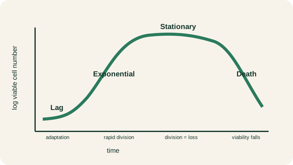
Closed-batch bacterial growth curve with lag, exponential (log), stationary and death phases.
SimpleMed original educational diagram
Reviewed by: Dr. Marcus Judge
- 139
