

Next Lesson - Introduction to Gene Mutation
Core
Epigenetic Regulation
Nearly every nucleated cell contains the same DNA sequence, yet a neurone, hepatocyte and lymphocyte use very different sets of genes. The difference is not a new genome for each tissue. It is controlled access to the shared genome. Epigenetic regulation changes how genes are used without changing the order of DNA bases. This lesson begins where Gene Expression ends: transcription machinery can use a regulatory region only when it is accessible. A useful analogy is a reference book: the DNA sequence supplies the text, while bookmarks, closed pages and access notes determine which passages can be read in a particular cell.
The epigenome includes chemical marks on DNA, modifications of histone proteins and the proteins that interpret or reorganise those marks. These features help a differentiated cell remember its identity after mitosis. They are also dynamic. Developmental signals, ageing, disease and environmental exposures can alter them. That does not mean every acquired mark passes to children. Much of the epigenome is reset during formation of gametes and early development, whereas some specialised parent-of-origin marks must be erased and then established again.
Three roles keep the topic organised. Writers add a mark, erasers remove it and readers bind the marked site and recruit other proteins. The mark is therefore not an isolated switch. Its effect depends on its genomic position, the protein that reads it and the surrounding chromatin. This is why statements such as "methylation always switches genes off" are too crude.
Do not confuse an epigenetic change with a mutation. A mutation changes the DNA sequence; an epigenetic state changes how an unchanged sequence is used. Either can alter phenotype and persist through mitosis, but an epigenetic state can, in principle, be reversed without repairing DNA bases. That potential reversibility does not mean every mark is short-lived. Differentiated cells can maintain regulatory states for many years.
DNA Methylation
DNA methylation adds a methyl group to DNA, commonly to cytosine in a cytosine-phosphate-guanine, or CpG, sequence. CpGs are unevenly distributed. Clusters called CpG islands are often found near gene promoters. When a promoter CpG island becomes heavily methylated, transcription is usually reduced. Methyl groups may hinder some transcription-factor interactions and attract methyl-CpG-binding proteins, which recruit complexes that compact chromatin.
DNA methyltransferases provide the writing activity. Some establish new patterns during development; maintenance enzymes copy the pattern after DNA replication by recognising that the parental strand is methylated. This copying gives daughter cells a molecular memory. A liver cell can therefore preserve much of its liver-specific expression programme as it divides, even though each new DNA molecule began with one newly synthesised strand.
Position matters. Promoter methylation is commonly associated with repression, but methylation elsewhere in a gene does not carry one universal meaning. Unmethylated promoters are not automatically active either: the required transcription factors and accessible chromatin must also be present. DNA methylation is best treated as one layer of regulation that works with histones, regulatory proteins and three-dimensional chromosome organisation.
Histones and Chromatin
Nuclear DNA wraps around histone octamers to form nucleosomes. Nucleosomes package the genome, but their arrangement also controls access. Loosely organised euchromatin is generally more accessible to transcription machinery. Compact heterochromatin is generally less accessible. ATP-dependent chromatin-remodelling complexes can slide, remove or restructure nucleosomes, exposing one regulatory sequence while concealing another.
Histone tails can be acetylated, methylated and modified in other ways. Histone acetyltransferases add acetyl groups to lysines. This reduces positive charge and creates binding sites for reader proteins, often favouring a more open, transcriptionally active state. Histone deacetylases remove acetyl groups and often support compaction and repression. Histone methylation is more context-dependent: its effect changes with the amino-acid residue and the number of methyl groups. It should not be memorised as simply activating or repressing.
These mechanisms cooperate. A transcription factor may recruit a histone-modifying enzyme, accessible chromatin may permit transcription, and DNA methylation may then help stabilise a silent state. The reverse can also occur during a change in cell programme. Epigenetic marks influence probability and accessibility; they do not replace the sequence-specific logic described in Gene Expression.
Genomic Imprinting
Most autosomal genes are expressed from both inherited copies. An imprinted gene is different: expression depends on parental origin, so only the maternal or only the paternal allele is normally active in a particular tissue. The two DNA sequences may be intact. What differs is the epigenetic mark placed during egg or sperm formation.
Imprinting-control regions coordinate marks across clusters of genes. The life cycle has a strict order:
- Old parent-of-origin marks are erased in developing germ cells.
- New marks are established according to whether those germ cells become eggs or sperm.
- After fertilisation, the embryo maintains the maternal and paternal marks through somatic cell division.
- In the next generation's germ line, the marks are erased and reset again.
Because one allele is normally silent, loss of the active parental copy can leave no functional expression. A deletion is one route. Uniparental disomy, or UPD, is another: both copies of a chromosome or chromosome segment come from one parent and none from the other. UPD often has no consequence because most genes are not imprinted. In an imprinted region, however, two correctly formed copies can carry the same silent parent-of-origin programme.
Prader-Willi and Angelman Syndromes
Prader-Willi and Angelman syndromes show why the parent matters as much as the chromosome. Both involve the imprinted region at 15q11-q13, but they lose different active programmes.
- Prader-Willi syndrome: paternally expressed genes are lost. A paternal deletion can remove them, while the maternal copies remain normally silent. Maternal UPD can produce the same functional result because both chromosome 15 copies carry a maternal imprint. An imprinting-control defect can also silence the paternal programme.
- Angelman syndrome: maternal UBE3A function is lost in neurones, where the paternal copy is normally silenced. Causes include a maternal deletion, a pathogenic variant in maternal UBE3A, an imprinting defect or paternal UPD.
The pairing rule is worth learning mechanistically rather than as two unrelated syndromes: no active paternal programme gives Prader-Willi syndrome; no active maternal neuronal UBE3A gives Angelman syndrome. The clinical patterns differ because the affected genes and tissues differ. This is a parent-of-origin expression problem, not a change in the basic definition of dominant or recessive inheritance.
X-inactivation
Cells with two X chromosomes require dosage compensation because two fully active X chromosomes would otherwise supply roughly twice the expression of many X-linked genes compared with a cell carrying one X. Early in embryonic development, one X is largely inactivated in each somatic cell. The XIST long non-coding RNA is produced from the chromosome chosen for inactivation, coats that X and recruits silencing machinery. The condensed inactive X can be seen as a Barr body.
Choice is usually random with respect to parental origin. Once made, it is maintained through the descendants of that cell. A person with two X chromosomes is therefore a mosaic: some cell clones express the maternal X and others the paternal X. This differs from genomic imprinting, where a particular parental allele is consistently selected according to its origin. Not every gene is silenced; genes in pseudoautosomal regions and some other X-linked genes escape inactivation.
Random choice does not guarantee a perfect 50:50 pattern. Chance, selection between cell populations or a structural X-chromosome change can produce skewed X-inactivation. A heterozygous carrier of an X-linked variant may then show more features if the X carrying the functional allele is inactive in a large proportion of the relevant tissue. Such a manifesting carrier demonstrates that mosaic proportions and tissue distribution can influence phenotype.
This dosage compensation follows chromosome segregation. Review how meiotic nondisjunction produces aneuploid gametes in Mitosis and Meiosis, then use X-inactivation here to explain why an extra X chromosome does not simply double every X-linked transcript.
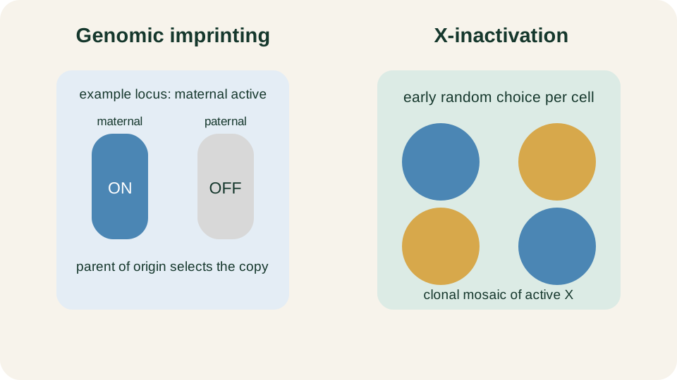
At this example locus the maternal copy is active; other imprinted loci select the paternal copy. Random X-inactivation instead produces neighbouring clones with different active X chromosomes.
SimpleMed original educational diagram
Mitochondrial Inheritance and Heteroplasmy
Mitochondria contain their own circular DNA, and each cell contains many mitochondrial DNA, or mtDNA, molecules. Human embryos normally receive their mitochondria through the egg. A mother with an mtDNA variant can transmit it to children of any sex, whereas an affected father does not normally transmit his mtDNA variant. This maternal pattern applies to pathogenic variants in mtDNA. Disorders of nuclear genes that support mitochondrial function can instead follow autosomal or X-linked inheritance.
If all mtDNA copies are alike, the state is homoplasmy. A mixture of usual and altered mtDNA is heteroplasmy. When mitochondria replicate and cells divide, mtDNA copies are distributed among daughter cells. The altered proportion can therefore differ between siblings, between tissues in one person and over time. A blood sample may not represent the proportion in muscle, brain or another affected tissue.
Cells can often compensate while the altered proportion remains below a functional threshold. Once the load crosses that threshold, energy production becomes inadequate and dysfunction emerges. The threshold is not one universal percentage. It depends on the variant, tissue and energy demand. High-demand tissues may reveal impairment sooner, while another tissue carrying the same variant proportion remains functional.
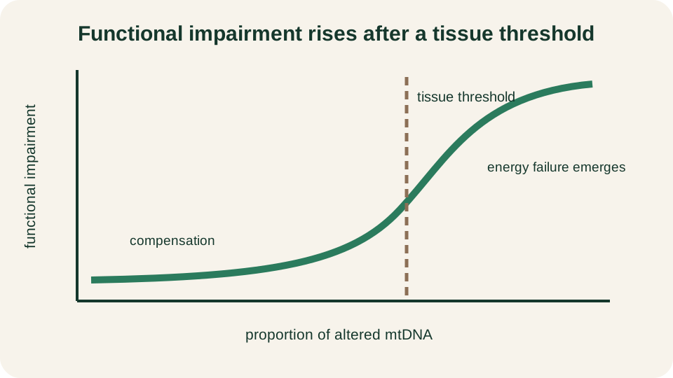
Heteroplasmy becomes clinically important when altered mtDNA exceeds the functional reserve of a particular tissue.
SimpleMed original educational diagram
Maternal transmission therefore gives the pedigree direction, but heteroplasmy and threshold effects shape severity. For conventional pedigree patterns, return to Genotype, Phenotype, and Inheritance. Classes of DNA sequence change remain in Introduction to Gene Mutation. Here the central rule is expression: epigenetic marks choose access, imprinting chooses a parental copy, X-inactivation chooses an X within each cell, and heteroplasmy determines how much altered mtDNA a tissue must accommodate.
Reviewed by: Dr. Marcus Judge
- 140
