

Next Lesson - Gastrointestinal Secretion, Digestion and Absorption
Core
Lobule Architecture and Opposing Flows
The liver is a dual-inflow chemical plant. About three quarters of its blood arrives in the portal vein, carrying absorbed nutrients, hormones and some gut-derived solutes. About one quarter arrives in the hepatic artery as oxygenated arterial blood. Those streams mix at the periphery of each microscopic unit, cross a short exchange bed and leave through hepatic venous branches. Regional gross anatomy belongs with the foregut lesson; the functional unit here is the classical hepatic lobule.
Orient this integrated physiology with Anatomy and Physiology of the Foregut, Anatomy and Physiology of the Midgut and Anatomy and Physiology of the Hindgut. Those regional lessons locate organs and vessels; this lesson follows lobular exchange, bile and exocrine pancreatic secretion across the same system.
Each lobule is classically drawn as a hexagon of hepatocyte plates (cords) radiating toward a central vein. At each corner sits a portal triad: a terminal portal venule, a terminal hepatic arteriole and a bile ductule, with lymphatics and nerves. Between the plates run sinusoids, specialised capillaries with discontinuous, fenestrated endothelium so plasma bathes the basolateral hepatocyte face more freely than in a continuous capillary. The gap between endothelium and hepatocyte is the space of Disse, filled with microvilli and matrix that support exchange without a classical basement membrane.
Kupffer cells, the liver's fixed macrophages, sit mainly in the sinusoidal lumen and clear debris, aged cells and gut-derived particles. Hepatic stellate (Ito) cells live in the space of Disse and store retinoids; under chronic injury they can become matrix-secreting myofibroblasts. For normal physiology the point is simpler: sinusoids are an exchange surface, not a sealed tube.
Blood and bile travel in opposite directions. Mixed portal and arterial blood enters at the triad, flows along sinusoids toward the central vein and exits into the hepatic venous system. Bile is secreted from the apical (canalicular) poles of neighbouring hepatocytes into blind-ended bile canaliculi. Those canaliculi form a lattice between cells and drain outward to portal triad ductules, then into larger intrahepatic ducts and the extrahepatic biliary tree. Memorise the spatial rule: blood portal-to-central; bile canalicular-to-portal. That opposition places newly formed bile at the periphery where it can leave without fighting venous outflow, and it means hepatocytes nearer the portal inflow see fresher blood than those nearer the central vein.
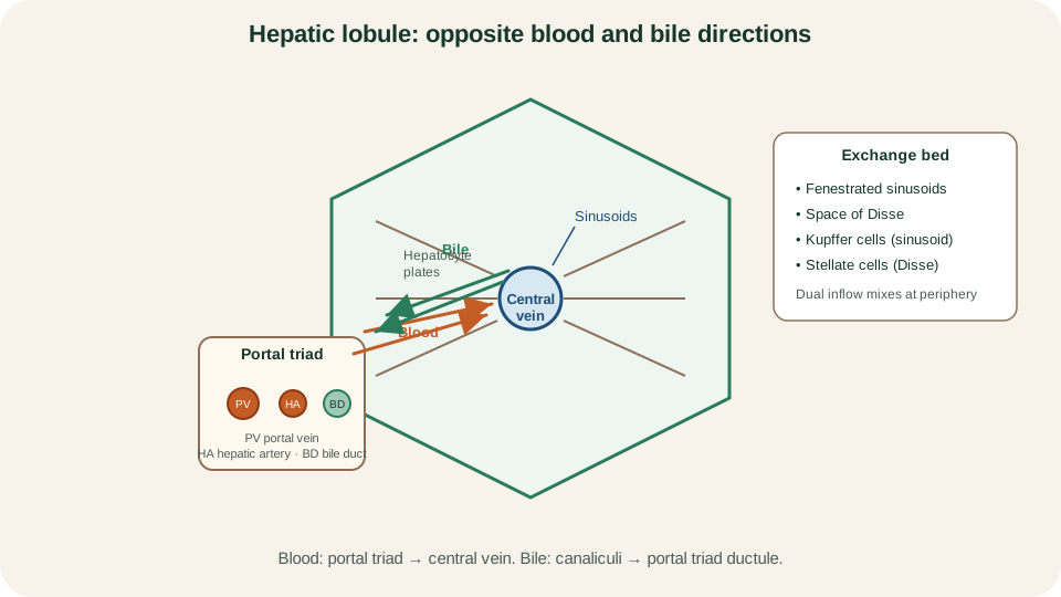
Classical lobule: dual inflow at the portal triad, sinusoidal flow toward the central vein, and bile flow in the opposite direction toward the triad ductule.
SimpleMed original educational diagram
Functional Zonation of Hepatocytes
Because blood enters at the portal triad and leaves at the central vein, oxygen, nutrients and many hormones form gradients across each plate of hepatocytes. The functional (acinar) description names three zones along that path. Zone 1 is periportal, nearest the dual inflow. Zone 3 is pericentral, nearest the central vein. Zone 2 is intermediate. These are continuous gradients, not watertight compartments, so metabolic maps are tendencies rather than absolute exclusive assignments.
Zone 1 hepatocytes usually see the highest oxygen and the first claim on many portal substrates. Simplified teaching maps place oxidative metabolism, gluconeogenesis, urea-cycle activity and bile-acid handling relatively here. Zone 3 cells receive blood already partly extracted upstream, so oxygen is lower; glycolysis, lipogenesis and selected cytochrome P450 activities are often emphasised there. Zone 2 bridges the two and shifts with perfusion, fasting and disease load.
The gradient already predicts common injury patterns without becoming a disease catalogue: centrilobular (zone 3) injury when a toxin needs bioactivation or hypoxia hits the last cells on the oxygen line; periportal injury when the first cells on the portal stream are exposed most heavily. Do not force every pathway into a single zone. Ask where substrate and oxygen arrive, and which enzymes are relatively concentrated along that path.
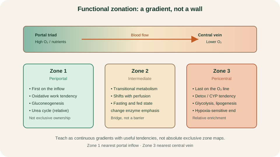
Acinar zones 1 to 3 follow the portal-to-central gradient; metabolic labels are relative tendencies, not exclusive maps.
SimpleMed original educational diagram
Synthesis, Metabolism and Detoxification
Hepatocytes are polarised secretory cells: basolateral face to sinusoids, apical face to canaliculi. That polarity lets one cell export albumin into blood and bile salts into bile. Organise hepatic work by purpose, not as a pathway catalogue.
Synthesis for plasma includes albumin (the main oncotic and carrier protein), most coagulation factors, regulatory proteins such as protein C, protein S and antithrombin, and many binding globulins. Factor VIII is a useful cell-type exception: it is produced mainly by liver sinusoidal endothelial cells rather than hepatocytes. Falling albumin and a rising prothrombin time, interpreted with vitamin K status, test synthetic function rather than mere enzyme leak from damaged cells.
Carbohydrate handling keeps blood glucose usable between meals. After a carbohydrate load the liver stores glycogen and can route surplus carbohydrate toward lipid; in fasting it releases glucose by glycogenolysis and gluconeogenesis. Lipid handling includes packaging of very-low-density lipoprotein, biliary export of cholesterol and phospholipid, and fatty-acid oxidation when fuel is needed. Amino-acid handling supports protein synthesis and nitrogen disposal: surplus amino groups enter the urea cycle, converting ammonia to urea that the kidney can excrete. Without that conversion, nitrogenous waste would accumulate in the circulation.
Storage is selective. Glycogen is the short-term carbohydrate store. Stellate cells hold much of the body's vitamin A. The liver also holds iron and copper within regulated systems and performs 25-hydroxylation of vitamin D before the kidney completes activation. Fat-soluble vitamins reach hepatocytes via chylomicron remnants and related lipoproteins after intestinal absorption.
Detoxification and xenobiotic handling use two classical phases. Phase I reactions (often cytochrome P450-mediated oxidation, reduction or hydrolysis) introduce or expose polar groups. Phase II reactions conjugate the product to glucuronate, sulphate, glutathione or related partners, increasing water solubility for biliary or renal excretion. Bilirubin conjugation is itself a phase II example. Reduced glutathione stores matter because some reactive intermediates become toxic when conjugation capacity is overwhelmed. The same machinery clears many drugs, hormones and endogenous waste; it is not a separate organ bolted onto the lobule.
Bilirubin from Haem to Pigment
Bilirubin is the coloured waste of haem breakdown. Senescent erythrocytes and other haem proteins are processed mainly in macrophages of the spleen, liver and marrow. Haem oxygenase opens the ring to biliverdin; biliverdin reductase yields unconjugated bilirubin. Unconjugated bilirubin is poorly water-soluble and travels in plasma bound to albumin, which keeps free pigment low under normal loads.
Hepatocytes take up unconjugated bilirubin at the basolateral membrane and conjugate it mainly with glucuronic acid via UDP-glucuronosyltransferase (UGT1A1). Conjugated bilirubin is more water-soluble and is actively exported across the canalicular membrane into bile. Small amounts of conjugated pigment can appear in plasma and are filtered by the kidney when levels rise, which is why dark urine can accompany conjugated hyperbilirubinaemia in clinical stories taught later. Classification of jaundice into pre-hepatic, hepatic and post-hepatic patterns belongs in pathology; the physiology question here is the sequence itself.
In the gut, bacteria deconjugate and reduce bilirubin to urobilinogen. Most of that load is further converted to stercobilin, which colours the faeces. A fraction of urobilinogen is reabsorbed, returns via the portal vein and is either re-excreted by the liver or passed to the kidney and oxidised to urobilin, contributing to urine colour. When bile fails to reach the intestine, stools pale and urobilinogen generation falls; when haem turnover is high, more unconjugated pigment is presented to an otherwise intact conjugating system. Always track solubility and route: albumin carriage, hepatic conjugation, biliary export, then bacterial conversion.
Bile Acids, Gallbladder and Enterohepatic Circulation
Bile is more than a bilirubin drain. Hepatocytes synthesise primary bile acids from cholesterol and conjugate them with glycine or taurine to form bile salts that remain ionised at intestinal pH. Canalicular transporters, including the bile salt export pump, secrete bile salts into canaliculi. Their osmotic pull draws water and generates much of canalicular bile flow; a bile-acid-independent fraction is driven by other solutes such as glutathione. Cholangiocytes lining the ducts modify bile further, adding a bicarbonate-rich fluid under secretin stimulation, so ductal flow is not a passive pipe.
Between meals, resistance at the sphincter of Oddi directs much of the bile into the gallbladder, where mucosa reabsorbs water and electrolytes and concentrates organic constituents. A fatty meal releases cholecystokinin (CCK) from duodenal I cells. CCK contracts the gallbladder and helps relax the sphincter of Oddi so concentrated bile enters the duodenum. There, bile salts emulsify dietary fat and form micelles that carry monoglycerides, fatty acids and fat-soluble vitamins to the enterocyte surface. Detailed micelle-to-chylomicron chemistry continues in the next lesson on digestion and absorption.
Most conjugated bile salts are not lost in the faeces. They are actively reabsorbed in the terminal ileum, return in portal blood and are efficiently extracted by hepatocytes for reuse. Only a few percent of the pool is lost each cycle and replaced by new hepatic synthesis. That closed loop is the enterohepatic circulation. Interrupt the loop at synthesis, canalicular secretion, biliary delivery, ileal uptake or portal return, and the same physiological map predicts steatorrhoea risk or compensatory increases in synthesis. Vagal tone and secretin support the bicarbonate and volume side of biliary and pancreatic duct flow during a meal, while CCK dominates gallbladder emptying and enzyme-side pancreatic signalling.
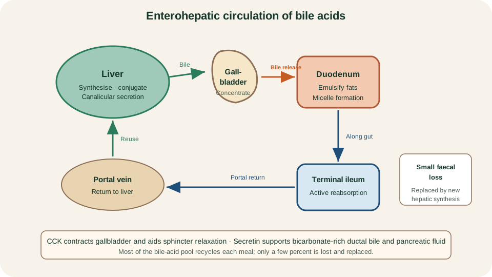
Enterohepatic circulation: hepatic synthesis and canalicular secretion, gallbladder storage, CCK-driven release, terminal-ileal reuptake and portal return.
SimpleMed original educational diagram
Exocrine Pancreas: Acini and Ducts
The pancreas is two organs in one body. The endocrine islets regulate systemic fuel and belong in endocrinology. The exocrine pancreas builds the bulk of the gland as secretory acini draining into a duct tree that joins the biliary stream at the ampulla before entering the duodenum. Exocrine output has two complementary products: a concentrated enzyme mix from acinar cells and a large volume of alkaline fluid from duct cells.
Acinar cells synthesise digestive enzymes on rough endoplasmic reticulum, package them in zymogen granules and release them by exocytosis into the acinar lumen. Proteases leave mainly as inactive zymogens (trypsinogen, chymotrypsinogen, procarboxypeptidases, proelastase). Pancreatic amylase and lipase are secreted in catalytically active forms because their substrates are not normally exposed within pancreatic tissue. Lipase still needs colipase at the intestinal fat-water interface; colipase is released as procolipase and activated in the duodenum by trypsin. CCK, released when fat and protein enter the upper small bowel, is the dominant hormonal drive to acinar enzyme secretion; vagal cholinergic input amplifies the same pathway, especially in the cephalic and early intestinal phases.
Duct cells secrete water and bicarbonate, raising pancreatic juice pH so enzymes can work and so acidic gastric chyme is neutralised in the duodenal lumen. Secretin, released from S cells when acidic chyme lowers duodenal pH, is the principal hormonal stimulus for this alkaline fluid. CFTR-dependent chloride and bicarbonate transport in duct epithelium underpins the volume of secretion; when duct flushing fails, enzymes linger where they should not. Functionally, remember the pair: CCK and vagus for enzymes; secretin for bicarbonate and water. Both secretions meet in the duct system and arrive together at the major duodenal papilla.
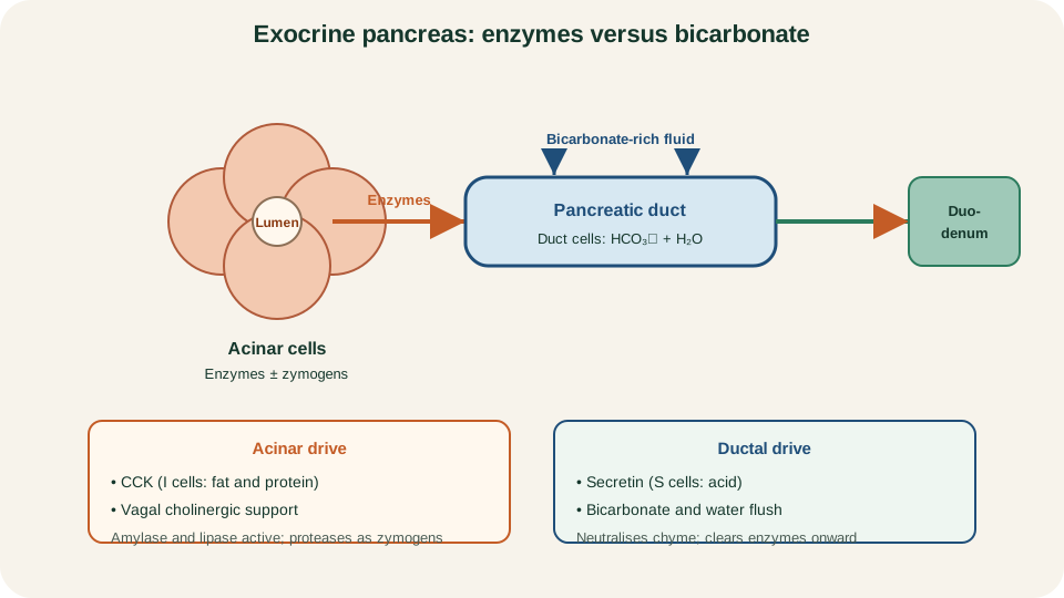
Acinar enzyme output versus ductal bicarbonate and water: CCK and vagal drive for enzymes, secretin for alkaline fluid.
SimpleMed original educational diagram
Zymogen Activation and Safeguards
Proteases must not digest the gland that made them. Activation is therefore delayed until the enzymes reach the intestinal lumen. Duodenal enterocytes display enteropeptidase (enterokinase) on the brush border. Enteropeptidase cleaves trypsinogen to trypsin. Trypsin then activates the remaining protease zymogens and can also activate more trypsinogen, creating a cascade once a small starter amount of trypsin appears. Amylase and lipase do not require this cascade; the critical safety design sits with the proteases.
Several layered protections keep premature trypsin low inside the pancreas. First, packaging: zymogens are stored in granules and segregated from lysosomal hydrolases under normal conditions. Second, secretory trypsin inhibitor (SPINK1) can bind and quench small amounts of trypsin that appear too early. Third, ductal bicarbonate and fluid flush enzymes rapidly into the duodenum, limiting residence time in the duct tree. Acute pancreatitis becomes intelligible as failure of this package: premature trypsinogen activation inside the gland starts a proteolytic cascade, local inflammation and autodigestion. Disease classification and management stay in pathology; the physiology lesson is why inactive packaging, an intestinal trigger enzyme, SPINK1 and duct flushing exist together.
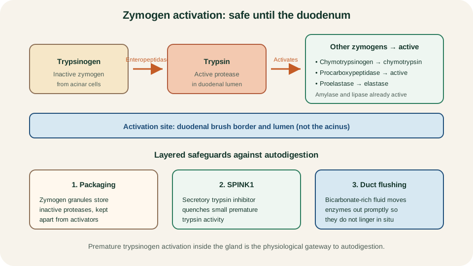
Enteropeptidase starts trypsin formation in the duodenum; trypsin activates other zymogens. Packaging, SPINK1 and duct flushing limit premature activity.
SimpleMed original educational diagram
The next article, Gastrointestinal Secretion, Digestion and Absorption, follows a mixed meal through luminal enzymes, micelles and enterocyte transporters once bile and pancreatic juice have done their preparatory work.
Reviewed by: Dr. Marcus Judge
- 135
