

Next Lesson - Upper Gastrointestinal Pathology
Core
Following a Mixed Meal
Think of one plate that holds starch, protein and fat, with water and a few micronutrients. Digestion is not a single chemical bath. At each station ask three questions: what is being cut, which enzyme or physical process acts, and what finally crosses the enterocyte. Motility mixes and propels the meal, as covered in Gastrointestinal Motility and the Enteric Nervous System. Bile salts and pancreatic juice that arrive in the duodenum are produced and controlled as taught in Liver, Biliary and Exocrine Pancreatic Physiology. This lesson follows the substrates themselves from mouth to blood or lymph.
The map is causal. The mouth increases surface area and starts starch. The stomach denatures protein and begins proteolysis. The duodenum receives acid chyme, bicarbonate, bile and pancreatic enzymes so luminal digestion can finish safely. The jejunum and ileum present a huge brush-border surface with transporters that take monosaccharides, amino acids, small peptides and, after micelle delivery, lipid building blocks. The colon reclaims water and electrolytes that escaped proximal absorption. Regional gross anatomy stays in the foregut, midgut and hindgut lessons; the physiology here is the hand-off sequence for one meal.
Place that sequence onto the regional maps in Anatomy and Physiology of the Foregut, Anatomy and Physiology of the Midgut and Anatomy and Physiology of the Hindgut. The links prevent regional anatomy and integrated transport physiology from becoming separate facts.
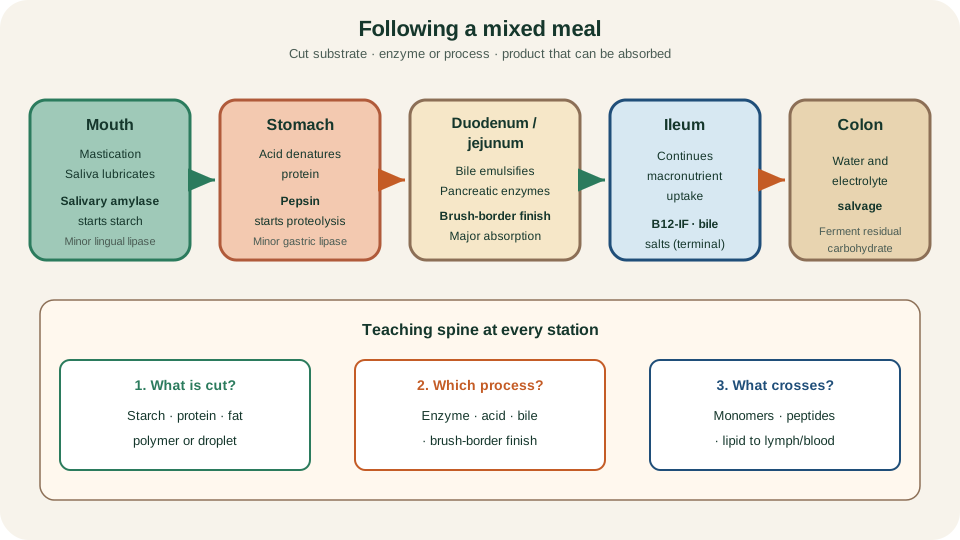
Meal journey by segment: surface-area and early enzyme work, gastric protein attack, duodenal enzyme and bile mixing, small-bowel absorption, then colonic water salvage.
SimpleMed original educational diagram
Cephalic, Gastric and Intestinal Phases
Secretion is phased so enzymes and acid appear when food is expected or present, without flooding an empty lumen. The cephalic phase is anticipatory. Sight, smell, taste and thought of food, acting through the vagus, increase salivary flow and prepare gastric acid and pepsinogen output before the bolus arrives. It is neural preparation, not yet full chemical feedback from the gut lumen.
The gastric phase is driven by what is in the stomach. Wall distension and the sensing of peptides and amino acids stimulate gastrin release from antral G cells. Gastrin supports acid secretion from parietal cells and promotes pepsinogen release; local and vagovagal reflexes amplify the same programme. Acid itself later restrains gastrin, so the phase is self-limiting once the meal is ground and emptied.
The intestinal phase is dominated by duodenal signals once acidic, fatty or hypertonic chyme arrives. Secretin from S cells, released when luminal pH falls, drives bicarbonate-rich fluid from pancreatic ducts and biliary epithelium. Cholecystokinin (CCK) from I cells, released by fat and protein digestion products, stimulates pancreatic enzyme secretion, contracts the gallbladder and helps relax the sphincter of Oddi so bile and juice enter the lumen. The same nutrient signals slow gastric emptying so the small bowel is not overwhelmed. Do not treat the intestinal phase as simply another strong driver of gastric acid; its main job is to coordinate accessory secretions and pace delivery. Motility detail remains with the ENS lesson; enzyme packaging and zymogen safety remain with the liver and pancreas lesson.
Mouth and Stomach Preparation
Mastication reduces particle size and mixes food with saliva. Saliva lubricates the bolus for safe swallowing and supplies salivary amylase, which begins starch digestion by cleaving internal alpha-1,4 glycosidic bonds to yield maltose, maltotriose and limit dextrins. That work continues until acid inactivates the enzyme in the stomach. Lingual lipase, and later gastric lipase, make only a minor contribution to triglyceride hydrolysis in adults; they matter more as early surface attack than as the main fat pathway.
In the stomach, acid does three useful things for digestion of a mixed meal: it creates a hostile environment for many swallowed microbes, it denatures protein so peptide bonds become more accessible, and it supports activation of pepsinogen to pepsin. Pepsin starts internal proteolysis, producing large peptides rather than free amino acids ready for absorption. Carbohydrate digestion largely pauses in the acid environment. Intrinsic factor, also secreted by parietal cells, travels onwards for later B12 handling rather than enabling gastric B12 absorption. Parietal-cell pharmacology and acid-suppressing drugs belong in their dedicated articles; here the point is functional preparation of the meal for the duodenum.
Carbohydrate Digestion and Transport
Once chyme is neutralised in the duodenum, pancreatic amylase continues starch breakdown. Its products are still oligosaccharides and disaccharides, not free glucose ready for bulk absorption. Final liberation of monosaccharides is a brush-border job. Maltase-glucoamylase, sucrase-isomaltase and lactase cleave maltose, maltotriose, sucrose, limit dextrins and lactose into glucose, galactose and fructose at the apical membrane. Dietary fibre that human enzymes cannot hydrolyse continues distally and may be fermented by colonic bacteria; that is not enterocyte absorption of the parent polymer.
Monosaccharide entry is transporter-specific. SGLT1 cotransports glucose or galactose with sodium across the apical membrane, powered by the basolateral Na+/K+-ATPase sodium gradient. GLUT5 facilitates fructose entry at the apex. All three monosaccharides leave the enterocyte toward the interstitium and portal blood mainly through basolateral GLUT2. Remember the division of labour: luminal enzymes cut polymers; brush-border enzymes finish disaccharides; SGLT1 and GLUT5 take monomers in; GLUT2 sends them out.
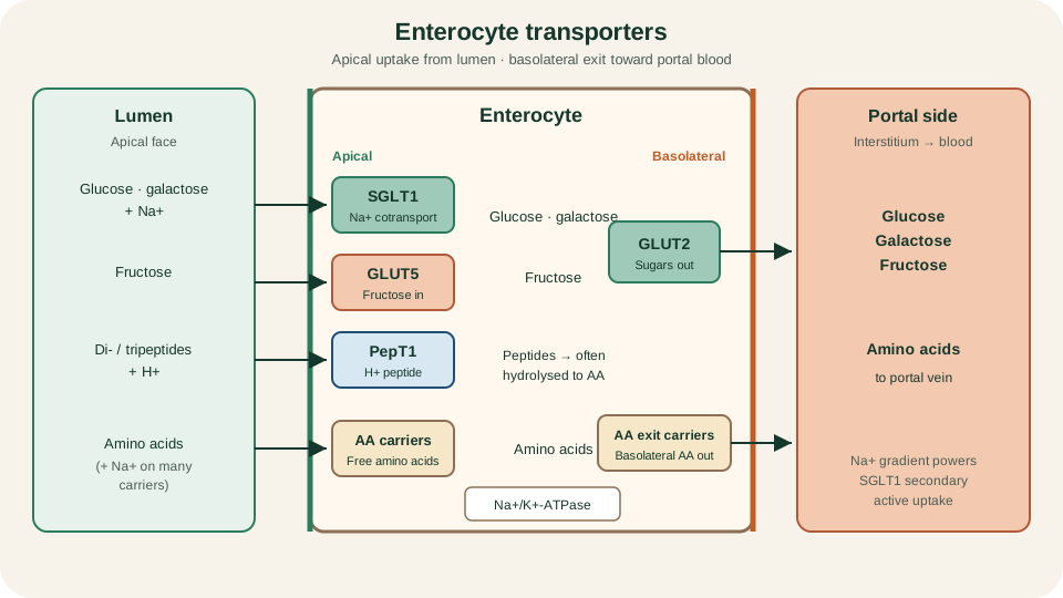
Apical uptake map: SGLT1 for glucose and galactose with sodium, GLUT5 for fructose, PepT1 for di- and tripeptides with H+, and representative amino-acid carriers; basolateral GLUT2 exports monosaccharides and basolateral amino-acid carriers export amino acids toward portal blood.
SimpleMed original educational diagram
Protein Digestion and Peptide Uptake
Protein digestion is staged. Pepsin in the stomach starts the process. In the duodenum, enteropeptidase on the brush border activates trypsinogen to trypsin; trypsin then activates other pancreatic protease zymogens. Endopeptidases (trypsin, chymotrypsin, elastase) cut internal bonds; carboxypeptidases trim free amino acids from the carboxyl end. The lumen therefore fills with free amino acids plus oligopeptides rather than only single amino acids.
Absorption uses two complementary routes. Free amino acids ride several apical carriers, many sodium-coupled, with overlapping specificities for neutral, cationic or anionic side chains. Dipeptides and tripeptides take a high-capacity path through PepT1, which couples peptide entry to the inward H+ gradient. Inside the enterocyte, cytosolic peptidases often complete hydrolysis to free amino acids. Absorbed products leave across the basolateral membrane into interstitial fluid and then portal blood. Intact dietary protein does not need to cross as whole molecules for nutrition; the teaching product is amino acids and small peptides processed for systemic use.
Fat: Emulsification, Micelles and Chylomicrons
Dietary triglyceride is insoluble in water. Bile salts emulsify large fat droplets into smaller ones, increasing the surface available to pancreatic lipase and colipase. Lipase yields mainly 2-monoglycerides and free fatty acids; cholesterol and fat-soluble vitamins travel with the lipid phase. Those products are packaged into mixed micelles: small, bile-salt-stabilised aggregates that ferry long-chain fatty acids, monoglycerides, cholesterol and vitamins A, D, E and K through the aqueous unstirred layer to the brush border.
Critical geometry: micelles do not enter the enterocyte intact. Lipid leaves the micelle at the apical membrane and is taken up; bile salts remain largely in the lumen for reuse until terminal-ileal reclaim. Inside the cell, long-chain products are re-esterified to triglyceride in the smooth endoplasmic reticulum, assembled with phospholipid, cholesterol and apolipoprotein B-48 into chylomicrons, and released into lacteals rather than portal capillaries. Short- and medium-chain fatty acids can reach portal blood more directly with less obligatory chylomicron packaging. Systemic lipoprotein metabolism after chylomicrons enter blood is taught with lipid transport articles, not here.
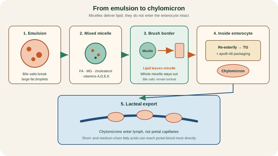
Fat path: emulsion to mixed micelle, monomer uptake at the brush border without whole-micelle entry, re-esterification and apoB-48 chylomicron assembly, then lacteal export.
SimpleMed original educational diagram
Water, Electrolytes and Regional Micronutrients
Several litres of fluid enter the gut each day from diet and secretions; most is reabsorbed so stool remains formed. Water is not pumped by a dedicated water ATPase. It follows net solute absorption osmotically through transcellular and paracellular routes that vary by segment. Sodium uptake includes nutrient cotransport (as with SGLT1) and other segment-specific pathways; chloride and water follow when net solute leaves the lumen. The small bowel absorbs the bulk of the fluid load; the colon salvages remaining water and electrolytes. Avoid fixed-volume claims that every meal always transfers the same number of litres; the principle is solute-led recovery with distal fine-tuning.
Location matters for selected micronutrients. Iron is absorbed chiefly in the duodenum and proximal jejunum after reduction of ferric iron and apical uptake of ferrous iron, with regulated basolateral export. Folate is taken up mainly in the proximal small bowel. Vitamin B12 is freed from food protein, bound first to haptocorrin, then transferred to intrinsic factor after pancreatic proteases digest haptocorrin; the intrinsic-factor-B12 complex is absorbed by receptor-mediated endocytosis in the terminal ileum. Conjugated bile salts are actively reclaimed in the terminal ileum for enterohepatic return, as mapped in the previous lesson. Major macronutrient absorption is predominantly small-bowel work along the jejunum and ileum, not a single exclusive centimetre. These are predominance statements, not absolute bans on minor uptake elsewhere.
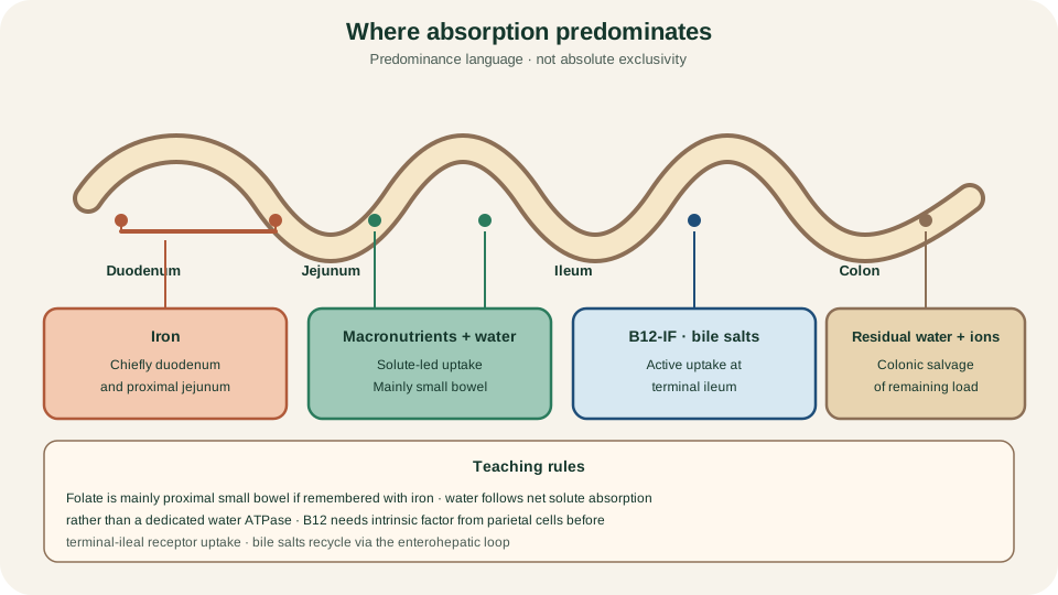
Predominant locations: proximal iron, small-bowel macronutrients with bulk solute-led water, terminal-ileal B12-intrinsic-factor and bile-salt reclaim, residual water and ion colonic salvage.
SimpleMed original educational diagram
Two brief failures illuminate the normal map. If lactase is deficient, lactose remains an osmotically active, fermentable sugar in the lumen, drawing water and generating gas after dairy. If coeliac injury flattens villi, surface area and brush-border enzyme capacity fall, so several absorption steps fail together. Diagnosis and treatment stay in clinical articles; the physiology lesson is that missing enzymes or missing surface both break the cut-and-cross sequence.
The next lesson, Upper Gastrointestinal Pathology, applies these mechanisms when structure or regulation fails in the upper gut.
Reviewed by: Dr. Marcus Judge
- 139
