

Next Lesson - Liver, Biliary and Exocrine Pancreatic Physiology
Core
Four Layers of the Gut Wall
From mouth to anus the hollow gut is built as a layered tube. Motility is not a free-floating muscle property; it is the coordinated work of those layers, paced electrically and steered by nerves that live inside the wall. Every pattern you meet later either mixes luminal contents with enzymes and mucosa or moves them onwards.
From lumen outward the four classical layers are:
- Mucosa: epithelium for absorption and secretion, lamina propria, and a thin muscularis mucosae that can ripple the mucosal surface.
- Submucosa: connective tissue, vessels, lymphatics and the submucosal (Meissner) plexus, which mainly steers secretion, local blood flow and short mucosal reflexes.
- Muscularis externa: usually an inner circular and outer longitudinal smooth-muscle layer, with the myenteric (Auerbach) plexus between them. This stack is the principal motor engine of the gut.
- Serosa or adventitia: a peritoneal covering where the organ is intraperitoneal, or connective adventitia where it is retroperitoneal or extraperitoneal.
Regional anatomy, arterial supply and peritoneal relations stay in Anatomy and Physiology of the Foregut, Anatomy and Physiology of the Midgut and Anatomy and Physiology of the Hindgut. Here the question is functional: which layer generates force, which houses which nerve network, and how those parts produce mix-or-move patterns.
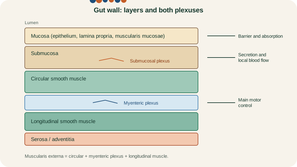
Gut-wall layers with both enteric plexuses: submucosal plexus in the submucosa; myenteric plexus between circular and longitudinal muscle.
SimpleMed original educational diagram
Enteric Plexuses and Autonomic Modulation
The enteric nervous system is the gut's intrinsic nervous system. It contains sensory neurons, interneurons and motor neurons arranged mainly in two ganglionated networks. The myenteric plexus lies between circular and longitudinal muscle and is the dominant controller of wall motility along almost the full length of the gut. The submucosal plexus lies in the submucosa of the intestines and mainly regulates epithelial secretion, local vasomotor tone and mucosal folding rather than long-range propulsion.
Intrinsic circuits can organise local reflexes without continuous orders from the brain or spinal cord. A classic example is the peristaltic reflex (sometimes called the law of the intestine): stretch or chemical stimulation of a segment excites ascending pathways that contract muscle oral to the stimulus and descending pathways that relax muscle aboral to it. Excitatory motor neurons often use acetylcholine and tachykinins; inhibitory motor neurons often use nitric oxide and vasoactive intestinal peptide. Those transmitters act on smooth muscle partly through intermediate cells such as interstitial cells of Cajal, rather than only through classical neuromuscular junctions.
Autonomic nerves do not replace the ENS; they modulate it. Parasympathetic pathways (vagus to much of the foregut and midgut; pelvic splanchnic nerves to the distal colon, rectum and anal canal) are generally facilitatory for motility and secretion, acting largely by synapsing on enteric neurons. Sympathetic pathways (thoracolumbar preganglionic outflow via prevertebral ganglia) are generally inhibitory to motility and divert blood flow, again mainly by acting on enteric circuits and sphincters. Sight, smell and emotion therefore change gut behaviour through extrinsic nerves, but the wall still executes the detailed motor programme locally.
Smooth Muscle, ICC and Slow Waves
Most gut motility uses unitary smooth muscle: cells are electrically coupled and can behave as a syncytium. Superimposed on that syncytium are interstitial cells of Cajal (ICC), specialised pacemaker and intermediary cells that generate and propagate slow waves, the rhythmic oscillations of membrane potential that set the maximum frequency of contraction in each region.
Slow-wave frequency is region-specific (for teaching purposes, about three cycles per minute in the stomach and higher in the proximal small intestine, falling more distally). Slow waves depolarise muscle toward threshold, but they are not the same as a full action potential. Forceful phasic contraction appears when additional depolarisation produces spike potentials (action potentials) on the crest of a slow wave, allowing substantial calcium entry. Neural and hormonal input shift the baseline and the likelihood of spikes. That is why a slow wave can pass with little mechanical effect at rest, yet the same rhythm supports strong contraction after a meal or under excitatory neural drive.
Ileus illustrates the distinction in reverse: pacemaker slow waves may continue while spike activity and effective propulsive contractions fail, so the electrical clock ticks without useful mechanical work. Always ask two questions of any recording or clinical story: is the slow-wave rhythm present, and are spikes recruiting contraction on those waves?
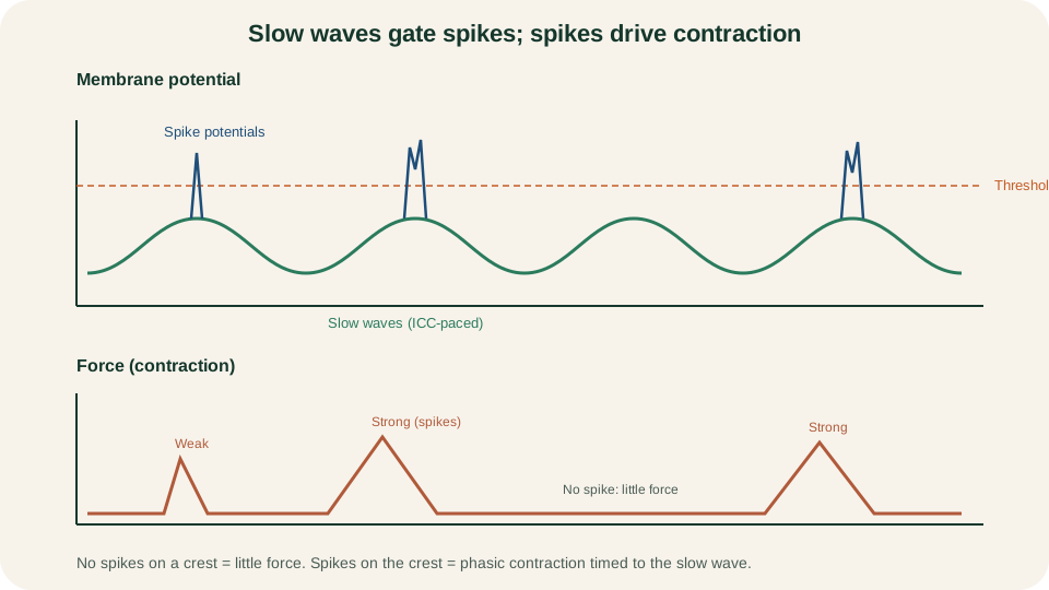
Slow waves set timing; spike potentials on the crest cross threshold and couple to contraction. Not every slow wave produces spikes.
SimpleMed original educational diagram
Segmentation and Peristalsis
Two mechanical patterns dominate post-prandial gut behaviour. Segmentation is rhythmic, localised contraction of circular muscle that divides a segment into temporary compartments. Contents are churned back and forth against the mucosa and enzymes with little net aboral progress. The small intestine uses segmentation heavily for mixing and absorption time; the colon uses related haustral mixing for water and electrolyte salvage.
Peristalsis is a propagating wave that moves contents aborally. Circular muscle contracts oral to a bolus while the aboral segment relaxes, and longitudinal muscle shortens and widens the receiving segment. Primary oesophageal peristalsis is swallow-triggered; secondary waves clear residual material after distension. In the intestine, short peristaltic runs advance chyme after mixing. Mass movements in the colon are high-amplitude propulsive events that shift larger volumes toward the rectum.
Keep the functional rule sharp: segmentation mixes; peristalsis propels. Both can coexist in the same region at different times of the digestive cycle, and both still depend on ENS patterning built on the slow-wave scaffold.
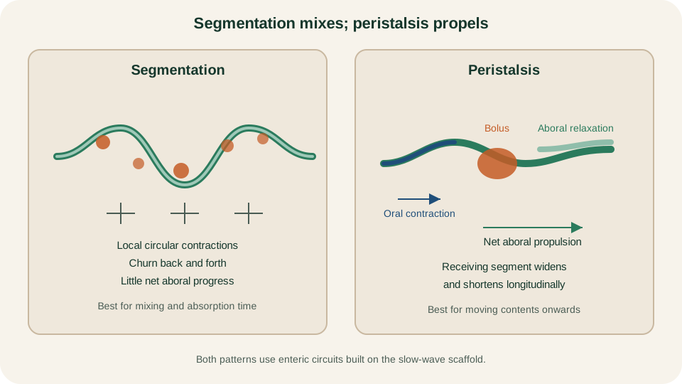
Segmentation mixes contents in place; peristalsis pairs oral contraction with aboral relaxation to move a bolus onwards.
SimpleMed original educational diagram
Migrating Motor Complex
Between meals the stomach and small intestine run a different programme: the migrating motor complex (MMC), a cyclic interdigestive pattern often nicknamed the intestinal housekeeper. It clears residual debris, secretions and bacteria toward the colon and is interrupted when a substantial meal is taken, after which fed segmentation and shorter peristaltic activity take over.
Classically the MMC is described in successive phases repeating about every 90 to 120 minutes in fasting humans:
- Phase I: motor quiet, little propulsion.
- Phase II: irregular intermittent contractions.
- Phase III: a burst of intense, regularly spaced contractions that originate in the antrum or duodenum and migrate distally; this is the clearing front.
- Phase IV: a brief transition back toward quiet.
Control is mixed. Enteric circuits are essential for intestinal phases; motilin is linked especially to gastric-origin phase III activity, and other mediators can shift duodenal-origin activity. Vagal influence is more important for the gastric MMC than for the small-bowel cycle. The teaching contrast that matters is temporal: fasting MMC housekeeper versus fed mixing and short-range propulsion. Do not describe the MMC as the main pattern during active digestion of a meal.
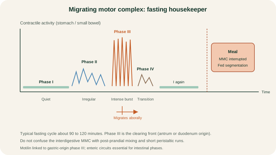
Fasting MMC cycle with quiet phase I, irregular phase II, intense migrating phase III and brief phase IV; feeding interrupts the cycle.
SimpleMed original educational diagram
Swallowing, Gastric Emptying and Defaecation
Regional programmes reuse the same wall machinery with different extrinsic triggers. Swallowing begins as a coordinated oropharyngeal sequence under brainstem control, then continues as oesophageal peristalsis with timed lower oesophageal sphincter (LOS) relaxation so the bolus can enter the stomach. The LOS is normally contracted between swallows; inhibitory enteric and vagal pathways relax it in advance of the peristaltic wave.
The stomach must first accommodate. Proximal reservoir relaxation includes a swallow-linked receptive component and further relaxation as the meal distends the stomach, coordinated largely through vagovagal and enteric inhibitory pathways. This accepts volume without a large rise in wall tension. Distal stomach then grinds and sieves solids with antral contractions against a pylorus that allows only small particles and liquid to pass. Emptying rate is paced by duodenal feedback: acid, fat and hypertonicity slow gastric emptying so the small bowel is not overwhelmed. Liquids usually leave faster than solids; high-fat meals empty more slowly than carbohydrate-rich ones of similar volume.
Defaecation couples colonic delivery with anorectal reflexes. Rectal filling stretches the wall, triggers the rectoanal inhibitory reflex (internal anal sphincter smooth muscle relaxes via enteric pathways) and generates the urge to defaecate. The external anal sphincter and puborectalis are striated and under voluntary and somatic pudendal control, so continence is preserved until a socially appropriate moment. Parasympathetic sacral pathways facilitate colonic and rectal contraction and internal sphincter relaxation when defaecation proceeds. Mass movements and the gastrocolic response after meals help load the rectum; detailed colonic pathology belongs elsewhere.
When Motility Circuits Fail
A few classic lesions illuminate the normal map without turning this page into a disease atlas. Achalasia reflects loss of inhibitory myenteric neurons in the oesophagus, so the LOS fails to relax adequately and oesophageal body peristalsis fails, trapping food above a closed sphincter. Hirschsprung disease is congenital aganglionosis of a distal bowel segment: missing myenteric and submucosal ganglia leave a permanently tonically narrowed segment that cannot participate in the peristaltic reflex, with dilation proximal to the transition zone. Ileus is temporary failure of effective propulsive contractions despite an often intact slow-wave rhythm, commonly after peritoneal irritation or major surgery. Vomiting is not ordinary reverse peristalsis alone; it is a brainstem-coordinated motor programme with reverse giant contractions in the small bowel, gastric relaxation and coordinated respiratory and sphincter events. Anti-emetic and prokinetic drug actions belong in the dedicated pharmacology articles rather than here.
The next lesson, Liver, Biliary and Exocrine Pancreatic Physiology, turns from wall motor programmes to the secretions that those programmes mix and carry.
Reviewed by: Dr. Marcus Judge
- 145
