

Next Lesson - Anatomy and Physiology of the Foregut
Core
Surface Regions and Wall Layers
The anterior abdominal wall has little bony support. Flexibility lets the trunk flex and rotate and allows the cavity to expand with a meal, pregnancy or ascites, but the same flexibility creates sites where pressure can force tissue outward. Surface maps keep later anatomy language consistent. A vertical median line and a horizontal transumbilical line give four quadrants. Two midclavicular vertical lines plus subcostal and intertubercular horizontal planes give nine regions, including the right and left hypochondrium, epigastrium, lumbar flanks, umbilical region, iliac fossae and hypogastrium.
From superficial to deep the anterolateral stack is skin; Camper fascia (fatty) and Scarpa fascia (membranous); three flat muscles with investing fascia and aponeuroses; transversalis fascia; extraperitoneal fat; and parietal peritoneum. Peritoneal folds and mesenteries are taught with gut development rather than repeated here: see Development of the Gastrointestinal Tract. The practical lesson is order. Any structure that leaves the abdomen, including the gonads during descent, must pass this stack, so the inguinal canal is best understood as a layered tunnel rather than a hole drawn on skin.
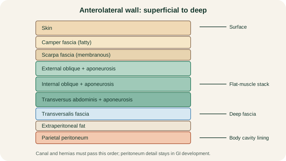
Layer order of the anterolateral abdominal wall, superficial to deep, ending at transversalis fascia and parietal peritoneum.
SimpleMed original educational diagram
Flat Muscles of the Anterolateral Wall
Three large paired flat muscles form the lateral and much of the anterior wall. The external oblique is most superficial. Its fibres run inferomedially from the lower ribs, like hands in pockets. Near the midclavicular line the muscle becomes an aponeurosis that continues to the linea alba. The free inferior edge of that aponeurosis thickens between the anterior superior iliac spine and the pubic tubercle as the inguinal ligament, the floor of the inguinal canal and a major surface landmark of the groin.
Deep to external oblique lies the internal oblique. Its fibres run roughly orthogonally, superomedially from the thoracolumbar fascia, iliac crest and lateral inguinal ligament. It rotates and flexes the trunk with its partner and contributes both muscle and aponeurosis to the canal roof and, medially with transversus, to the conjoint tendon.
Deepest of the three is transversus abdominis, with near-horizontal fibres suited to compressing the abdominal contents and bracing the lumbar spine. Its aponeurosis joins the other two in building the rectus sheath. Together the three layers act as a pressure jacket: coughing, straining or lifting raises wall tension and reduces the chance that viscera will herniate through natural openings. Where those openings already exist, the same pressure explains why hernias often appear or enlarge during Valsalva-type effort.
Medially, rectus abdominis runs vertically from the pubic crest and symphysis to the costal margin and xiphoid. Tendinous intersections segment the muscle and explain the common six-pack appearance. A small pyramidalis may sit anterior to the lower rectus within the sheath; it is often absent and is not needed for the canal story.
Rectus Sheath and the Arcuate Line
The rectus sheath is the fibrous envelope around rectus abdominis, built from the aponeuroses of the three flat muscles as they reach the midline. Midline fusion produces the linea alba. The composition of the sheath is not uniform from costal margin to pubis, and the change is named for the arcuate line, usually about one third of the distance from umbilicus to pubic crest on the deep surface of the wall.
Above the arcuate line, the external oblique aponeurosis and the anterior lamina of the internal oblique form the anterior sheath. The posterior lamina of the internal oblique and the transversus aponeurosis form a true posterior sheath, with transversalis fascia deep to that. Rectus is therefore enclosed front and back by strong aponeurotic layers.
Below the arcuate line, all three aponeuroses pass anterior to rectus. Posteriorly, only transversalis fascia (and then peritoneum) remains. The posterior wall is therefore thinner and less resistant to expanding haematoma or certain hernia paths in this lower zone. Superior and inferior epigastric vessels run on the deep surface of rectus within the sheath and anastomose near the umbilicus, linking subclavian and external iliac territories.
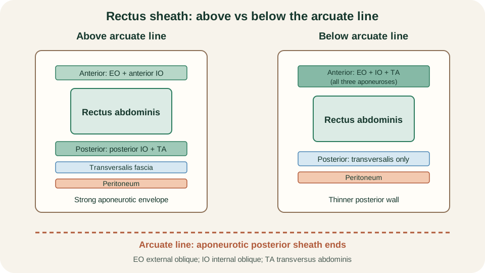
Rectus sheath construction above versus below the arcuate line: full posterior aponeurotic sheath superiorly, transversalis fascia only inferiorly.
SimpleMed original educational diagram
Neurovascular Supply
Segmental thoracoabdominal nerves from roughly T7 to T12, plus contributions near L1, supply the anterolateral wall. They travel in the plane between internal oblique and transversus abdominis, the same neurovascular plane used for a transversus abdominis plane block. Lower fibres of internal oblique and transversus, including the conjoint tendon, receive iliohypogastric and ilioinguinal input from the lumbar plexus. Ilioinguinal and the genital branch of genitofemoral also relate closely to the inguinal canal; full spermatic-cord inventory belongs in regional male anatomy rather than this wall lesson.
Arterial supply is multi-source. Superiorly, the superior epigastric artery continues from the internal thoracic after the musculophrenic branch is given off. Inferiorly, the inferior epigastric and deep circumflex iliac arteries arise from the external iliac just before it becomes femoral. Superficial epigastric and superficial circumflex iliac branches of the femoral artery supply more superficial lower wall. Lateral wall receives lower intercostal, subcostal and lumbar vessels running in the same plane as the nerves. Lymph from skin above the umbilicus drains largely toward axillary nodes; skin below drains to superficial inguinal nodes. Deep drainage follows the deep vessels toward parasternal, external iliac and lumbar nodes.
The inferior epigastric vessels are the spatial landmark that separates direct from indirect inguinal hernia later in this article. Learn them as a deep vascular landmark medial to the deep ring, not as an isolated vascular fact.
Inguinal Canal: Rings, Walls and Contents
During development the gonad descends, guided by the gubernaculum, and a peritoneal diverticulum (processus vaginalis) accompanies the path. In the male the testis usually reaches the scrotum; in the female the analogous path relates to the round ligament. The result in both sexes is an oblique passage through the lower abdominal wall: the inguinal canal, about four to six centimetres long in the adult, running inferomedially just above the inguinal ligament. The canal is therefore a designed weakness: it must transmit cord or round ligament, yet it is arranged so that raised wall tension narrows rather than gapes the tunnel.
The deep (internal) inguinal ring is an opening in transversalis fascia, roughly above the midpoint of the inguinal ligament and lateral to the inferior epigastric vessels. The superficial (external) inguinal ring is a triangular defect in the external oblique aponeurosis, superolateral to the pubic tubercle. Between them the canal has four walls built from layers you already know:
- Anterior wall: external oblique aponeurosis, reinforced laterally by internal oblique fibres.
- Posterior wall: transversalis fascia, strengthened medially by the conjoint tendon (fused lower internal oblique and transversus).
- Roof: arching fibres of internal oblique and transversus abdominis.
- Floor: inguinal ligament, reinforced medially by the lacunar ligament.
Contents differ by sex. In males the canal transmits the spermatic cord (with ductus deferens, vessels, lymphatics and coverings derived from wall layers) plus the ilioinguinal nerve. In females it transmits the round ligament of the uterus, accompanying vessels and the ilioinguinal nerve. Detailed cord coverings and examination technique are out of scope here; the mechanism question is which wall layer contributes each covering and which opening starts or ends the path.
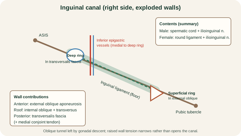
Exploded inguinal canal: deep ring lateral to inferior epigastric vessels, superficial ring near the pubic tubercle, and walls from the flat-muscle stack.
SimpleMed original educational diagram
Hesselbach Triangle and Hernia Paths
An inguinal hernia is abdominal content that leaves the cavity through a weakness related to the canal. Two classical paths share the superficial ring as a possible exit but enter the wall differently, and the inferior epigastric vessels are the dividing landmark.
Hesselbach (inguinal) triangle is the medial inguinal region bounded by the lateral edge of rectus abdominis, the inguinal ligament, and the inferior epigastric vessels. Its floor is largely transversalis fascia of the posterior canal wall. A direct inguinal hernia pushes straight through this triangle, medial to the inferior epigastric vessels, usually because of acquired posterior-wall weakness. It may still reach the superficial ring, but it does not begin at the deep ring.
An indirect inguinal hernia enters the canal at the deep ring, lateral to the inferior epigastric vessels, and travels along the canal. It often reflects a patent processus vaginalis or a widened deep ring pathway and is therefore congenital in mechanism even when presentation is later. Both types can present as a groin swelling that is more obvious on standing or straining. Clinical examination that distinguishes them, assesses reducibility and looks for femoral disease belongs in the separate hernia OSCE lesson rather than here.
Femoral hernia is the brief contrast you need for spatial sense. The femoral canal lies medial in the femoral sheath, below the inguinal ligament and lateral to the lacunar ligament, with the femoral vein immediately lateral to the canal. A femoral hernia therefore passes below the inguinal ligament into the upper thigh, not through the inguinal canal. The narrow femoral ring explains the higher risk of strangulation relative to many inguinal hernias, which is a mechanical consequence of space, not a management algorithm for this page.
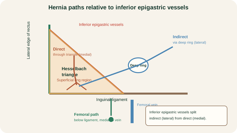
Indirect path through the deep ring lateral to the inferior epigastric vessels; direct path through Hesselbach triangle medial to those vessels; femoral path below the inguinal ligament.
SimpleMed original educational diagram
The next regional lesson, Anatomy and Physiology of the Foregut, moves inside the cavity to the organs the wall protects.
Reviewed by: Dr. Marcus Judge
- 159
