

All done here! Check out our other subjects!
Core
An upper gastrointestinal (GI) bleed is bleeding from the foregut, which means the oesophagus, stomach or duodenum.
Major upper GI bleeds have two major causes:
- Peptic Ulceration - A peptic ulcer is a sore in the lining of the stomach or duodenum. It can erode into an artery and cause a large volume of bleeding. Duodenal ulcers commonly involve the gastroduodenal artery (a large artery lying close to the duodenum). Gastric ulcers may involve nearby branches of the gastric arteries.
- Oesophageal Varices - Varices are swollen veins in the lower oesophagus. They form when the pressure in the portal vein is raised (portal hypertension). This is often caused by cirrhosis or by a clot (thrombosis) in the portal or hepatic (liver) veins. The raised pressure opens up portosystemic anastomoses, which are connections between the portal veins and the veins of the general circulation. The swollen veins can rupture and cause a large volume of bleeding.
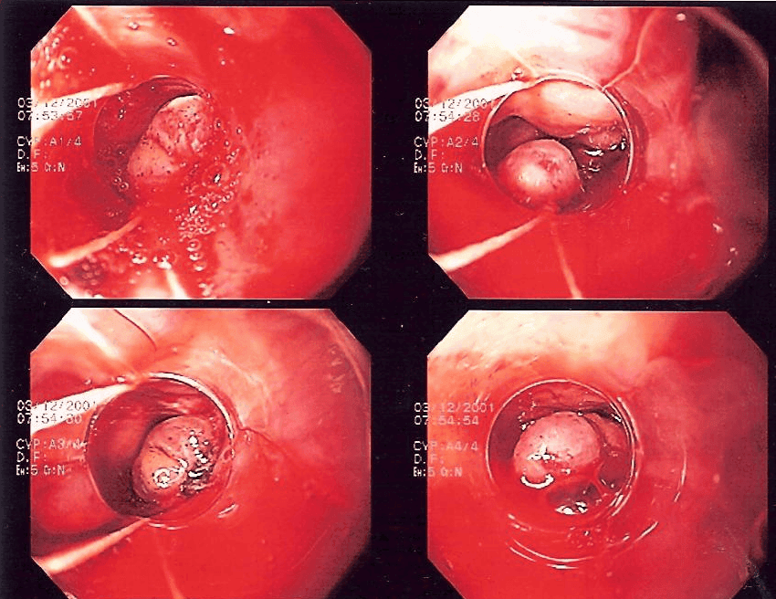
Image - Band ligation of oesophageal varices
Creative commons source by Samir [CC BY-SA 4.0 (https://creativecommons.org/licenses/by-sa/4.0)]
Patients often present with haematemesis (vomiting blood). The vomit can be bright red, or brown or black, and may look like coffee granules. A patient who has lost a lot of blood is likely to be pale and clammy. Upper GI bleeds can cause large volumes of blood to be lost. Therefore, management starts with resuscitation, using fluid/blood replacement guided by the full clinical picture, because giving too much blood can be as harmful as giving too little. A risk score (the Blatchford score) is used at first assessment. Patients then need endoscopy (a camera passed through the mouth) to find and stop the source of bleeding. This is done immediately after resuscitation if the patient is unstable and the bleeding is severe, and within 24 hours of admission for all other patients. If the cause is oesophageal varices, the bleeding is usually stopped by band ligation (tying off the swollen veins with tight rubber bands, using an endoscope). A Transjugular Intrahepatic Portosystemic Shunt (TIPS) is considered if band ligation does not control the bleeding. A TIPS is a channel made inside the liver that links the portal vein to a hepatic vein, which lowers the pressure in the portal system. Terlipressin (a drug that narrows the blood vessels supplying the gut, which reduces portal venous pressure) is offered as soon as variceal bleeding is suspected, before endoscopy. Patients with suspected variceal bleeding are also offered antibiotics straight away to prevent infection. If the cause is peptic ulceration, the ulcer is treated at endoscopy (for example, with clips). Proton pump inhibitors (PPIs, drugs that reduce stomach acid) are not given before endoscopy. They are offered afterwards if the ulcer shows signs of recent bleeding. Patients who re-bleed are offered a repeat endoscopy. If an unstable patient re-bleeds after endoscopic treatment, interventional radiology (a radiologist treats the bleeding vessel through a catheter) is offered. The patient is referred urgently for surgery if interventional radiology is not promptly available.
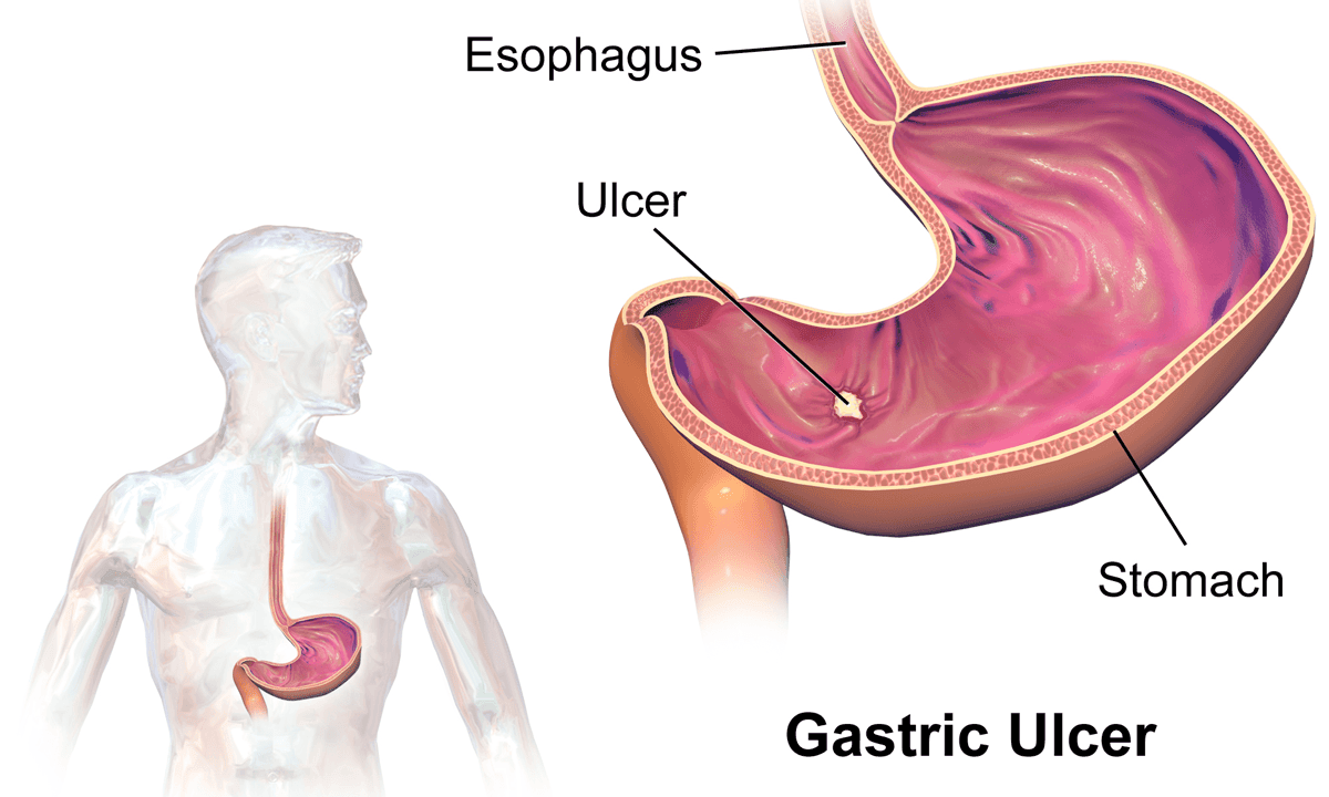
Diagram - Common location of gastric ulcers
Creative commons source by BruceBlaus [CC BY-SA 4.0 (https://creativecommons.org/licenses/by-sa/4.0)]
Ruptured Abdominal Aortic Aneurysm (AAA)
An AAA is a permanent pathological dilation (a lasting, abnormal widening, 3 cm or more across) of the abdominal aorta. More than 90% of AAAs originate below the renal arteries. AAAs are usually asymptomatic (they cause no symptoms). Large AAAs may cause nausea, urinary frequency or back pain by pressing on nearby structures.
Risk factors of this condition include:
- Male sex
- Genetic predisposition
- Increasing age
- Smoking
If an AAA is to rupture the results are disastrous. In one review of studies, about a third (32%) of patients with a ruptured AAA died before they reached hospital, and about 8 in 10 (81%) died overall. Death can come so quickly because the aorta is the largest artery in the body and carries blood at high pressure, so a rupture causes very rapid blood loss.
Those that make it to primary care may present with severe abdominal pain (with or without flank and groin pain), back pain, pulsatile mass in the abdomen, transient hypotension, and sudden cardiovascular collapse. A pulsatile mass is a clinical clue. The diagnosis is made with an immediate bedside ultrasound scan (USS) of the aorta. If the scan shows an AAA, or it is unavailable or unclear and an AAA is still suspected, the case is discussed immediately with the regional vascular service. A CT scan of the aorta with contrast dye (CT aortogram) is considered if the patient is being evaluated for repair. Some patients go straight to repair without waiting for a CT scan.
Treatment of a ruptured AAA is emergency repair, either by open surgery or by an endovascular stent graft. During transfer to a regional vascular service, a restrictive approach to intravenous fluids may be used. This is called permissive hypotension: the blood pressure is allowed to stay lower than normal. The mortality rate is very high. In order to prevent this clinical emergency, there is an NHS screening programme for AAAs.
In England, all men are invited for an ultrasound scan (USS) of their abdominal aorta in the year they turn 65. Wales and Northern Ireland also invite men at 65. The scan is done once unless an aneurysm is found. It allows any enlargement of the abdominal aorta to be picked up early and watched carefully. The larger the aneurysm, the more likely it is to rupture, so scans become more frequent as it grows.
- If AAA measures between 3.0-4.4 cm, the patient is invited back for yearly USS.
- If AAA measures between 4.5-5.4 cm, the patient is invited back for USS every 3 months.
- If AAA is 5.5 cm or larger, the patient is referred to a vascular surgeon (a surgeon who operates on blood vessels) for more tests and possible surgical intervention.
Surgical management of AAAs is either open surgery to clamp the aorta and suture in a synthetic graft, or insertion of an endograft (stent) via the femoral artery.
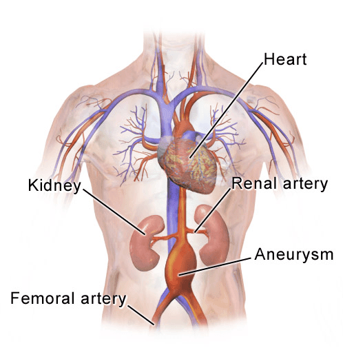
Diagram - Location of abdominal aortic aneurysm
Creative commons source by BruceBlaus [CC BY-SA 4.0 (https://creativecommons.org/licenses/by-sa/4.0)]
Peritonitis is the inflammation of the peritoneum lining the abdominal cavity, and it can be divided into two categories:
- Primary Peritonitis - also known as spontaneous bacterial peritonitis (SBP). This is an infection of ascitic fluid (fluid that has collected in the abdominal cavity, often because of liver disease) with no surgically treatable source of infection inside the abdomen. It is most commonly seen in patients with end-stage liver disease.
- Secondary Peritonitis - this is inflammation of the peritoneum caused by a problem in a nearby structure. The structure may be inflamed, perforated (it has a hole in it) or have gangrene (tissue that has died after losing its blood supply). It may lie inside the abdominal cavity (intra-abdominal) or behind the peritoneum (retroperitoneal, for example the kidneys or pancreas). Common causes include peptic ulcer disease, appendicitis, diverticulitis (inflamed pouches in the wall of the colon), abdominal surgery, ruptured ectopic pregnancy (a pregnancy growing outside the uterus) and ovarian cyst.
Patients usually present with pyrexia (fever), vomiting and severe abdominal pain. The pain is unremitting, which means it is constant and does not ease. It causes the patient to lie very still with their knees flexed. They often breathe very shallowly to avoid further irritation of the peritoneum.
Drawing off a sample of ascitic fluid with a needle (an ascitic tap) can confirm the diagnosis. A neutrophil count greater than 250 cells/mm3 in the fluid is diagnostic for spontaneous bacterial peritonitis (or primary peritonitis), as long as there is no surgically treatable source of infection inside the abdomen.
Treatment includes control of the infectious source (if secondary peritonitis), antibiotics and maintenance of organ function (which may require intensive care). In secondary peritonitis, antibiotics are chosen to cover the bowel bacteria that are usually involved. These include Gram-negative bacteria (bacteria that stain pink on a Gram stain, such as Escherichia coli, or E. coli) and anaerobes (bacteria that live without oxygen). In spontaneous bacterial peritonitis, antibiotics are started immediately. The bacteria are most often E. coli, streptococci and enterococci. Patients whose creatinine (a blood test of kidney function) is raised or rising are also given an albumin infusion (a protein solution given into a vein).
Small bowel obstruction is a mechanical or functional problem that inhibits normal movement of gut contents through the small bowel.
Causes can be split into three categories:
Extrinsic Causes - pathology outside the bowel that restricts its function e.g. intra-abdominal adhesions (the most common cause), compression by an external tumour, incarcerated hernias or volvulus of the small intestine. Adhesions are bands of scar tissue between organs, often after surgery. An incarcerated hernia is a hernia where bowel is trapped and cannot be pushed back. Volvulus is a twisting of the bowel around its mesentery, the fold of tissue that attaches it to the back wall of the abdomen.
Intrinsic Causes - pathology within the bowel that restricts its function e.g. inflammation from Crohn’s disease (a long-term inflammatory bowel condition), a tumour of the small bowel or intussusception (the telescoping of one portion of the intestine into an adjacent section, which primarily occurs in children).
Intraluminal Causes - pathology of the lumen (the hollow space inside the bowel) e.g. gallstone ileus (a gallstone that has passed into the small intestine and physically blocks it, so the obstruction is mechanical) or the presence of a foreign body in the lumen.
Risk factors for small bowel obstruction are:
- Previous abdominal/gynaecological surgery (adhesions)
- Previous radiation of the abdomen (adhesions)
- Frequent raising of intra-abdominal pressure e.g. weightlifting (hernias)
- History of Crohn’s disease
Patients commonly present with intense abdominal pain that is often diffuse across the abdomen, and colicky (coming and going in waves) with cramping in ~3-5 minute intervals. Other features include vomiting, which comes early if the obstruction is high up (proximal) and later if it is lower down (distal). The vomit is often bilious (green, containing bile) but may be feculent (smelling of faeces) depending on the location of the obstruction. There is also abdominal distension (a swollen abdomen) and late absolute constipation (passing neither faeces nor wind). Do not rule out small bowel obstruction just because the patient is still opening their bowels early on. The bowel below the obstruction may still be emptying.
A CT scan of the abdomen is the first line imaging choice when small bowel obstruction is suspected as it often allows the identification of the cause of obstruction. The exception to this rule is suspected bowel perforation (a hole in the bowel wall). One clue is pain that is out of proportion to the findings on examination and does not ease. In that case emergency surgical management may be performed without a CT scan. Classically, an erect chest x-ray was used to look for pneumoperitoneum (free air in the abdominal cavity) as a sign of perforation. It is used less often today because it can miss free air, and CT is more sensitive. A normal x-ray does not rule out perforation.
Small bowel obstruction is considered a gastrointestinal emergency because of the high risk of complications such as dehydration, metabolic alkalosis (the blood becoming too alkaline, from vomiting stomach acid), sepsis, bowel ischaemia/necrosis and perforation.
Management of small bowel obstruction is cause dependent. Adhesional obstruction (obstruction caused by adhesions) is first treated without surgery. This applies if there are no signs of peritonitis, strangulation (the blood supply to the bowel is cut off) or ischaemia. Treatment is nothing by mouth (nil per os), a nasogastric tube (a tube passed through the nose into the stomach) to drain the bowel, and intravenous fluids and electrolytes. A trial of non-operative treatment can safely continue for up to 72 hours. Surgery is needed if there are signs of peritonitis, strangulation or ischaemia, or if non-operative treatment does not work.
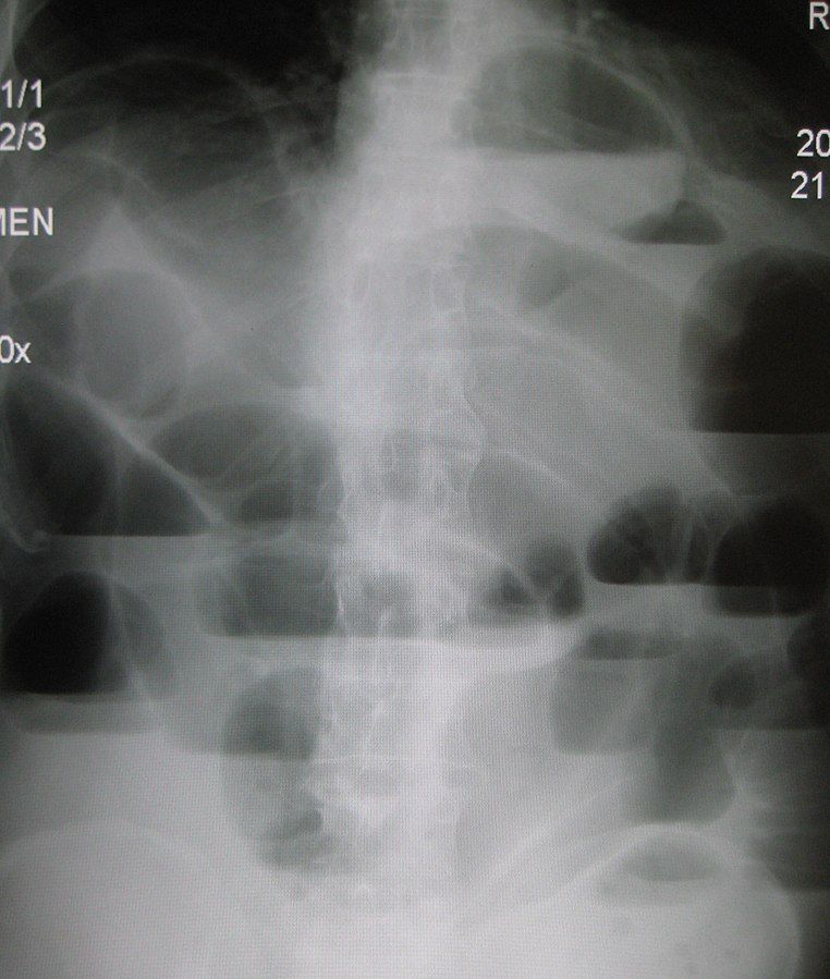
Image - An erect abdominal x-ray showing small bowel obstruction
Creative commons source by James Heilman, MD [CC BY-SA 4.0 (https://creativecommons.org/licenses/by-sa/4.0)]
Large bowel obstruction is a mechanical or functional problem that inhibits normal movement of gut contents through the large bowel.
Causes can be split into two categories:
- Mechanical causes e.g. colon malignancy (cancer; the most common cause), diverticular disease (small pouches in the colon wall), volvulus or inflammatory bowel disease (Crohn’s disease and ulcerative colitis). Volvulus is a twisting of the colon around its mesentery (the fold of tissue that attaches the bowel to the back wall of the abdomen). It most commonly occurs in the sigmoid colon (the S-shaped last part of the colon) but also occurs in the caecum (the first part of the large bowel).
- Non-mechanical causes e.g. colonic pseudo-obstruction (the colon stops working but nothing is blocking it) or toxic megacolon (a severely widened colon, usually with serious inflammation).
Risk factors of large bowel obstruction are:
- High fibre diet (a risk factor for volvulus; sigmoid volvulus has a relatively high incidence in African, Asian and Middle Eastern countries, where high fibre diets are eaten)
- Low fibre diet (diverticular disease)
- Increasing age
Patients commonly present with intense abdominal pain that is often diffuse and colicky with cramping in 15-20 minute intervals. Other presentations can include abdominal distension (a swollen abdomen), absence of flatus (not passing wind), absolute constipation (passing neither faeces nor wind), and late-stage vomiting. With the bowel blocked and faeces building up, the bowel secretes large volumes of electrolyte-rich fluid into itself. This fluid is lost from the circulation, which is often called ‘third spacing’. This can result in dehydration and electrolyte imbalance. Symptoms may come on gradually if the underlying cause is of a progressive nature, e.g. colon cancer, but may also come on suddenly if the cause is acute, e.g. volvulus.
First-line diagnostic imaging is a CT scan of the abdomen as it often allows the identification of the location and the cause of the obstruction. The exception to this rule is suspected bowel perforation (a hole in the bowel wall). Clues include pain that is out of proportion to the findings on examination and does not ease, or a patient who is peritonitic (showing signs of peritonitis, such as a very tender abdomen). In that case an emergency surgical intervention may be undertaken without a CT scan. An erect chest x-ray can show free air in the abdominal cavity, but it can miss it, and CT is more sensitive. A normal x-ray does not rule out perforation. In stable patients, endoscopy (a camera passed through the anus) can take a biopsy (a tissue sample) of a mass causing the obstruction. Endoscopy can also treat sigmoid volvulus by untwisting the bowel (detorsion) and decompressing it. Surgery to remove the sigmoid colon (sigmoid colectomy) is still recommended during the same admission.
Large bowel obstruction is considered a gastrointestinal emergency because of the high risk of complications such as dehydration, electrolyte imbalance, sepsis, bowel ischaemia/necrosis and perforation.
Most cases are managed surgically.
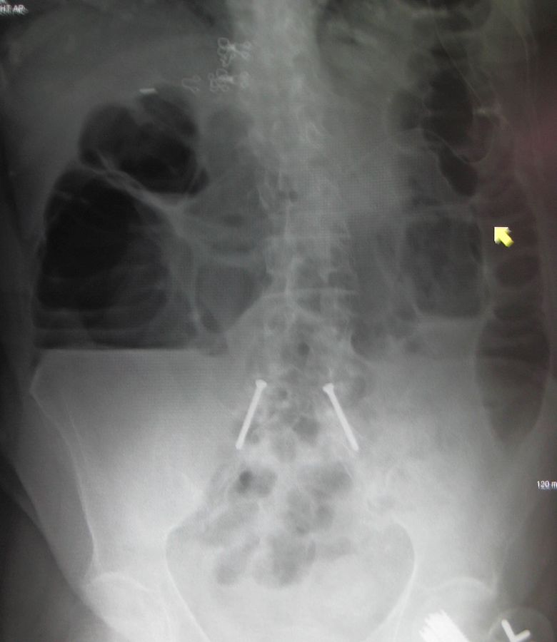
Image - Abdominal x-ray showing large bowel obstruction
Creative commons source by James Heilman, MD [CC BY-SA 4.0 (https://creativecommons.org/licenses/by-sa/4.0)]
Abdominal X-Rays and Gut Structures
Plain abdominal x-rays are rarely ordered because they show much less detail than a CT of the soft tissue structures that make up most of the abdominal cavity. One of the few indications for a plain abdominal x-ray is bowel obstruction. In obstruction, an abdominal x-ray is quick to obtain and can identify whether the obstruction is in the small or large bowel.
There are a number of features that can help differentiate between the small and large intestine on plain film x-ray:
- The small bowel is narrower than the large bowel - the diameter of the small bowel should not exceed 3 cm, whereas the large bowel should not exceed 6 cm, or 9 cm in the caecum (the first part of the large bowel). Bowel wider than these limits is dilated.
- The small bowel is often more central in the abdominal cavity, with the large bowel peripherally around the abdomen. This may be less reliable with very dilated bowel loops, as can be seen in obstructions (the main indication for plain film abdominal x-rays).
- The plicae circulares traverse the whole diameter of the small bowel, whereas the haustra in the large bowel do not traverse the whole diameter. Plicae circulares (also called valvulae conniventes) are folds in the lining of the small bowel. Haustra are the pouch-like segments of the large bowel wall.
While the interpretation of abdominal x-rays is outside the scope of this article, it is important to understand the clinical applications for the anatomical knowledge explained in this article.
Acute mesenteric ischaemia is described as the symptomatic reduction in the blood supply to the gastrointestinal tract.
Causes include:
- Acute arterial occlusion (~70% of cases) - a sudden blockage of an artery, e.g. an embolism (a clot that has travelled from elsewhere, often the heart) in the superior mesenteric artery (SMA), or a thrombosis (a clot forming in the artery itself).
- Non-occlusive (~20% of cases) - e.g. low cardiac output.
- Mesenteric Venous Thrombosis (~5-10%) - e.g. a systemic coagulopathy (a disorder of blood clotting that makes clots more likely) due to malignancy.
Risk factors for this condition include:
- Female sex
- History of peripheral vascular disease
- Atrial fibrillation (an irregular heart rhythm that can cause clots to form in the heart and travel to the gut arteries)
- Cardiovascular risk factors such as raised cholesterol, smoking and familial hyperlipidaemia (an inherited tendency to high levels of fats in the blood)
Patients typically present with sudden-onset, severe abdominal pain that is disproportionate to clinical findings. The pain is often central or periumbilical (around the belly button) and may be accompanied by nausea and vomiting. As ischaemia progresses, peritonism (signs of peritoneal irritation, such as tenderness and guarding) and systemic features (signs of illness affecting the whole body) may develop.
Diagnostic investigations include blood tests (to look for metabolic acidosis, meaning the blood has become too acidic, or raised lactate levels). No blood test is accurate enough to rule ischaemia in or out, so a normal result does not exclude it. CT angiography (a CT scan with contrast dye that shows the blood vessels) should be done as soon as possible when mesenteric ischaemia is suspected.
Management of acute mesenteric ischaemia is either thrombolysis/angioplasty or surgical resection of the ischaemic bowel depending on the severity of the ischaemia, but mortality is high.
Edited by: Dr. Maddie Swannack
Reviewed by: Dr. Thomas Burnell and Dr. Marcus Judge
- 938



