

Next Lesson - Gastrointestinal Emergencies
Core
The gastrointestinal tract contains a microbiome (the community of microorganisms that lives there). The stomach is an acidic environment with few bacteria, the proximal gut also has relatively few bacteria, and the colon is an anaerobic environment containing approximately 1011 bacteria per gram of contents. The microbiome has many advantages. It helps useful bacteria to compete with harmful bacteria for nutrients. It produces short-chain fatty acids, which may help to lower cholesterol and regulate satiety (the feeling of being full), and which provide energy to colonocytes (the cells lining the colon). It also produces nutrients such as vitamin K.
Gastroenteritis and enterocolitis are terms used to describe inflammation (-itis) in the stomach (gastro-), intestines (entero-) or colon (col-). This inflammation is usually caused by an infection, but there are non-infectious causes such as food sensitivity, medications or inflammatory bowel disease (long-term inflammation of the gut). These non-infectious causes present very similarly to infectious gastroenteritis or enterocolitis, but have no causative organism.
Generally, gastroenteritis or enterocolitis will present with some combination of nausea, vomiting, diarrhoea, abdominal pain and fever. Not all organisms cause all of these symptoms, but this is the common picture. Enterocolitis (inflammation of the small and large intestines) does not include the stomach, but it can still present with vomiting.
Below are the common organisms that cause gastroenteritis and enterocolitis.
Image - Bacteria in the gastrointestinal tract
Public Domain Source [Public domain]
Salmonella (non-typhoidal and typhoid)
Gram-negative bacillus (a rod-shaped bacterium that stains pink on a Gram stain).
Non-typhoidal Salmonella (for example S. Enteritidis and S. Typhimurium) causes gastroenteritis. Salmonella Typhi causes typhoid (enteric) fever, a more serious illness that spreads through the body.
Spreads via faeco-oral route (from faeces to mouth, for example through contaminated food or water).
Incubation period (the time from infection to the first symptoms): usually 12 to 48 hours for non-typhoidal Salmonella (range 4 to 120 hours). For typhoid fever, the mean incubation period is 7 to 21 days, and the vast majority of cases develop symptoms within 28 days.
In typhoid fever, Salmonella Typhi enters enterocytes (the cells lining the small intestine) via endocytosis and then travels to the submucosa (the layer under the lining) of the intestines. Here the bacteria encounter macrophages, which carry them to the reticuloendothelial system (the network of immune cells that clear germs from the body), where they multiply. The bacteria then cause lymphoid hyperplasia (enlargement of lymph tissue) and re-enter the gut to continue the cycle.
Non-typhoidal infection presents with fever, abdominal pain, and diarrhoea, which is usually non-bloody but can contain blood. Typhoid fever starts more slowly, with a sustained fever, headache, malaise (feeling generally unwell) and abdominal pain.
In healthy individuals, non-typhoidal Salmonella infections are self-limiting (they get better without treatment) within 4-7 days and do not usually need treatment with antibiotics. Supportive treatment with fluid and electrolyte replacement may be required. Antibiotics are considered, after specialist advice, for people at risk of invasive infection (infection that spreads beyond the gut). This includes people who are immunocompromised (have a weakened immune system), people with a haemoglobinopathy (an inherited condition affecting haemoglobin) and babies under 6 months old.
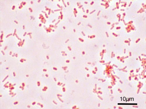
Image - Gram stain of Salmonella Typhimurium
Creative commons source by Y tambe [CC BY-SA 3.0 (https://creativecommons.org/licenses/by-sa/3.0)]
Gram-negative bacillus (‘S’ shaped).
Spreads via faeco-oral route.
Incubation period of usually 2 to 5 days (range 1 to 10 days).
Campylobacter jejuni penetrates the intestinal mucus layer and releases cytotoxins (toxins that damage cells). This disrupts the epithelial lining of the intestines (the layer of cells that lines the gut).
Presents with fever, abdominal pain, and diarrhoea (which may be bloody).
In healthy individuals, Campylobacter infections are self-limiting, and most cases resolve within about 1 week. Supportive treatment with fluid and electrolyte replacement may be required. Antibiotics are not usually needed for mild symptoms. Consider early treatment with an antibiotic such as clarithromycin if symptoms are severe or the person is immunocompromised, starting it within 3 days of the illness beginning. Severe symptoms include a high fever, bloody diarrhoea or high-output diarrhoea (a very large volume of watery stool).
Complications can include Guillain-Barré syndrome (a rare condition in which the immune system attacks the nerves, causing weakness).
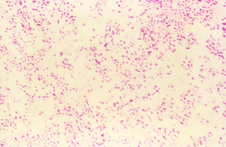
Image - Gram stain of Campylobacter jejuni
Public Domain Source [Public domain]
Gram-negative bacillus.
Spreads via faeco-oral route.
Incubation period of usually 1 to 4 days.
Highly virulent (very good at causing disease).
Shigella bacteria invade colonocytes, multiply and then invade neighbouring cells, killing them and forming abscesses in the intestinal wall.
Presents with diarrhoea that is often bloody and may contain mucus, with abdominal cramps. Illness can be milder with Shigella sonnei, where only 10 to 50% of cases have blood in the stool.
In healthy individuals, shigellosis (infection with Shigella bacteria) is self-limiting within 7 days. Supportive treatment with fluid and electrolyte replacement may be required. Antibiotics are not usually needed for mild illness. Seek specialist advice on antibiotic treatment if symptoms are severe or the person is immunocompromised. Severe symptoms include a high fever, bloody diarrhoea or high-output diarrhoea (a very large volume of watery stool). Some strains are resistant to several antibiotics, so antibiotics should be used carefully and in line with current guidance.
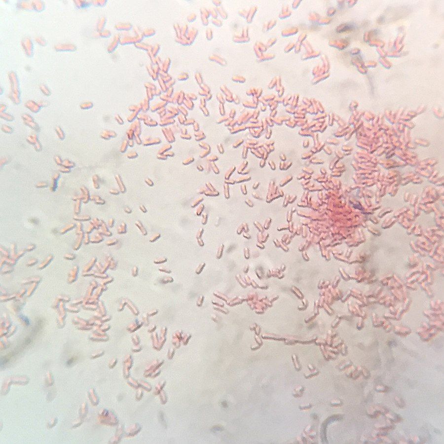
Image - Gram stain of Shigella flexneri
Creative commons source by B. Domangue [CC BY-SA 4.0 (https://creativecommons.org/licenses/by-sa/4.0)]
Enterotoxigenic Escherichia coli
Gram-negative bacillus.
Spreads via faeco-oral route.
Incubation period: most cases begin within about 10 to 50 hours of infection.
Most common cause of ‘Traveller’s diarrhoea’.
E. coli attaches to enterocytes (the cells lining the small intestine) and produces enterotoxins (toxins that act on the gut). These cause hypersecretion (excess release) of chloride ions into the gut lumen. Water follows the osmotic gradient (it moves towards the higher concentration of ions) into the lumen, leading to diarrhoea.
Presents with abdominal pain and non-bloody diarrhoea, which is often watery. Fever, nausea and vomiting may also occur.
In healthy individuals, infections with enterotoxigenic E. coli (ETEC, the type that makes enterotoxins) are self-limiting within 4-7 days and do not require treatment with antibiotics. Supportive treatment with fluid and electrolyte replacement may be required.
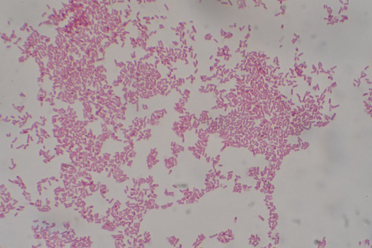
Image - Gram stain of Escherichia coli
Creative commons source by Dr Graham Beards [CC BY-SA 4.0 (https://creativecommons.org/licenses/by-sa/4.0)]
Clostridioides difficile (formerly Clostridium difficile)
Gram-positive bacillus (a rod-shaped bacterium that stains purple on a Gram stain).
Anaerobic and spore-forming (it makes tough resting forms called spores). Spores are very difficult to remove from an environment.
Spreads via faeco-oral route.
C. difficile infections often occur following antibiotic therapy, particularly with broad-spectrum antibiotics (antibiotics that act against many types of bacteria). This is because C. difficile can live harmlessly as part of the normal gut bacteria, and competes for survival in this environment. When the antibiotics wipe out the rest of the normal gut flora, C. difficile colonises the gut in the normal flora’s absence and releases enterotoxins/cytotoxins.
Presents with diarrhoea and abdominal cramping.
Treatment involves the removal of the causative antibiotic, fluid/electrolyte replacement and oral vancomycin or fidaxomicin. Antibiotics used to treat C. difficile are given orally so they reach the gut.
Complications include pseudomembranous colitis (inflammation of the colon with plaques on its lining), toxic megacolon (a dangerously widened colon) and sepsis.
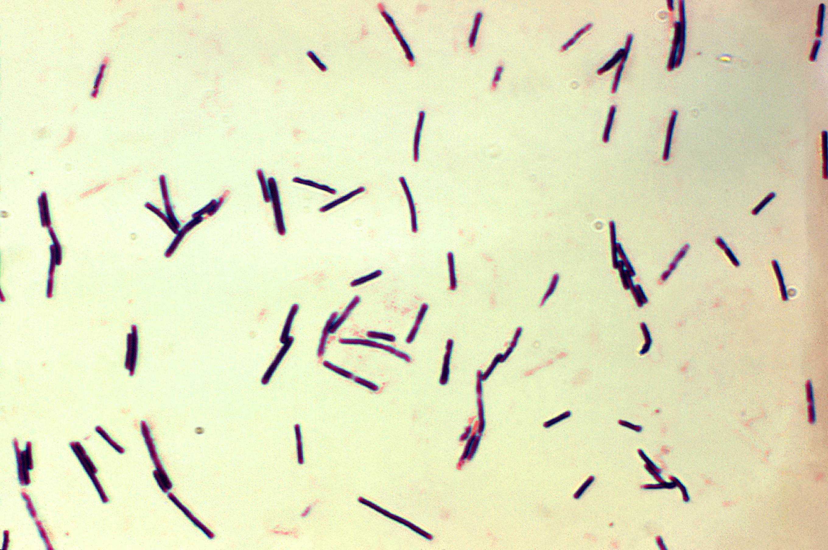
Image - Gram stain of a related species, Clostridium perfringens
Public Domain Source by Don Stalons [Public domain]
Double-stranded RNA virus (a virus whose genetic material is RNA made of two strands).
Main cause of viral gastroenteritis in young children. Most cases occur in children aged 6 months to 2 years. Rotavirus infection is much less common in adults.
An oral rotavirus vaccine is part of the UK routine childhood vaccination schedule (given at 8 and 12 weeks old), and cases have fallen since it was introduced.
Presents with fever, vomiting and diarrhoea.
In healthy individuals, rotavirus infections are self-limiting. Vomiting usually settles within 1 to 3 days and diarrhoea within 5 to 7 days, but the illness can last up to 2 weeks. Supportive treatment with fluid and electrolyte replacement may be required.
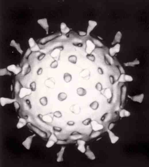
Image - Computer reconstructed image of a rotavirus particle that has reacted with monoclonal antibodies (identical, laboratory-made proteins that all stick to the same target)
Creative commons source by Graham Beards, edited by Dr. Laura Hansell [CC BY-SA 3.0 (https://creativecommons.org/licenses/by-sa/3.0)]
RNA virus.
Incubation period of 24-48 hours.
Most common cause of gastroenteritis in England and Wales. Norovirus infection can occur in adults or in children. Outbreaks are very common in semi-closed places such as schools, hospitals and nursing homes.
Causes brush border enzyme disruption (the brush border is the finger-like surface of the cells lining the gut, which carries digestive enzymes).
The large number of strains and the small dose required to produce symptoms result in norovirus being highly contagious.
Presents with fever, vomiting and watery diarrhoea.
In healthy individuals, norovirus infections are self-limiting within 3 days. Supportive treatment with fluid and electrolyte replacement may be required.
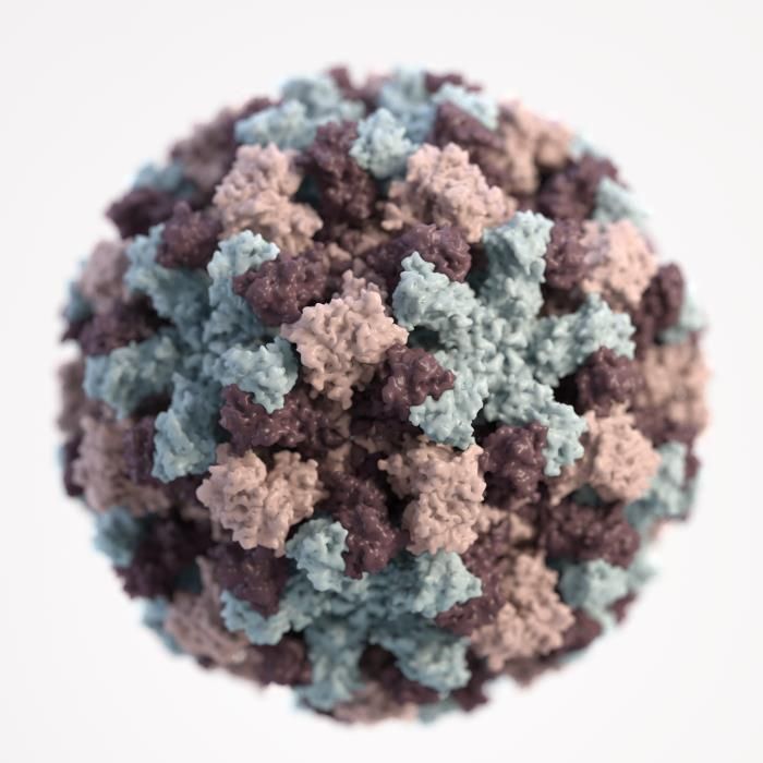
Image - A single norovirus virion (one virus particle)
Public Domain Source by the US Centers for Disease Control and Prevention (CDC) [Public domain]
Sporozoan protozoa (single-celled parasites that produce spore-like stages).
Spreads via faeco-oral route or via water.
The disease cycle has three stages. First, a person swallows (ingestion) an oocyst (a tough, egg-like stage) containing the parasite, for example in contaminated water or after contact with infected animals such as calves and lambs. Second, the parasite undergoes reproduction within the epithelial cells (the cells lining the gut) of the small intestine. Third, oocysts leave the body in the faeces (excretion) to continue the cycle.
Causes brush border enzyme dysfunction and increased chloride ion secretion.
Presents with watery diarrhoea.
In healthy individuals, Cryptosporidium infections are self-limiting and usually last about 2 weeks, but symptoms can persist for up to a month. Supportive treatment with fluid and electrolyte replacement may be required.
No specific treatment is licensed in the UK. People who are severely immunocompromised (for example, some patients with HIV) are at risk of serious, life-threatening illness. They need specialist advice on whether anti-parasitic treatment (medicines that kill parasites) is appropriate.
Flagellate (it moves using whip-like tails called flagella) protozoa (also called Giardia duodenalis or Giardia intestinalis).
Spreads via faeco-oral route or via water.
Incubation period of 1-2 weeks.
Giardia causes diarrhoea in children under 5 years in many parts of the world, and it also affects adults. In England, adults aged 30 to 39 years make up the largest age group of laboratory-confirmed cases (20.0% of laboratory reports in 2025).
The disease cycle involves ingestion of a cyst containing the parasite. Stomach acid and pancreatic enzymes release the parasite to multiply in the small intestine over 1-2 weeks. The parasite causes villous atrophy (flattening of the finger-like folds that absorb food) in the small intestine and is then excreted in the faeces to continue the cycle.
Presents with diarrhoea.
Treatment involves antimicrobials (medicines that kill microbes) such as metronidazole. Supportive treatment with fluid and electrolyte replacement may be required.
Amoeba protozoa.
Spreads via faeco-oral route or via water.
Endemic (always present) in countries with poor sanitation. Most UK cases are imported by travellers returning from these areas.
The disease cycle involves ingestion of a cyst containing the parasite. Then excystation (the parasite coming out of its cyst) occurs in the terminal ileum. This produces trophozoites (the active, feeding form of the parasite), which live mainly in the colon. They can invade the mucosa and cause inflammation (they may then spread to the liver). Cysts are then excreted in the faeces to continue the cycle.
Infection is most often asymptomatic (without symptoms) but may present with bloody diarrhoea. In rare cases, it may present with symptoms of a liver abscess such as fever and pain in the right upper quadrant (the top right of the abdomen).
Treatment involves antimicrobials (medicines that kill microbes), such as metronidazole followed by an anti-protozoal drug that acts inside the bowel, such as diloxanide. Specialist advice is needed. Supportive treatment with fluid and electrolyte replacement may be required.
Complications include toxic megacolon (a dangerously widened colon). Other complications are fulminant necrotising colitis (severe, fast-moving inflammation of the colon with tissue death) and amoebic liver abscess (a collection of pus in the liver).
Edited by: Dr. Maddie Swannack
Reviewed by: Dr. Thomas Burnell and Dr. Marcus Judge
- 1029



