

Next Lesson - The Endocrine Pancreas
Core
Thyroid Anatomy and Development
The thyroid gland lies anterior to the larynx and trachea, and inferior to the thyroid cartilage. The isthmus (the part of the thyroid gland that joins the two lobes together) extends from the 2nd to 3rd rings of the trachea. The thyroid gland is shaped like a bowtie.

Image: Shows the position and gross anatomy of the thyroid gland
Creative commons source by Don Bliss (illustrator) [CC BY-SA 4.0 (https://creativecommons.org/licenses/by-sa/4.0)]
The thyroid gland first appears as an epithelial proliferation (a thickening of the lining cells) on the floor of the pharynx between 3 and 4 weeks of gestation (pregnancy). To migrate to its final position, it descends as a diverticulum (an outpouching of tissue) through the thyroglossal duct, in front of the hyoid bone (the small U-shaped bone above the thyroid cartilage). The thyroglossal duct connects the thyroid gland to the tongue, but by birth this duct should have degenerated.
Failure of this duct to degenerate can lead to conditions such as a thyroglossal duct cyst, seen as a lump on the midline of the neck. The lump moves upwards on tongue protrusion because the duct is still connected to the base of the tongue.
It is important to remember that the thyroid gland and the parathyroid glands are two separate glands. The parathyroid glands are usually four small glands around the thyroid. However, this number can vary between individuals. They contain parathyroid principal cells (also known as chief cells), which produce parathyroid hormone, an essential hormone for calcium homeostasis. The parathyroid gland will be further discussed in the ‘calcium homeostasis’ article of the endocrinology series.
Thyroid Structure and Function
The thyroid gland consists of follicular cells and parafollicular cells. The follicular cells are arranged in spheres called thyroid follicles and produce thyroglobulin. Thyroglobulin is stored outside the cells, in the central cavity (lumen) of each follicle, as a substance called colloid. The follicular cells surround the colloid. Colloid acts as the source of tyrosine residues (the tyrosine amino acids within thyroglobulin) needed for the production of thyroid hormones. Colloid is classed as an extracellular substance (outside the cells), even though it is within the thyroid gland, because it lies in the lumen of the follicle and not inside the follicular cells. The parafollicular cells produce calcitonin, a hormone involved in calcium homeostasis.
The Production and Regulation of Thyroid Hormones
Thyroid hormones (and their precursors, the molecules they are made from) are the only hormones in the body that contain iodine. The thyroid gland holds about 70-80% of the body’s iodine. Dietary iodine, most commonly found in fish and dairy, must be reduced to iodide (the ion I-) before it can be absorbed from the small intestine into the blood. The iodide is then carried in the blood to the thyroid. There it is taken up by thyroid epithelial cells through a sodium-iodide symporter (a membrane protein that moves two Na+ ions into the thyroid cell together with one I-). Iodine deficiency, once considered eliminated in the UK, has re-emerged, particularly among women of reproductive age. Its effects on the thyroid are discussed later in this article.
The two main thyroid hormones are called triiodothyronine (T3) and tetraiodothyronine (thyroxine - T4). Thyroglobulin produced by the thyroid gland acts as a scaffold for the formation of these hormones. The two hormones are made through the linkage of two molecules of iodinated tyrosine (tyrosine with iodine atoms attached) on the thyroglobulin scaffold. T4 has four iodine atoms and is 3,3',5,5'-tetraiodothyronine. T3 has three iodine atoms and is 3,5,3'-triiodothyronine. The numbers give the positions of the iodine atoms on the two aromatic rings (the ring-shaped parts of the molecule).
The basic steps of T3 and T4 synthesis are outlined below:
- Thyroglobulin is synthesised in the follicular cells and secreted through exocytosis into the lumen of the thyroid follicle.
- Iodide is transported into the thyroid epithelial cells through the 2Na+:I- transporter, against its concentration gradient, and is secreted into the lumen of the thyroid follicle.
- Iodide undergoes oxidation to produce an iodinating species (a reactive form of iodine that can attach to tyrosine), catalysed by thyroid peroxidase (an enzyme) in the presence of H2O2 (hydrogen peroxide).
- Side chains of tyrosine found in the thyroglobulin are iodinated to form either mono- or diiodotyrosine (MIT or DIT), catalysed by thyroid peroxidase.
- MIT and DIT then couple to form T3 and T4 within the thyroglobulin, catalysed by thyroid peroxidase:
- MIT + DIT → T3
- DIT + DIT → T4
- Thyroglobulin, with the newly formed thyroid hormones attached to its backbone, is brought back into the thyroid follicular cells via pinocytosis (the cell engulfing fluid and dissolved material in small vesicles).
- Thyroglobulin is broken down and the thyroid hormones are released. Any uncoupled iodine residues are recycled.

Diagram: Shows thyroid hormone production: thyroglobulin is synthesised and exocytosed into the colloid (top left). Iodide is transported into the colloid to join with tyrosine on the thyroglobulin (left). The resulting MITs and DITs are conjugated to form T3 or T4 attached to thyroglobulin (bottom left). The thyroglobulin is taken into the cell and undergoes proteolysis (breakdown of the protein) to liberate the thyroid hormones which are released into the blood (bottom right).
Creative commons source by Mikael Häggström [CC BY-SA 4.0 (https://creativecommons.org/licenses/by-sa/4.0)]
About 80% of the thyroid hormone made by the thyroid gland is T4. However, T3 is the biologically active form and is about three times as potent as T4. T4 is largely a prohormone (a precursor that is converted into the active form). Most of the T3 in the blood is made from T4 in peripheral tissues (tissues outside the thyroid gland). Thyroid hormones are hydrophobic (they do not dissolve well in water), so both T3 and T4 are transported in the blood predominantly bound to thyroxine-binding globulin.
Thyroid hormone secretion is regulated via negative feedback, which is outlined in the diagram below. The hypothalamus releases thyrotropin-releasing hormone (TRH). TRH stimulates the pituitary gland to release thyroid-stimulating hormone (TSH). TSH stimulates the thyroid gland to make and release T3 and T4. High levels of T3 and T4 then reduce the release of TRH and TSH.
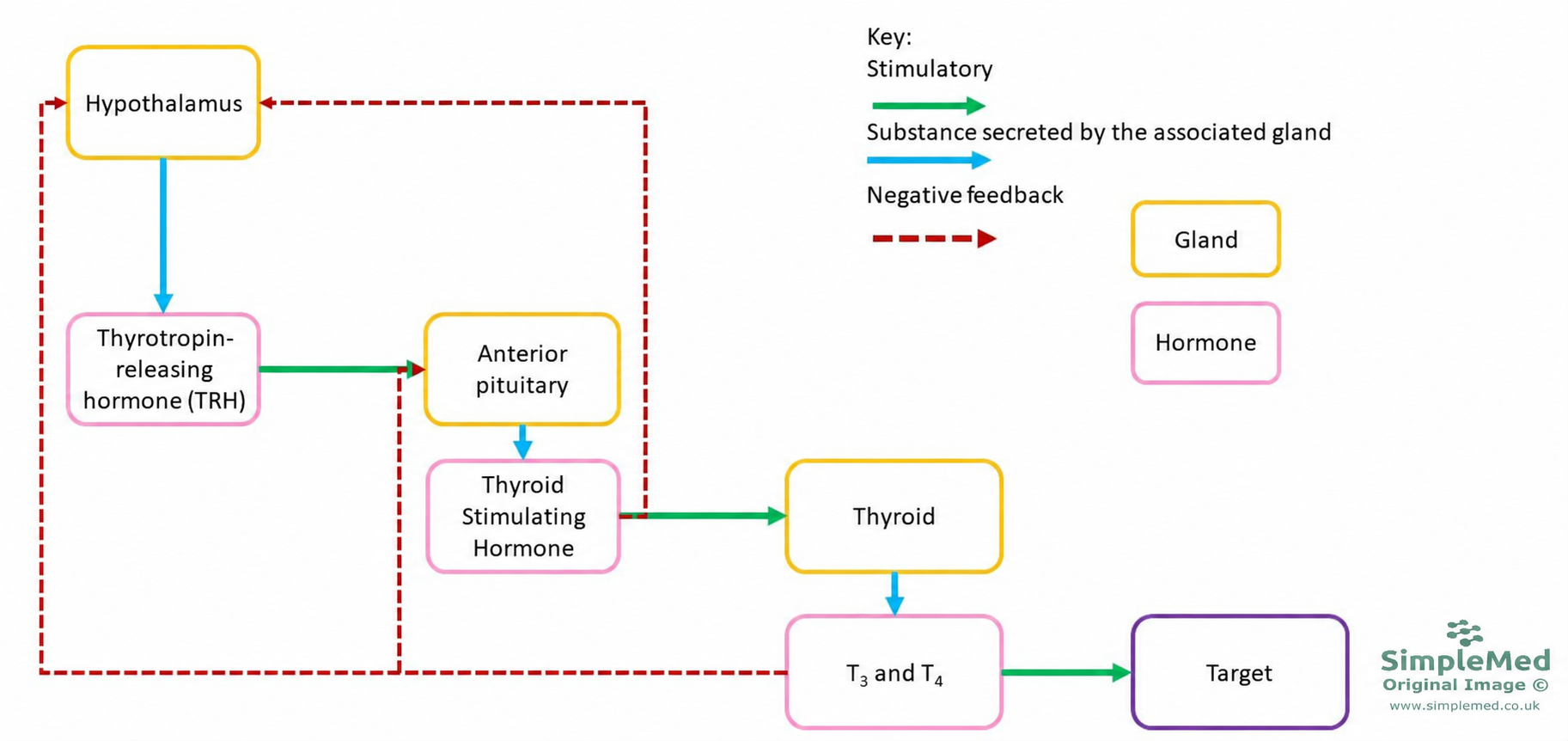
Image: Shows the regulation of thyroid hormone production
SimpleMed original by Dr. Jenny Hubball
Thyroid-stimulating hormone (TSH) is a glycoprotein hormone (a protein with sugar chains attached) composed of 2 non-covalently bound subunits (protein chains held together by weak bonds), one α and one β. The α-subunit is also found in follicle-stimulating hormone (FSH) and luteinising hormone (LH), so it is the β-subunit that makes TSH unique. TSH acts through GPCRs (G-protein-coupled receptors, which signal through the G proteins Gα-s and Gα-q). The processes that TSH stimulates are listed below. Together they increase the making and release of thyroid hormone and the growth of the gland:
- Iodine uptake.
- Iodine oxidation.
- Thyroglobulin synthesis.
- Thyroglobulin iodination.
- Colloid pinocytosis.
- Thyroglobulin proteolysis.
- Thyroid cell metabolism and growth.
Thyroid hormone receptors are nuclear receptor proteins that bind specific DNA sequences called thyroid hormone response elements. The receptors act as transcription factors (proteins that switch genes on or off), regulating gene expression. At positively regulated genes, DNA-bound receptors can recruit corepressors (proteins that switch a gene off) in the absence of thyroid hormone, causing transcriptional repression. Binding of T3 changes the receptor conformation (its shape), releases the corepressors and favours recruitment of coactivators (proteins that switch a gene on), increasing transcription. The effect depends on the target gene: thyroid hormone can also reduce transcription of some genes.
Examples of thyroid-hormone-regulated genes are PEPCK (phosphoenolpyruvate carboxykinase, an enzyme of gluconeogenesis), Ca2+ ATPase, Na+- K+ ATPase (the sodium-potassium pump), cytochrome oxidase (an enzyme of the respiratory chain), and 6-phosphogluconate dehydrogenase. The proteins encoded by these genes contribute to the effects of thyroid hormone outlined below.
Thyroid hormones affect virtually every cell in the body. Their main effects are on cellular differentiation and development and on metabolic pathways. Some of the main functions are outlined below:
- Increase basal metabolic rate and heat production in tissues by increasing the size and number of mitochondria and stimulating the synthesis of enzymes in the respiratory chain (the electron transport chain). The rate of oxygen uptake is not increased in the brain, spleen and testes.
- Stimulate metabolic pathways (mainly catabolic, meaning those that break molecules down).
- Stimulate lipolysis and β-oxidation of fatty acids (the breakdown of fat and of fatty acids to release energy).
- Stimulate insulin-dependent entry of glucose into cells.
- Increase gluconeogenesis (making new glucose) and glycogenolysis (breaking down glycogen to glucose).
- Increase the number of adrenoreceptors (receptors for adrenaline and noradrenaline) on cells, therefore having a sympathomimetic effect (an effect like that of the sympathetic nervous system).
Pathology of the Thyroid Gland
Goitre
A goitre is an enlargement of the thyroid gland. It has several causes, including conditions in which the gland is overstimulated. A goitre can occur in both hypo- and hyperthyroidism and can also occur when thyroid hormone levels are normal, so it is not a definitive feature of either condition. A physiological goitre (one caused by normal hormone changes) can occur during puberty, pregnancy, and the menopause.
The most common causes of goitre are:
- Iodine deficiency, which leads to reduced thyroxine levels (hypothyroidism) and therefore increased TSH release due to the feedback loop. The increased TSH causes thyroid cell maturation and proliferation, increasing the size of the gland and causing a goitre.
- Multinodular goitre (a goitre with several lumps, called nodules). Its cause is not fully understood. Multinodular goitre is the most common cause of a goitre in the UK. After a number of years this may become toxic multinodular goitre (a multinodular goitre whose nodules make too much thyroid hormone), causing the patient to develop hyperthyroidism. A multinodular goitre may also enlarge downwards (inferiorly) into the superior mediastinum (the upper part of the space between the lungs) to form a retrosternal goitre (one that lies behind the breastbone). The enlarged gland may then press on the windpipe, causing tracheal compression.
It is possible, but rare, for thyroid nodules to be malignant (cancerous). Thyroid cancers often do not cause a metabolic disturbance.

Image: Shows a goitre (an enlarged thyroid gland)
Creative commons source by Drahreg01 [CC BY-SA 3.0 (https://creativecommons.org/licenses/by-sa/3.0)]
Hypothyroidism
Hypothyroidism can have many causes, including:
- Failure of the thyroid gland
- TSH or TRH (thyrotropin-releasing hormone) deficiency, which causes central hypothyroidism
- Inadequate dietary supply of iodine
- Radioactive iodine intake
- Autoimmunity - Hashimoto’s disease
- Post-surgery complications
- Congenital defects
- Anti-thyroid medication
The general symptoms of hypothyroidism are weight gain, lethargy (tiredness), intolerance to cold, bradycardia (a slow heart rate), dry skin, alopecia (hair loss), hoarse voice, constipation, and slow reflexes. Clinically, a patient with primary hypothyroidism (where the problem is in the thyroid gland itself) would have a raised TSH with a low free T4 (the unbound, active part of T4) in their blood plasma. NICE advises measuring free T4 in the same sample when the TSH is above the reference range. TSH is elevated due to the lack of negative feedback from the thyroid hormones (think about the diagram pictured earlier).
Congenital hypothyroidism is hypothyroidism that is present from birth. The child may have growth failure, intellectual disability, poor bone development, slow pulse, muscle weakness, and gastrointestinal (GI) disturbances. It is checked for by the newborn blood spot test. About 80% of cases are due to thyroid dysgenesis (the gland does not develop normally). Other cases are due to missing enzymes needed to make thyroid hormone. Iodine deficiency, for example low iodine intake by the mother during pregnancy, is still an important cause worldwide. In adults, severe hypothyroidism can cause myxoedema, characterised by thick, puffy skin due to generalised non-pitting oedema (swelling that does not leave a dent when pressed), muscle weakness, slow speech, mental deterioration, and intolerance to cold. In very rare cases, severe hypothyroidism leads to myxoedema coma. This is a life-threatening emergency that needs treatment in hospital.
The normal treatment for hypothyroidism is oral levothyroxine (synthetic thyroxine, T4).
Hashimoto’s disease is an autoimmune disease resulting from destruction of the thyroid follicles, leading to hypothyroidism. In the UK, it is the most common cause of hypothyroidism. It is most common in women. When Hashimoto’s disease causes hypothyroidism, it is treated with oral levothyroxine (T4). T4 is given in preference to T3 (liothyronine), because T4 has a longer half-life (it takes longer to fall to half its level in the blood), even though T4 is less biologically active. NICE advises against routinely offering liothyronine.
Hyperthyroidism
Hyperthyroidism also has many causes, including:
- Autoimmunity - Graves’ disease
- Toxic multinodular goitre
- Solitary toxic adenoma (a single benign lump of thyroid tissue that makes too much thyroid hormone)
- Excessive thyroid hormone therapy
- Drugs (e.g. amiodarone, a drug used for abnormal heart rhythms)
- Functioning metastases of thyroid carcinoma (cancer that has spread and still makes thyroid hormone)
- Ectopic thyroid tissue (thyroid tissue in the wrong place). An example is struma ovarii, where thyroid tissue in an ovarian teratoma (a type of ovarian tumour) makes thyroid hormone.
The symptoms of hyperthyroidism are weight loss, irritability, heat intolerance, tachycardia (a fast heart rate), fatigue, increased bowel movements, tremor, hyperreflexia (brisk reflexes), breathlessness, loss of libido, and sweating. Another sign of hyperthyroidism can be staring eyes, in which the levator palpebrae superioris muscle is over-stimulated by the sympathetic nervous system, leading to the eyelids being drawn up causing ‘staring eyes’ and lid lag. Thyrotoxicosis is the clinical state caused by too much thyroid hormone in the blood, from any cause. Hyperthyroidism (an overactive thyroid gland) is one cause. Others include thyroiditis (inflammation of the thyroid) and taking too much thyroid hormone medicine. The clinical markers of primary hyperthyroidism are a low TSH with elevated free T4 and/or free T3 in the blood plasma.
The treatment for hyperthyroidism is antithyroid medication, which acts by ‘blocking’ the formation of thyroid hormone. This can be done through Carbimazole, an anti-thyroid drug that needs to be taken continuously, or in severe cases, by taking radioactive iodine, which ablates thyroid tissue and commonly results in hypothyroidism that then requires long-term thyroxine replacement.
Graves' disease is an autoimmune disease caused by the production of thyroid stimulating immunoglobulin (TSI), which continually stimulates thyroid hormone secretion outside of normal hormonal control, resulting in hyperthyroidism.
Toxic adenoma is a single adenoma which has developed in the thyroid and produces thyroxine.
TSH levels are the usual first blood test for suspected hypo-/hyperthyroidism in adults when pituitary disease is not suspected, as it is very rare for a pituitary adenoma (a benign tumour of the pituitary gland) to directly produce TSH. If the TSH is above the reference range, free T4 is measured in the same sample. If the TSH is below the reference range, free T4 and free T3 are measured in the same sample.
Most hypothyroidism and hyperthyroidism is primary. This means the problem lies in the thyroid gland itself and not in the pituitary gland or hypothalamus, which control TSH release. Secondary (central) disease, which starts in the pituitary gland or hypothalamus, is rare. In primary thyroid disease, if TSH is raised, it indicates that not enough thyroid hormone is being produced, and if TSH is reduced it indicates that too much thyroid hormone is being produced. This pattern is due to the negative feedback loop, where TSH concentrations are modified to control the output of the thyroid gland. It does not hold in secondary (central) hypothyroidism, which is caused by pituitary or hypothalamic disease. There, TSH is inappropriately low or normal (rarely raised) while free T4 is low.
Thyroid scintigraphy is an imaging test that uses a radioactive tracer to show how the thyroid gland takes up iodine or a similar ion. It is not the only imaging test. For adults with thyrotoxicosis, NICE advises considering a technetium scan if TSH receptor antibodies are negative, and considering ultrasound only if there is a palpable thyroid nodule (a lump that can be felt). Technetium-99m (a radioactive tracer, or radioisotope) is used for isotope scanning of the thyroid with a gamma camera (a camera that detects the gamma rays given off by the tracer), as it is taken up by the thyroid gland. Technetium-99m is the most commonly used medical radioisotope and has a physical half-life (the time taken for its radioactivity to halve) of about 6 hours, meaning the radiation exposure is relatively low. Technetium-99m can also be used for bone scans, myocardial perfusion imaging (imaging of blood flow to the heart muscle), and brain imaging.
Edited by Dr. Bethany Turner andDr. Maddie Swannack
Reviewed by: Dr. Marcus Judge
- 10264


