

Next Lesson - Development of the Gastrointestinal Tract
Core
Congenital Anomaly Is a Description
A congenital anomaly is a structural or functional difference that originates before birth. It may be recognised on antenatal imaging, at delivery or only later, as with some hearing, visual or metabolic effects. The word congenital describes timing of origin, not cause. A congenital condition can arise from a chromosome or gene change, an environmental exposure, an infection, maternal disease, mechanical constraint, or an interaction among several factors.
Teratology studies abnormal development produced by environmental conditions or agents. A teratogen is therefore not simply anything encountered during pregnancy, and exposure is not equivalent to injury. The agent must reach a susceptible tissue at a biologically relevant dose and time. Many exposed pregnancies are unaffected, while the same agent can produce different outcomes at different stages. Conversely, a recognised anomaly does not identify its cause from appearance alone.
This lesson supplies a mechanism-and-timing framework rather than a list of syndromes. Normal limb and body-wall patterning is taught in Development of the Limbs and Body Wall. Here the first question is different: did tissue form abnormally, become distorted, break down after normal formation, or trigger a cascade of secondary effects?
Four Developmental Mechanisms
A malformation arises from an intrinsically abnormal developmental process during formation of a structure. The organ may be absent, incomplete or patterned abnormally. Genetic and environmental causes can both disturb intrinsic morphogenesis, so malformation does not mean inherited. A deformation changes the shape or position of a part that initially formed normally, usually through mechanical constraint. Reduced amniotic fluid or limited uterine space can distort limbs without having specified their bones incorrectly.
A disruption is extrinsic breakdown of, or interference with, tissue that had been developing normally. Amniotic bands can constrict and damage a previously formed limb, producing an irregular distribution unlike a repeatable patterning defect. A sequence is not a fourth kind of tissue injury. It is a chain of secondary anomalies caused by one initiating malformation, deformation or disruption.
Oligohydramnios sequence shows the logic. Severe renal malformation can reduce fetal urine and therefore amniotic fluid. Low fluid then permits compression-related limb and facial deformation and restricts lung expansion, producing pulmonary hypoplasia. The later findings are not independent renal malformations. Reading the chain backwards identifies one primary event and prevents a sequence being confused with a syndrome, in which several features share an underlying cause but are not simply downstream of one initiating anomaly.
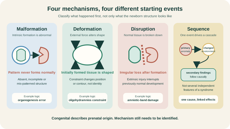
Classify the initiating event first: abnormal formation, altered shape, tissue breakdown, or downstream cascade.
SimpleMed original educational diagram
Teratology Is a Timing Problem
Core teratology principles turn an exposure history into a developmental question. Susceptibility depends on the maternal and conceptus genotypes, developmental stage, dose and duration, route and placental access, and the agent's mechanism. Outcomes include embryonic or fetal death, structural malformation, altered growth and functional impairment. These are not mutually exclusive, and risk is rarely described by dose alone.
During roughly the first two post-fertilisation weeks, before and around implantation, severe injury often causes loss while a surviving conceptus may develop without a major structural anomaly. This is the useful all-or-none heuristic: a small pool of relatively unspecialised cells can sometimes compensate, whereas extensive injury cannot. It is not a guarantee. Genetic damage, mosaicism or an agent persisting into later development can escape the simple rule.
Once cell populations are committed to particular organs, timing becomes anatomically predictive. Interference while a heart septum, limb field or palate is being constructed can alter structure. The same exposure after gross form is established may instead affect growth, renal function, brain maturation or hearing. Timing often changes the phenotype more dramatically than the agent's name.
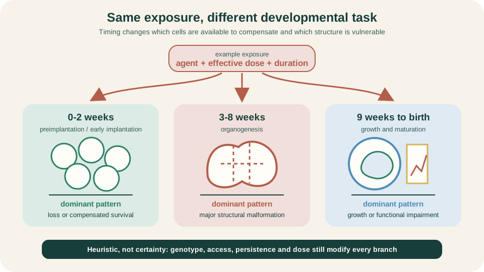
The exposure may be unchanged; the developmental task underway redirects the likely outcome.
SimpleMed original educational diagram
Critical Periods from Conception to Birth
The embryonic period of organogenesis, approximately post-fertilisation weeks three to eight, carries the broadest risk of major structural malformation. Organs do not develop simultaneously. The heart begins major morphogenesis early, limb patterning occupies a narrower later span, and palate formation continues after early facial patterning. A calendar week is therefore meaningful only when paired with the structure and process active then.
From post-fertilisation week nine to birth, the fetal period is dominated by growth and functional maturation, but vulnerability does not end. Brain organisation, neuronal migration, synapse formation, eye and ear function, renal physiology and genital maturation continue. Structural effects can still occur where morphogenesis persists, while growth restriction and functional impairment become relatively more prominent. A normal external form cannot prove normal later function.
Clinical pregnancy dating usually counts from the last menstrual period and is about two weeks ahead of post-fertilisation age. The diagram uses post-fertilisation age to match developmental events. Its bands are deliberately broad: critical periods overlap, individual development varies, and colour boundaries are not safe-versus-dangerous switches.
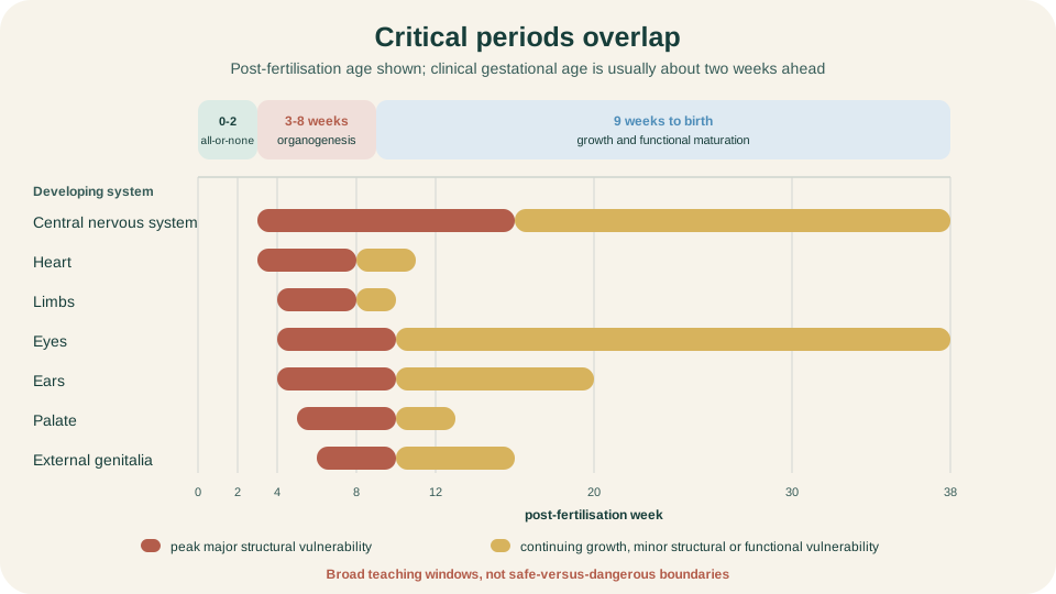
Critical periods overlap. Rust marks peak structural vulnerability; gold marks continuing growth or functional vulnerability.
SimpleMed original educational diagram
Placental Transfer and Drug Patterns
The placenta is an active transport and metabolic interface, not an impermeable shield. Many small, lipid-soluble, relatively un-ionised and unbound molecules cross by diffusion; transport proteins can move other compounds towards or away from fetal blood. Transfer also changes with maternal concentration, placental blood flow and gestational age. Detectable transfer proves exposure, not teratogenicity. Harm still requires a susceptible target, relevant timing and sufficient effective dose.
Selected medicines illustrate mechanism and timing. Isotretinoin can disturb retinoid signalling during organogenesis and produce a characteristic craniofacial, cardiac, thymic and central nervous system pattern. Valproate exposure is associated with major malformations including neural tube defects and with later neurodevelopmental effects, so its risk is not confined to one visible anatomical window. Current UK controls require specialist oversight and prevention of pregnancy exposure.
ACE inhibitors and angiotensin receptor blockers (ARBs) are contraindicated throughout pregnancy. A woman planning a pregnancy should be switched to an alternative antihypertensive before conception, and treatment should be stopped as soon as pregnancy is confirmed, preferably within 2 working days of the pregnancy being notified (NICE NG133). Separately from that blanket contraindication, these drugs produce a characteristic fetopathy that reflects the fetal renin-angiotensin system becoming functionally important from the second trimester onwards: they impair fetal renal function and reduce amniotic fluid, and the resulting oligohydramnios generates secondary pulmonary hypoplasia, skull ossification defects (hypocalvaria) and limb contractures. This is fetopathy and sequence logic rather than a classic early patterning defect. These examples teach principles, not prescribing. Starting, stopping or switching treatment during pregnancy requires current specialist risk-benefit guidance because uncontrolled maternal disease and abrupt withdrawal can also cause harm.
Infection, Maternal Disease and Environment
TORCH is a memory aid, not one mechanistic family. Toxoplasma, rubella, cytomegalovirus and syphilis can cross the placenta, but transmission probability, critical period and organ targets differ. Primary maternal toxoplasmosis acquired during or just before pregnancy can reach the fetus; earlier fetal infection, when it occurs, is more likely to be severe, especially for brain and eye. Early rubella carries prominent cardiac, ocular and hearing risks. Congenital CMV may affect growth, brain and hearing after primary, reinfection or reactivation events.
The letter O means other and cannot be treated as a fixed organism list. Syphilis, varicella, parvovirus B19 and Zika illustrate different pathways and outcomes. Herpes simplex is included in the traditional acronym, yet most neonatal HSV is acquired around delivery rather than through a classic transplacental malformation pathway. The acronym should prompt route, timing and target questions, not a memorised shared phenotype.
Maternal physiology is part of the developmental environment. Poorly controlled pre-existing diabetes around conception increases malformation risk; later placental and metabolic effects can alter growth. Alcohol can affect growth, face and the developing nervous system across overlapping windows. Hyperthermia, smoking, radiation and environmental contaminants each require agent, dose and timing context. Association after an exposure is not enough to infer causation in one pregnancy.
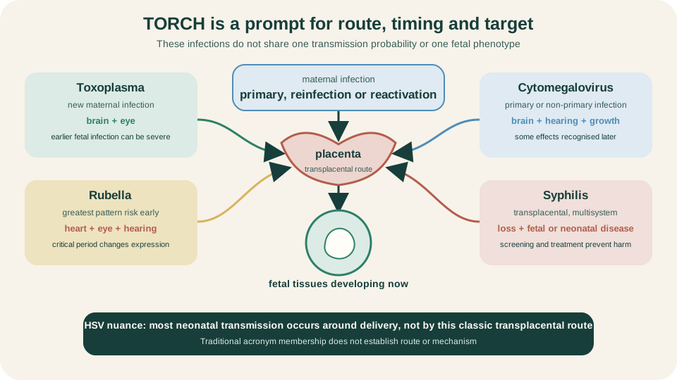
TORCH opens a route-and-timing enquiry. It does not imply one route, one critical period or one fetal pattern.
SimpleMed original educational diagram
Prevention before Organogenesis
Prevention must precede the developmental window it protects. Neural tube closure occurs early, often before pregnancy is recognised, so folic acid needs to be present before conception and through early pregnancy. Population fortification and daily supplementation reduce neural tube defects, but folate does not neutralise every teratogen or prevent every neural tube defect.
Preconception care also creates time to review medicines with the relevant specialist, optimise diabetes and other maternal disease, update rubella immunity before pregnancy where appropriate, and reduce alcohol, tobacco and harmful occupational or environmental exposure. During pregnancy, infection prevention, antenatal screening and prompt clinical assessment add further layers. The principle is anticipatory: move protection upstream rather than waiting for organogenesis to finish.
Screening and Diagnostic Detection
Screening sorts pregnancies by chance or looks for predefined features. Maternal serum markers, cell-free DNA pathways and ultrasound can identify higher-chance results or visible structural findings, but a screening result is not a fetal diagnosis. Detection depends on gestational age, test performance, the condition sought and whether the phenotype is visible. A lower-chance or normal scan cannot exclude every structural, genetic, infectious or functional condition.
Diagnostic testing asks a narrower question of sampled material. Chorionic villus sampling obtains placental tissue; amniocentesis obtains amniotic fluid containing fetal cells and other analytes. Chromosome, gene or infection assays can then diagnose the target within that method's scope. Even diagnostic tests do not explain every anomaly, and placental mosaicism can complicate some CVS results. The route is screening finding or prior risk, informed choice, appropriate diagnostic test, then interpretation.
Detection and causation remain separate. Ultrasound may show a limb reduction, but classification still asks whether it reflects intrinsic patterning, tissue disruption or a sequence. A laboratory diagnosis can establish a chromosome or infection finding without proving that every observed feature follows from it. Good counselling preserves uncertainty, test limits and parental choice rather than turning a probability into certainty.
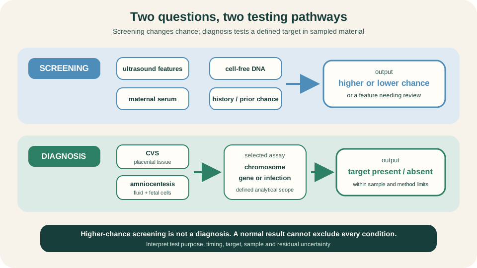
Screening changes probability. Diagnostic testing answers a defined question, within the limits of its sample and method.
SimpleMed original educational diagram
References and Further Reading
- Developmental mechanisms of congenital anomalies, CDC.
- Identifying Human Teratogens: An Update, Cassina et al.
- Placental Control of Drug Delivery, Griffiths and Campbell.
- Isotretinoin Capsule Information, FDA.
- Valproate reproductive risks, MHRA.
- Congenital disorders, WHO.
- About Folic Acid, CDC.
- Fetal Anomaly Screening Programme overview, NHS England.
Reviewed by: Dr. Marcus Judge
- 143
