

Next Lesson - Teratology and Congenital Malformations
Core
Limb Buds: Timing and Origin
Limb development becomes externally visible near the end of the fourth embryonic week. Upper-limb buds appear first, at about day 26 to 28 after fertilisation, and lower-limb buds follow roughly one or two Carnegie stages later. Each begins as a mesenchymal swelling under surface ectoderm on the ventrolateral body wall. Timing is approximate because embryos are staged by a set of features rather than by the calendar alone.
The bud has more than one lineage. Somatic lateral-plate mesoderm supplies the skeletal elements and much of the limb's connective tissue, including the connective framework that helps pattern tendons and muscles. Myogenic precursors leave the hypaxial part of nearby somites and migrate into the bud to form skeletal muscle. Peripheral axons then grow into this prepared territory. This lesson starts with those derivatives; The Basics of Embryology owns gastrulation, neurulation and somite formation.
Growth then changes the external silhouette quickly. The small bud elongates, flattens distally into a hand or foot plate, and develops digital rays before fingers or toes are separate. Upper-limb events usually lead equivalent lower-limb events by a short interval, so one embryo can show a more advanced hand plate than foot plate without implying abnormal lower-limb development. Calendar timing should always be read with the whole embryo's stage.
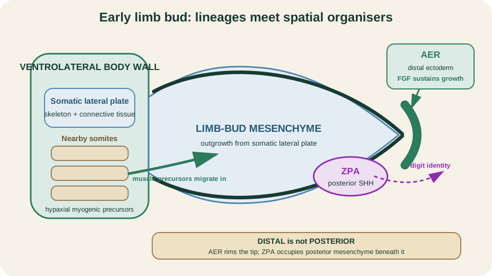
The early bud is a meeting place: body-wall mesenchyme grows out, the distal AER sustains growth and the posterior ZPA supplies directional information.
SimpleMed original educational diagram
Proximal-Distal Growth and the AER
The apical ectodermal ridge (AER) is a thickened strip of ectoderm along the distal rim. Its fibroblast growth factor signals keep underlying distal mesenchyme proliferative and sustain outward growth. As the bud lengthens, elements appear in a proximal-to-distal sequence: stylopod (humerus or femur), zeugopod (paired forearm or leg bones), then autopod (wrist or ankle and digits). Modern models combine distal FGF with other spatial cues; the historic progress-zone clock is useful context, not an uncontested complete explanation.
AER experiments reveal a powerful time rule. Removing distal ridge signalling early arrests outgrowth when only proximal structures have been established. Later disruption permits more proximal and intermediate elements but removes structures that still depended on continued distal growth. The failed level therefore follows the time of signal loss, not an arbitrary adult amputation plane.
Growth and identity are related but not interchangeable. FGF-dependent outgrowth supplies enough tissue and time to elaborate distal parts, while other signals tell that tissue whether it occupies anterior, posterior, dorsal or ventral territory. Continued length without a coherent positional map would not produce a normal limb, and correct positional signals cannot rescue a bud whose distal growth has stopped.
Three Patterning Axes
A usable limb needs position in three dimensions. The AER and its FGFs support the proximal-distal axis from girdle to digits. Posterior mesenchyme forms the zone of polarising activity (ZPA), where Sonic hedgehog (SHH) helps assign anterior-posterior identity from the preaxial thumb or great-toe side toward the postaxial little-digit side. Experimentally adding posterior signalling at the anterior border can create a second, mirror-orientated digit pattern.
Dorsal and ventral ectoderm set the third axis. Dorsal WNT7A induces LMX1B in underlying mesenchyme and promotes dorsal fates such as the nail-and-knuckle surface. Ventral EN1 restricts that dorsal programme, helping specify palm or sole. These organisers cross-regulate one another, so a limb is not built by three isolated switches. Growth must continue while positional information remains coherent.
Axis names refer to the early bud, before its later rotation. Anterior-posterior therefore means preaxial-to-postaxial position within the developmental field, not whichever adult surface happens to face forwards. Keeping the embryonic coordinate frame separate from the final anatomical position prevents errors when following the thumb, great toe, muscle masses and dermatomes into adult orientation.
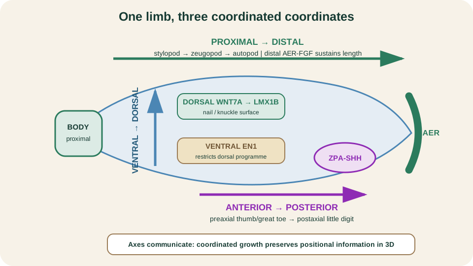
Three coordinated coordinates: AER-FGF sustains length, ZPA-SHH patterns the preaxial-to-postaxial field, and opposing ectodermal signals distinguish dorsal from ventral.
SimpleMed original educational diagram
Digit Formation and Separation
The distal autopod first broadens into a hand or foot plate. Condensations within it establish digital rays. Tissue between those rays is then removed and remodelled through programmed cell death, separating the emerging digits from distal toward proximal. Apoptosis does not create the rays; it sculpts the spaces after positional and skeletal patterning has begun.
This order makes two failure mechanisms distinct. If interdigital tissue is not removed adequately, adjacent digits remain joined, producing syndactyly of soft tissue or, when skeletal pattern is involved, bone. If the anterior-posterior positional field is altered, digit number and identity can change. Ectopic SHH-like polarising activity can generate extra digits in mirror arrangement rather than simply fusing correctly specified neighbours.
Programmed cell death is spatially controlled removal, not tissue injury spreading at random. Cells fragment and are cleared while the digital condensations are preserved. The result is a web that retreats between stable rays. That distinction matters when reading a defect: a normal set of rays linked by persistent tissue suggests a separation problem, whereas duplicated rays or a reversed sequence requires earlier positional-patterning logic.
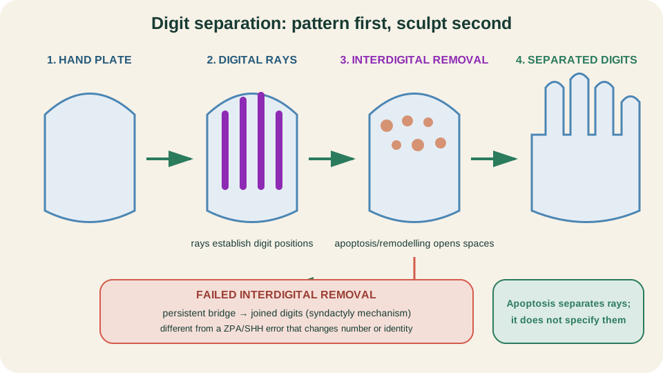
Pattern first, sculpt second: rays mark future digits before programmed interdigital removal opens the spaces between them.
SimpleMed original educational diagram
Muscle, Nerve and the Segmental Map
Somitic myogenic precursors enter each bud and organise into dorsal and ventral muscle masses. In the upper limb these broadly become extensor and flexor territories respectively before rotation changes their adult orientation. Ventral rami enter early, split into branches associated with the two muscle masses and form plexuses. A plexus mixes axons from several spinal levels into named peripheral nerves, but it does not erase their segmental origin.
The skin retains that history as dermatomes. Growth stretches each segmental territory from root level toward the distal limb, and rotation twists the map. Adult dermatomes therefore run in curved or spiral bands rather than straight transverse stripes. Nerves follow and serve tissues that are being patterned; they are not the primary signal that tells a humerus, digit or dorsal surface what identity to adopt.
Dorsal and ventral divisions of the plexus broadly preserve the early muscle-mass relationship even after named nerves recombine fibres. A single peripheral nerve can therefore contain axons from several roots, and one root can contribute to several peripheral nerves. Root-level sensory testing follows dermatomes; peripheral-nerve testing follows the later branch pattern. Both maps are true because they describe different stages of the same wiring history.
Limb Rotation and Adult Anatomy
Upper and lower limbs begin with comparable preaxial and postaxial borders but rotate in opposite directions. The upper limb rotates laterally. The thumb becomes lateral, the elbow faces largely posteriorly, flexor compartments lie mainly anteriorly and extensor compartments mainly posteriorly. The lower limb rotates medially. The great toe becomes medial, the knee faces anteriorly, extensor compartments lie mainly anteriorly and flexor compartments mainly posteriorly.
This is why homologous compartments do not face the same way in arm and leg. It also explains the spiral character of dermatomes and why the preaxial border ends at the thumb in the upper limb but at the great toe in the lower limb. Rotation reorients an already patterned limb; it does not swap its segmental roots or convert dorsal muscle lineage into ventral lineage.
Do not reduce rotation to a memorised arrow alone. Use three linked checks. After upper-limb lateral rotation the thumb is lateral and the extensor surface is mainly posterior. After lower-limb medial rotation the great toe is medial and the extensor surface is mainly anterior. If a proposed map violates one of those paired outcomes, its rotation direction has probably been reversed.
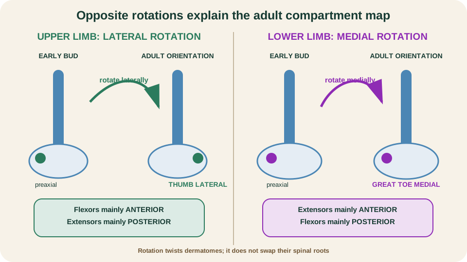
Opposite rotations explain the adult map: upper-limb extensors finish posteriorly, while lower-limb extensors finish anteriorly.
SimpleMed original educational diagram
Lateral Folding and Body-Wall Closure
The limb buds arise from a body wall that is itself moving. In transverse view, paired lateral folds carry somatic lateral-plate mesoderm and overlying ectoderm ventrally. Their right and left edges approach and meet at the ventral midline, helping transform a flat embryonic disc into a closed cylindrical body. The somatopleure becomes the primary wall around the intraembryonic coelom, while endoderm is internalised as gut tube. The coelom is enclosed, not erased.
Closure is deliberately incomplete at the umbilical ring, where the embryo retains a controlled connection to extraembryonic structures. Hypaxial myoblasts extend into the developing wall to form its skeletal musculature. This article owns wall closure as a morphogenetic mechanism. Earlier folding is reviewed in The Basics of Embryology; gut tube, physiological herniation and peritoneal detail remain in Development of the Gastrointestinal Tract.
The wall is therefore layered. Surface ectoderm supplies the external covering, somatic lateral plate supplies connective tissue and parietal lining, and migrating hypaxial myoblasts supply muscle. The paired folds must move, expand and unite while maintaining the intended openings. A defect can arise from inadequate fold movement, failed fusion, abnormal tissue growth or a later event at the umbilical ring; the surface appearance alone does not make those mechanisms identical.
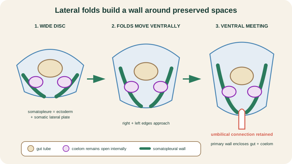
Lateral folding converts paired somatopleural sheets into a ventral wall around the gut and coelom, with a controlled opening at the umbilical ring.
SimpleMed original educational diagram
Failed-Step Mechanisms
Developmental defects become easier to localise when read backwards. A severe early reduction suggests failure of limb initiation or early AER-dependent outgrowth; a more distal reduction fits later loss of continued growth. Joined digits point toward failed interdigital removal, whereas extra or mirror-patterned digits point toward altered anterior-posterior positional information. These are mechanism examples, not a catalogue of syndromes or exposures.
Ventral wall defects also need precise language. Omphalocele reflects failure of physiologically herniated bowel to return through the umbilical ring and has a covering sac. Gastroschisis is usually a paraumbilical full-thickness wall defect with exposed bowel and no covering sac. Both identify trouble at the wall-umbilical interface, but they should not be presented as one identical midline-fold mechanism. Detailed gut movement belongs in the linked GI lesson, while exposure timing, teratology and malformation classification belong in the next lesson.
The same failed-step method prevents superficial associations. Ask first whether the problem is initiation, continued outgrowth, positional identity, tissue removal, rotation, innervation or wall closure. Then predict what should be absent, duplicated, joined or reorientated. Only after that should a named abnormality be attached. This sequence is more transferable than memorising lists and keeps this anatomy lesson separate from the teratology framework that follows.
References and Further Reading
- Formation of the Limb Bud, Gilbert.
- Generating the Proximal-Distal Axis of the Limb, Gilbert.
- Specification of the Anterior-Posterior Limb Axis, Gilbert.
- The Generation of the Dorsal-Ventral Axis, Gilbert.
- Embryology, Hand, Raszewski and Singh.
- Molecular Regulation of Limb Growth, Zeller et al.
- Paraxial Mesoderm: The Somites and Their Derivatives, Gilbert.
- Musculoskeletal System - Limb Development, UNSW Embryology.
- Embryology of the Abdominal Wall and Associated Malformations, Raitio et al.
Reviewed by: Dr. Marcus Judge
- 196
