

Next Lesson - Structure of Proteins
Core
The Cell Cycle Map
A proliferating somatic cell does not enlarge continuously and then split. It advances through ordered phases that separate growth, genome duplication and chromosome segregation. G1 is the first gap: mass increases, organelles are replenished and the cell samples mitogens, nutrients and contact signals before any DNA is copied. S phase replicates the nuclear genome once, producing sister chromatids tethered by cohesin. G2 is the second gap, used to finish mitotic protein synthesis and to confirm that replication is complete. M phase comprises mitosis and cytokinesis. The morphological stages from prophase to telophase are developed in Mitosis and Meiosis; this lesson concerns the decisions that allow entry into those stages and the programmes that abort an unsafe cycle.
G0 is not a failed G1. It is a resting state in which cyclin-CDK activity is suppressed and replication origins are not fired. Many differentiated cells occupy G0 for long periods yet retain the capacity to re-enter G1 when tissue demand returns, as hepatocytes do after substantial cell loss. Fully post-mitotic cells, including most mature neurons, remain outside the cycle permanently. The practical map is therefore five states, G0, G1, S, G2 and M, linked by irreversible commitments once DNA synthesis or chromosome segregation has begun.
That temporal separation is the first safety rule of tissue renewal. DNA is copied only in S. Chromosomes are pulled apart only in M. Checkpoints guard the transitions at which an error would become heritable in both daughters.
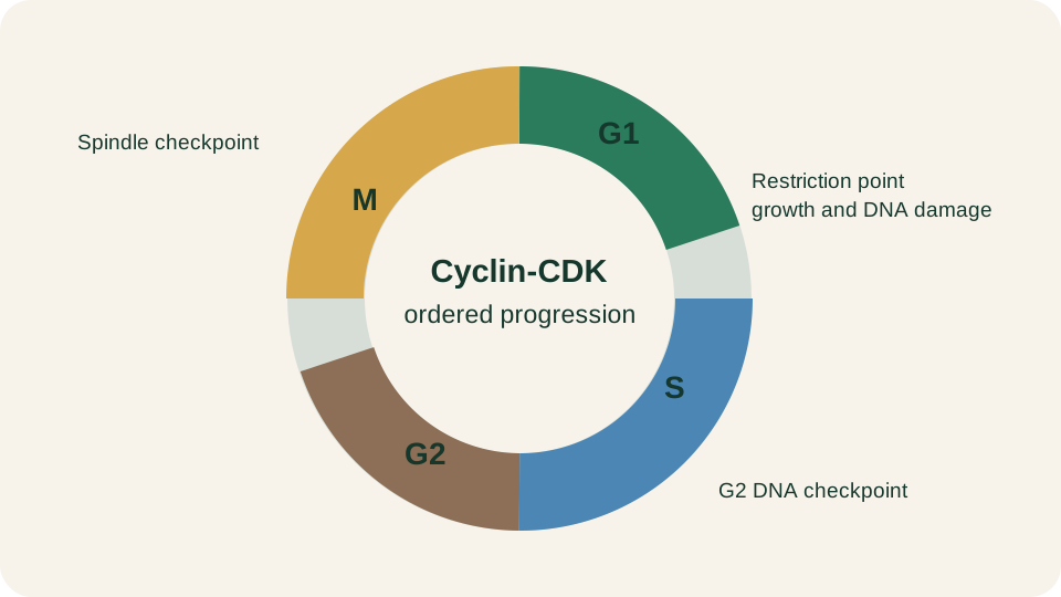
Ordered phases of the cell cycle with the restriction point and the DNA, replication and spindle checkpoints that gate unsafe progress.
SimpleMed original educational diagram
Cyclin-CDK Control
Forward motion is not the work of a single clock protein. It is produced by cyclin-dependent kinases (CDKs) that become catalytic only when bound to the correct cyclin. Cyclin abundance oscillates through transcription and ubiquitin-mediated proteolysis; CDK polypeptides are comparatively stable. The pairing therefore creates a temporary kinase whose substrate preference matches the needs of one phase.
A usable medical set of pairs is small. Cyclin D with CDK4 or CDK6 advances early G1 by phosphorylating the retinoblastoma protein (Rb). Progressive Rb phosphorylation releases E2F transcription factors that drive genes required for DNA synthesis. Cyclin E-CDK2 completes late G1 and licenses entry into S. Cyclin A-CDK2 supports S-phase progression and helps prevent re-firing of origins that have already been used. Mitotic entry depends on cyclin B-CDK1, which phosphorylates nuclear lamins, microtubule regulators and other mitotic effectors. Destruction of mitotic cyclins by the anaphase-promoting complex/cyclosome (APC/C) then permits exit from mitosis and reassembly of G1.
CDK output is sharpened by layers beyond cyclin binding. Inhibitory phosphorylation by Wee1 and activating dephosphorylation by Cdc25 create switch-like mitotic entry. CDK inhibitors of the INK4 family, which target CDK4/6, and CIP/KIP family, including p21 and p27, provide direct brakes. The first-pass principle is simple: the cyclin decides when a CDK can work; phosphorylation state and stoichiometric inhibitors decide whether that work is allowed.
Restriction and DNA Checkpoints
The restriction point in late G1 is the principal commitment decision in mammalian cells. Before it, mitogen withdrawal or strong contact inhibition can return the cell towards G0. After it, progression becomes largely independent of external growth factors and the cell is committed to complete the cycle unless a later checkpoint intervenes. Molecularly, the restriction point is crossed when cyclin D- and cyclin E-dependent CDKs have phosphorylated Rb deeply enough that E2F-driven S-phase gene expression becomes self-reinforcing.
DNA damage checkpoints operate mainly in G1 and G2; a related replication checkpoint monitors S phase. ATM responds principally to DNA double-strand breaks and signals strongly through Chk2. ATR responds principally to replication stress and stretches of single-stranded DNA, signalling strongly through Chk1. Their shared output is reduced CDK activity until the genome is again a safe template. In G1, stabilised p53 is central. Transcription of p21 inhibits G1 CDKs, enforcing arrest while repair pathways act. The enzymology of those pathways belongs in DNA Repair and Cancer. If damage is extensive or persistent, the same p53 programme can abandon temporary arrest in favour of apoptosis or permanent senescence rather than allowing an unrepaired genome into S phase.
During S phase, stalled forks and unreplicated DNA activate ATR-Chk1 signalling that slows late origin firing and protects fork stability. In G2, residual damage or incomplete replication prevents full activation of cyclin B-CDK1, blocking mitotic entry with an unfinished genome. These delays are not passive pauses. They are decision gates that trade time for fidelity before either DNA synthesis or chromosome segregation can fix an error in both daughters.
The Spindle Checkpoint
Once the cell is in mitosis the dominant risk changes. Incomplete replication gives way to unequal segregation. The spindle assembly checkpoint monitors kinetochore-microtubule attachment and inter-kinetochore tension. Unattached or incorrectly attached kinetochores generate a diffusible wait-anaphase signal that keeps the APC/C from ubiquitinating securin and cyclin B. Securin binds and inhibits separase, so cohesin remains intact while the checkpoint is active. Only when every chromosome is bi-oriented does the checkpoint silence. APC/C-mediated securin destruction releases separase, which cleaves cohesin so sister chromatids disjoin and anaphase begins.
Checkpoint failure produces aneuploidy: daughters with missing or extra chromosomes. Aneuploidy is not automatically lethal to the cell, but it is a potent source of genomic instability and a recurring feature of many solid tumours. The spindle checkpoint therefore sits between ordinary mitosis and the chromosomal chaos that later pathology lessons classify under neoplasia.
Intrinsic Apoptosis
When repair cannot restore a safe trajectory, programmed cell death is often preferable to division. Apoptosis is ordered, energy-dependent dismantling of the cell with preservation of plasma-membrane integrity until late. The intrinsic, or mitochondrial, pathway is the default route for genotoxic stress, oncogene activation and many developmental cues.
BCL-2 family proteins set the apoptotic threshold at the outer mitochondrial membrane. Anti-apoptotic members such as BCL-2 and BCL-XL restrain membrane permeabilisation. Effectors BAX and BAK, once activated, oligomerise and create pores. BH3-only sensors, including BIM, PUMA and NOXA, integrate stress signals and tip the balance towards permeabilisation by inhibiting the protectors or helping activate the effectors. Cytochrome c released into the cytosol binds APAF-1, assembling the apoptosome that activates initiator caspase-9. Caspase-9 cleaves and activates executioner caspases-3 and -7.
Executioners dismantle the nuclear lamina, enable CAD-mediated DNA cleavage, expose phosphatidylserine on the outer leaflet and package the corpse into membrane-bound apoptotic bodies. Neighbouring phagocytes recognise those eat-me signals and clear the fragments. Because contents are not spilled early, inflammation remains minimal compared with necrotic rupture. Apoptosis is therefore tissue housekeeping as much as cell suicide.
Extrinsic Apoptosis
The extrinsic pathway begins at the plasma membrane. Death receptors of the TNF receptor superfamily, including Fas and TRAIL receptors, bind cognate ligands and recruit adaptors such as FADD. The death-inducing signalling complex activates initiator caspase-8 and, in some cells, caspase-10. In type I cells, caspase-8 can process executioners directly. In type II cells, caspase-8 cleaves BID to tBID, which engages mitochondria and amplifies the signal through cytochrome c release. The two routes are therefore not sealed compartments. They converge on the same executioner caspases and the same apoptotic morphology of shrinkage, chromatin condensation and apoptotic-body formation followed by phagocytic clearance.
Immune-mediated killing of infected or transformed targets often uses the receptor route, whereas genotoxic stress more often uses the intrinsic route. The examination point is the logic chain: ligand to receptor to initiator caspase, optional mitochondrial amplification, shared executioners, silent clearance.
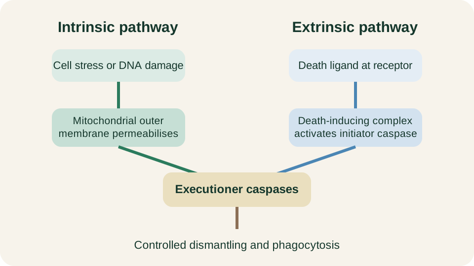
Intrinsic mitochondrial and extrinsic death-receptor pathways converge on initiator then executioner caspases, producing apoptotic bodies for phagocytic clearance.
SimpleMed original educational diagram
When Control Fails
Controlled proliferation and controlled death are complementary defences of tissue integrity. Cells that ignore the restriction point continue to cycle without adequate mitogen permission. Cells that disable p53 lose a major coupling between DNA damage and either CDK inhibition through p21 or apoptotic elimination, so mutations can be fixed through S phase and mitosis. Overexpression of anti-apoptotic BCL-2 family members raises the threshold for mitochondrial permeabilisation; damaged cells survive signals that should have removed them. Loss of death-receptor signalling or caspase function blunts extrinsic clearance.
None of these lesions creates a tumour alone. Each removes a quality-control step that would otherwise limit clonal expansion of an altered genome. Checkpoint failure allows error-prone genomes to be copied and segregated. Apoptotic failure allows those genomes to persist when death would have been safer than repair. Together they convert the cell cycle from a programme of safe renewal into a vehicle for propagating damage. The clinical taxonomy of dysplasia, invasion and metastasis is developed in Introduction to Neoplasia. The mechanistic reading for this lesson is narrower and sufficient: cyclin-CDK engines drive the cycle, checkpoints decide whether progress is safe, and apoptosis removes the cell when safety cannot be restored.
Reviewed by: Dr. Marcus Judge
- 159
