

Next Lesson - Membrane Transport and Intracellular Calcium Regulation
Core
Receptor Classes and Time Course
Receptor location and molecular work predict how quickly a cellular response appears and how long it lasts. Membrane receptors sample the extracellular space, whereas intracellular receptors bind ligands that have crossed the plasma membrane. The membrane barrier and its signalling logic are developed in Cell Membrane and Signalling.
A practical timescale map follows from effector mechanism. Ligand-gated ion channels change membrane potential in milliseconds because ions flow as soon as the channel opens. G-protein-coupled receptors (GPCRs) usually act through heterotrimeric G proteins and second messengers on a seconds-to-minutes scale; their cascade detail belongs in G-Protein Coupled Receptors. Enzyme-linked receptors, the focus of this lesson, alter existing proteins by phosphorylation or by generating cyclic nucleotides, again typically within seconds to minutes. Intracellular receptors for steroid, thyroid, vitamin D and retinoid ligands act principally as ligand-regulated transcription factors. New mRNA and protein must be made, so the physiological onset is commonly measured in hours rather than seconds.
Modification of existing proteins is usually faster than altered transcription and translation. For an unfamiliar hormone or cytokine, receptor location and effector chemistry predict tempo before pathway detail is memorised.
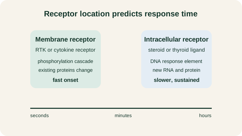
Typical response tempo separates ion channels in milliseconds, enzyme-linked membrane receptors in seconds to minutes, and intracellular transcriptional receptors over hours.
SimpleMed original educational diagram
Receptor Tyrosine Kinases
Receptor tyrosine kinases (RTKs) are single-pass transmembrane proteins with an extracellular ligand-binding region and a cytosolic tyrosine kinase domain. Many growth factors, including EGF, PDGF and FGF, use this architecture. Many RTKs are restrained in an inactive state until ligand binding promotes dimerisation or higher-order clustering. For many receptors, phosphorylation of the activation loop by a partner kinase helps activate the catalytic domain. Phosphorylation between receptor chains is called trans-autophosphorylation. This is not a universal activation mechanism: EGFR kinase domains form an asymmetric dimer in which one partner allosterically activates the other by changing its conformation. EGFR can still phosphorylate receptor tyrosines to create docking sites, but its initial kinase activation should not be described as obligatorily reciprocal activation-loop phosphorylation.
Those phosphotyrosines are not decoration. They create docking sites for proteins that carry SH2 or PTB domains. Bound partners include adaptors such as Grb2, enzymes such as phospholipase C-γ and phosphatidylinositol 3-kinase (PI3K), and further scaffolds that assemble local signalling complexes at the membrane. The RTK therefore converts a brief extracellular binding event into a temporary intracellular docking board. Which pathways fire depends on which partners are recruited in that cell type, not on a single universal cascade shared by every RTK.
Classical growth-factor RTKs usually dimerise after ligand binding, but the insulin receptor is already a covalent tetramer and activates by conformational change. Protein tyrosine phosphatases reverse the docking code, so without continued kinase input the signal collapses.
Insulin as a Worked Example
The insulin receptor is the pre-clinical prototype of an enzyme-linked receptor with clear branch logic. It is synthesised as a precursor that is cleaved and assembled into a preformed disulphide-linked α2β2 heterotetramer comprising two αβ half-receptors. The α subunits bind insulin outside the cell; the β subunits span the membrane and carry the tyrosine kinase domains. Ligand therefore does not recruit two separate monomers from the membrane sea. Insulin binding rearranges the preformed complex and activates the β-subunit kinases.
Autophosphorylation of the β chains creates binding sites for insulin receptor substrate (IRS) proteins. IRS polypeptides are scaffolds rather than the final effectors. Once tyrosine-phosphorylated by the receptor, they present docking sites for SH2-domain proteins, notably the adaptor Grb2 and the regulatory subunit of class I PI3K. From that single membrane event the cell can open both a mitogenic Ras-MAPK branch and a metabolic PI3K-Akt branch. Tissue context decides the dominant readout: adipocytes and myocytes emphasise glucose uptake and storage, whereas other cells may couple the same receptor family more strongly to growth programmes.
Remember the preformed α2β2 RTK, the IRS scaffold and the split into MAPK versus PI3K outputs. Endocrine-axis control and diabetes pharmacology belong elsewhere.
MAPK and PI3K
The Ras-Raf-MEK-ERK cascade is the core mitogen-activated protein kinase (MAPK) branch used by many RTKs. A common entry is Grb2 bound to phosphotyrosine, carrying the guanine nucleotide exchange factor Sos. Sos activates membrane-associated Ras by promoting GDP release so that GTP can bind. Ras-GTP recruits and helps activate Raf family kinases. Raf phosphorylates MEK, and MEK phosphorylates ERK. Active ERK phosphorylates cytosolic targets and also enters the nucleus to modify transcription factors. The cascade is a three-tier kinase relay: each step can phosphorylate many downstream molecules, so a modest number of active receptors can produce a large ERK output.
The PI3K-Akt branch has different chemistry and a different first job. Class I PI3K phosphorylates PIP2 to PIP3 in the inner leaflet of the plasma membrane. PIP3 recruits PH-domain proteins, including Akt (protein kinase B) and its upstream activating kinases. Once activated, Akt phosphorylates targets that promote survival, growth and, in insulin-responsive tissues, redistribution of glucose transporters and engagement of anabolic enzymes. Lipid phosphatases such as PTEN reverse PIP3 to PIP2 and are therefore hard brakes on this branch.
MAPK is often a growth and gene-expression route; PI3K-Akt often supports survival and metabolism. Cells run both with cross-talk. For insulin, PI3K-Akt dominates acute metabolic adjustment, while Ras-MAPK contributes more to longer-term transcription and growth.
JAK-STAT and Guanylyl Cyclase
Not every cytokine receptor is itself a kinase. Many cytokine receptors have no intrinsic enzymatic domain. Instead they associate non-covalently with Janus kinases (JAKs). Ligand-induced receptor clustering brings JAKs close enough to activate one another by phosphorylation. Active JAKs then phosphorylate tyrosines on the receptor tails. Those phosphotyrosines dock STAT transcription factors, which are themselves phosphorylated by JAKs, dimerise and move to the nucleus to regulate gene expression. The route is therefore membrane-started but transcriptionally finished.
An RTK carries its own kinase domain; a cytokine receptor borrows activity from an associated JAK. Both create phosphotyrosine docking sites, but catalytic ownership differs. JAK-STAT is a direct cytokine-to-nucleus module, although its transcription-dependent physiological output is slower than post-translational RTK effects.
Guanylyl cyclases generate the second messenger cGMP rather than phosphotyrosine codes. Membrane receptor guanylyl cyclases, typified by natriuretic peptide receptors, bind extracellular ligand and convert GTP to cGMP through an intracellular catalytic domain. Soluble guanylyl cyclase in the cytosol is activated by nitric oxide that has diffused from a neighbouring cell. In both cases cGMP activates protein kinase G and can modulate ion channels and phosphodiesterases. The shared idea is enzyme-linked production of a diffusible cyclic nucleotide, terminated when phosphodiesterases hydrolyse cGMP.
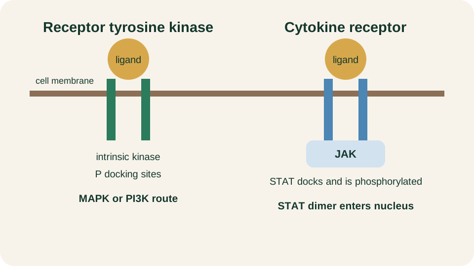
Intrinsic RTK kinase domains compared with JAK-associated cytokine receptors that phosphorylate STAT transcription factors at the membrane.
SimpleMed original educational diagram
Intracellular Receptors
Lipophilic hormones and related ligands do not need a membrane enzyme to enter the signalling logic. Steroid hormones, thyroid hormone, 1,25-dihydroxyvitamin D and retinoids cross the plasma membrane and bind intracellular receptors of the nuclear receptor superfamily. Some receptors reside mainly in the cytoplasm in complexes with chaperones and translocate after ligand binding. Others are already nuclear and are switched from repressive to activating states when ligand arrives. In either case the ligand-receptor complex binds specific DNA sequences termed hormone response elements (HREs) and recruits co-activators or co-repressors that remodel local chromatin and transcription.
The physiological tempo follows the mechanism. Even when ligand is present, new proteins appear only after transcription and translation. Responses such as altered enzyme complement, transporter expression or developmental gene programmes therefore unfold over hours. That slower onset is not a defect; it is appropriate for set-point changes in metabolism, differentiation and long-term endocrine tone. Non-genomic actions of some steroids exist at membranes, but the examination core remains ligand-regulated transcription at HREs.
Steroid, thyroid, vitamin D and retinoid pathways share the intracellular-receptor and HRE pattern despite different upstream physiology. The core sequence is lipophilic ligand, receptor binding, DNA response element, transcriptional change and delayed proteome remodelling.
Amplification and Termination
Signalling would be useless if one occupied receptor produced only one modified protein. Amplification is built into enzyme-linked routes. A single active RTK can phosphorylate many substrate molecules. Each kinase in a MAPK cascade can modify many MEK or ERK molecules. One active guanylyl cyclase can generate many cGMP molecules. Amplification lets sparse extracellular ligands control abundant intracellular targets, but it also makes termination essential. Without off-switches, every brief binding event would become a permanent cellular state.
Termination uses several complementary tools. Protein phosphatases remove the phosphates that created docking sites and activated kinases, restoring the pre-signal covalent state. Lipid phosphatases such as PTEN clear PIP3. Phosphodiesterases destroy cGMP and, in GPCR territory covered elsewhere, cAMP. GTP-binding proteins such as Ras hydrolyse GTP to GDP, with help from GTPase-activating proteins, and thereby leave the active conformation. Receptor internalisation by endocytosis removes receptors from the cell surface, after which they may be recycled or degraded. Negative feedback, including ERK-driven expression of inhibitors or STAT-induced suppressors of cytokine signalling, further limits prolonged output.
Receptor location tells you whether a ligand acts from outside or enters the cell. Effector mechanism identifies the first productive step. Enzyme-linked receptors rapidly rewire existing proteins; intracellular receptors produce slower, durable proteome change. This map predicts response speed, amplification and the required off-switches.
Reviewed by: Dr. Marcus Judge
- 143
