

By Dr. Thomas Burnell and Dr. Bethany Turner
Next Lesson - Cardiovascular Drugs
Core
This article is split into different sections:
- Arrhythmias
- Heart block
- Ischaemia and injury
- Electrolyte disturbances
- Afterdepolarisations
Many arrhythmias (abnormal heart rhythms) originate outside of the normal conduction system of the heart, arising from ectopic foci (small areas of heart muscle that fire on their own) in the atria or ventricles. However, some arrhythmias originate within the normal conduction system. Sinus tachycardia (a fast rhythm) and sinus bradycardia (a slow rhythm) both start in the sinoatrial node, the heart's natural pacemaker.
An easy rule to help determine where a rhythm is originating from is to look at the width of the QRS complex. If it is narrow, this usually suggests that the rhythm is originating above the ventricles and travelling via the normal conduction pathway. If the QRS is broad, this often suggests a ventricular origin or abnormal ventricular conduction, although this rule is not absolute.
There are three main ways that an arrhythmia can start. These are an ectopic focus (abnormal automaticity, where cells fire spontaneously), triggered activity (afterdepolarisations, covered at the end of this article) or a re-entry loop.
Ectopic beats are impulses that are generated by an area (focus) in the myocardium, not the sinoatrial (SA) node. Ectopic impulses can be generated by a small area of highly excitable myocardium which can spontaneously depolarise and cause a wave of depolarisation. Some ectopic beats are benign. Others can indicate an underlying heart problem that needs further assessment. Ectopic beats can also lead to faster rhythms (tachycardias).
The features seen on an ECG depend on where the ectopic impulse originates:
- Atrial ectopics - an abnormally shaped P wave (the ECG wave made when the atria depolarise) that appears early and is usually followed by a QRS complex due to the impulse being conducted to the ventricles. Atrial ectopic beats can trigger atrial fibrillation, which is sustained by interacting focal and re-entrant mechanisms.
- Atrioventricular (AV) junctional ectopics - these ectopics can activate the ventricles by travelling via the His-Purkinje system (the fast-conducting fibres that spread the impulse through the ventricles), therefore giving a normal QRS. Impulses can also retrogradely activate the atria (the impulse travels backwards from the AV node to spread across the atria). This gives an inverted P wave. However, the QRS complex can mask it, because the two events happen at the same time.
- Ventricular ectopics - premature impulses can arise in ventricular myocardium or Purkinje cells. They usually produce a broad QRS because ventricular activation differs from the normal rapid, coordinated His-Purkinje sequence and includes slower spread through myocardium. A compensatory pause (a longer gap before the next normal beat) is common when the sinus node is not reset and the next sinus impulse reaches ventricles that are still refractory (not yet able to respond). A pause is not obligatory.
In the conduction system of the myocardium, there are areas in the pathway where electrical impulses can split and travel down two paths. This usually isn't a problem because when the two impulses meet each other again, they cancel each other out. This allows the myocardium to contract in an even and efficient way.
However, problems arise when there is damage to areas of myocardium that disrupt the normal pathway of electrical impulses, or there are structural abnormalities. In the case of re-entry loops, there is an area of myocardium that is damaged and causes a unidirectional conduction block. Impulses can pass through the damaged tissue in one direction only. Therefore an impulse that is blocked in one direction can still travel backwards (retrogradely) through the damaged tissue.
If the surrounding myocardium has recovered by the time the impulse arrives, the impulse in a re-entry loop can travel back on itself and take alternative routes through the myocardium. This sets up a circuit of continuous, abnormal electrical activity. The diagram below shows this.
If there are re-entry loops, patients can have:
- AV nodal re-entry - if there are 'fast' and 'slow' pathways in the AV node (the electrical relay between the atria and the ventricles), there can be a re-entry loop which can cause a supraventricular tachycardia (a fast rhythm that starts above the ventricles).
- Atrioventricular re-entry - an accessory pathway (an extra electrical connection between the atria and ventricles) leads to a re-entry loop. An example is Wolff-Parkinson-White syndrome.
- Atrial flutter can be caused by a re-entry loop in the atria.

Diagram - Compares the pathways of electrical impulses in normal and damaged myocardium. Note how in the damaged myocardium, the impulse cannot travel down the right limb (purple arrow) due to damage, however, it can travel up the right limb (blue arrow). This is a unidirectional block of conduction. The impulse shown by the blue arrow can now spread across the myocardium causing contraction
SimpleMed original by Dr. Bethany Turner
In atrial tachycardia the heart rate is above 100 beats per minute (bpm). At faster rates, P waves may become difficult to identify on the ECG and can be obscured by preceding T waves. Atrial tachycardia is usually caused by an ectopic focus within the atrial myocardium. This is distinct from AV nodal re-entry tachycardia, which is due to a re-entry circuit involving the AV node and is classified separately as a supraventricular tachycardia.
Causes include digoxin toxicity, ischaemic heart disease, rheumatic heart disease.

Diagram - Atrial Tachycardia on an ECG. The P and T waves occur at the same time and have merged to form one wave
SimpleMed original by Dr. Bethany Turner
Atrial flutter is caused by a re-entry loop in the atria.
The AV node usually conducts only some of the rapid atrial impulses in flutter. How many impulses get through depends on refractoriness (how quickly the AV node recovers after each impulse), autonomic tone (the balance of nervous system activity) and medicines. 1:1 conduction can occur, for example with sympathetic stimulation or an accessory pathway. It causes a very fast ventricular rate and can make the patient seriously unwell. A common pattern is 2:1 conduction, with two atrial flutter waves for each ventricular QRS complex; other fixed ratios or variable conduction are possible. Flutter waves can produce a sawtooth baseline on the ECG (a regular zigzag between the QRS complexes).
The re-entry loop sends off impulses at around 300 beats per minute. With the common 2:1 pattern, the heart rate is therefore about 150 beats per minute. Other ratios, such as 3:1 or 4:1, or variable conduction, give different rates.
Causes include: hypertension, ischaemic heart disease, hyperthyroidism, alcoholism, cardiomyopathy.

Diagram - Atrial Flutter on an ECG. Note how there are 2 flutter waves for every QRS complex
SimpleMed original by Dr. Bethany Turner
AF is initiated and maintained by interacting focal and re-entrant mechanisms. Triggers often arise near the pulmonary veins, while electrical and structural remodelling of the atria helps sustain disorganised activation. Coordinated atrial contraction is lost and the atria quiver.
The AV node conducts atrial impulses inconsistently because of its refractory behaviour (it needs time to recover after each impulse), usually producing irregular R-R intervals (uneven gaps between the R waves, the tall peaks of the QRS complexes). There are no distinct, repeating P waves, only irregular atrial activity. QRS complexes are generally narrow when ventricular conduction is otherwise normal. Bundle branch block (delayed conduction down one of the two main branches to the ventricles), rate-related aberrancy (abnormal conduction caused by a fast rate) or pre-excitation (conduction through an accessory pathway) can produce broad complexes, so a normal QRS width is not required for AF.
The pulse is usually irregularly irregular. The ventricular rate may be fast, within the usual resting range or slow, depending on AV conduction and treatment. AF can occur at any ventricular rate, including rates below 100 bpm. Pulse strength may vary as changing filling intervals alter stroke volume, and some ventricular beats may be too weak to produce a pulse that can be felt at the wrist.
In AF, the quivering atria essentially cause stasis of blood as the atria are less efficient at pumping blood into the ventricles. This can result in thrombus formation. A thrombus in the left atrium is more common as blood can sit in the left auricle (the small pouch of the left atrium, also called the left atrial appendage) and clot. The clot can easily embolise (break off and travel in the blood) to the brain and cause an ischaemic stroke.
Causes include: dilated left atrium, hypertension, ischaemic heart disease, hyperthyroidism, alcohol, cardiomyopathy.

Diagram - Atrial Fibrillation on an ECG. Note the wavy baseline and irregular R-R intervals
SimpleMed original by Dr. Bethany Turner
Ventricular tachycardia is a fast rhythm that starts in the ventricular myocardium. After a myocardial infarction it is usually caused by a re-entry circuit around the scar, but an ectopic focus can also cause it. VT is identified as three or more consecutive ventricular ectopic beats at a fast rate (usually above 100 beats per minute). These are seen as broad QRS complexes on the ECG. VT is a dangerous rhythm as it can cause cardiac arrest (pulseless VT) or degenerate into ventricular fibrillation.
Causes include: myocardial infarction, ischaemic heart disease, hypertrophic/dilated cardiomyopathy.
In ventricular fibrillation there is chaotic ventricular depolarisation. It is thought to be maintained mainly by several re-entry circuits (waves of electrical activity that wander or spin around the ventricles), rather than by numerous ectopic foci. The exact mechanism is still debated, and focal activity may also play a part. As a result, there is no co-ordinated contraction of the ventricles. This results in an immediate loss of cardiac output, and the patient will go into cardiac arrest.
VF is commonly associated with myocardial infarction. It can also follow torsades de pointes (discussed next).

Diagram - Ventricular tachycardia degenerating into ventricular fibrillation. The left-hand side shows ventricular tachycardia. Note the wide QRS complexes of the ectopic beats, and the fact there are at least three in a row. The right-hand side shows ventricular fibrillation. Note the chaotic trace with no normal ECG features
SimpleMed original by Dr. Bethany Turner
Torsades de pointes is a polymorphic ventricular tachycardia. This means that there is a rapid ventricular rhythm in which the QRS complexes vary in shape and axis.
Causes include: medicines that prolong the QT interval (the time from the start of the QRS complex to the end of the T wave, which reflects ventricular depolarisation and repolarisation), such as some anti-arrhythmic drugs (medicines that treat abnormal heart rhythms), antibiotics, antipsychotics (medicines that treat psychosis) and antihistamines. Electrolyte abnormalities, such as low plasma potassium (hypokalaemia) or low magnesium (hypomagnesaemia), can also cause a long QT interval. Some people have an inherited long QT syndrome. Patients can also have a long QT interval with no identifiable cause.

Diagram - Torsades de Pointes on an ECG
SimpleMed original by Dr. Bethany Turner
First degree AV block is caused by delayed conduction through the AV node resulting in a consistently prolonged PR interval (the PR interval is the time from the start of the P wave to the start of the QRS complex). It is longer than 0.2 seconds, which is more than 5 small squares on ECG paper running at the standard 25 mm per second. First degree AV block can develop into second or third degree.
Causes include ischaemic heart disease, increased vagal tone (more activity of the vagus nerve, which slows the heart) and AV nodal blocking drugs such as beta blockers (medicines that slow the heart rate).

Diagram - First Degree AV Block on an ECG. Note the lengthened but constant distance between the P wave and QRS complex
SimpleMed original by Dr. Bethany Turner
There are two types of second degree AV block: Mobitz type I and Mobitz type II.
- Mobitz type I - the PR interval becomes progressively longer until one of the QRS complexes is dropped. The PR interval is shortest in the beat after the dropped QRS complex, and the cycle then repeats.

Diagram - Second Degree AV Block, Mobitz Type 1 on an ECG
SimpleMed original by Dr. Bethany Turner
- Mobitz type II - the PR interval is constant (it does not lengthen before the block), but occasionally the P wave is not conducted so a QRS complex is dropped. This type of second degree heart block is dangerous as it has a higher chance of progression to third degree heart block.

Diagram - Second Degree AV Block, Mobitz Type II on an ECG
SimpleMed original by Dr. Bethany Turner
In 2:1 block, every second P wave is not conducted. It is a type of second degree AV block that cannot be classed as Mobitz type I or II on the ECG. This is because there are no two conducted beats in a row, so the PR intervals cannot be compared to see whether they lengthen or stay the same.
Third degree AV block is when there is no impulse transmission between the atria and ventricles and therefore they work independently. There is no relationship between the P wave and QRS.
A pacemaker below the block takes over as an escape rhythm. Escape rhythms occur when the conduction system is blocked, and so the heart has to generate its own backup rhythm. If the escape rhythm starts in the AV junction (the AV node and the bundle of His, the first part of the His-Purkinje system), the QRS complex is usually narrow and the rate is usually about 35 to 60 bpm. If it starts in the ventricles, the QRS complex is wide and the rate is usually slower, about 20 to 50 bpm, because the impulse is not travelling via the His-Purkinje system. Escape rhythms are slow and can be unreliable, so the blood pressure may not be maintained. Patients with third degree heart block need urgent assessment and monitoring. If there are adverse signs (shock, fainting, heart failure or myocardial ischaemia), urgent treatment is needed, including pacing (using a device to make the heart beat with small electrical impulses). Most patients eventually need a permanent pacemaker (an implanted device that keeps the heart beating at a safe rate).

Diagram - Third Degree AV Block on an ECG
SimpleMed original by Dr. Bethany Turner
ST Elevation Myocardial Infarction (STEMI) and Non-ST Elevation Myocardial Infarction (NSTEMI)
STEMI and NSTEMI will both be talked about in more detail in the article on Acute Coronary Syndromes.
A STEMI is an acute myocardial infarction presenting with a characteristic ST-elevation pattern on the ECG. It often reflects acute coronary occlusion (a blocked coronary artery) and requires urgent assessment for reperfusion (restoring blood flow to the heart muscle). The ECG label does not prove full-thickness necrosis (death of the whole thickness of the heart wall): the final depth of injury depends on factors including duration of ischaemia, collateral flow and treatment.
For a person presenting with acute STEMI, assess eligibility for coronary reperfusion immediately and deliver reperfusion as quickly as possible if they are eligible. These actions are guided by NICE NG185, recommendations 1.1.1 and 1.1.3; they do not depend on waiting for later Q-wave evolution.
The following describes one possible ECG evolution, not a sequence every STEMI follows. Q waves may be absent, and the timing and persistence of ST-T changes vary. Troponin (a protein released when heart muscle cells are damaged) is assessed using serial measurements and the local assay pathway; treatment should not wait for a predetermined ECG sequence.
- Acutely there is an ST elevation and the T wave becomes taller - hyperacute T wave.
- Over time a pathological Q wave may develop. The R wave decreases in height and the ST elevation is still present.
- Pathological Q waves must be distinguished from small physiological septal q waves. Assess their duration and depth in the specific leads showing them, alongside the rest of the ECG and the clinical findings; a single size rule applied to every lead is insufficient.
- The pathological Q wave is denoted with a capital Q rather than the normal small q as the wave is larger than normal.
- Troponin I or T may rise within hours, with the timing of detection depending on the assay and presentation. Serial results are interpreted alongside symptoms and ECG findings.
- T wave inversion may appear during evolution, and a developing Q wave may deepen.
- ST elevation often decreases as the infarct evolves; T wave inversion may persist.
- Later, ST and T wave changes may improve. A Q wave can persist, and R wave height may remain reduced, but these findings are variable.
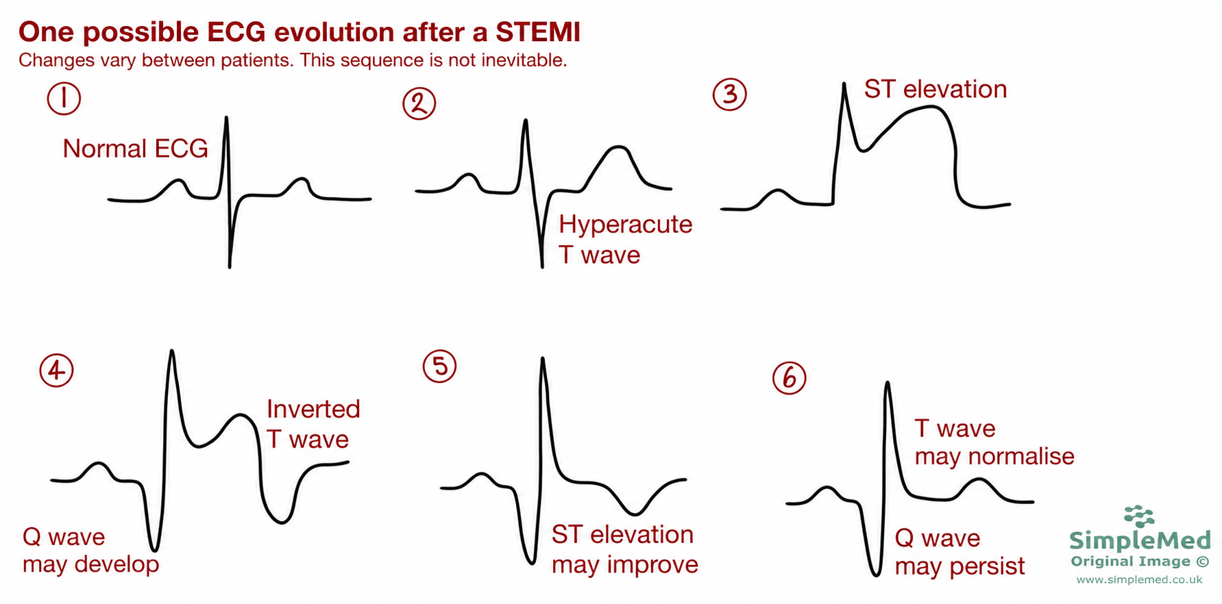
Diagram - One possible ECG evolution after a STEMI. The sequence varies between patients: Q waves may develop or persist, ST elevation may improve, and T waves may normalise. These changes do not establish the depth of the infarct.
Adapted from the SimpleMed original by Dr. Bethany Turner; revised diagram, 1 October 2026.

Diagram - A 12-lead ECG of an anteroseptal STEMI (affecting the front of the heart and the wall between the ventricles). This is shown by the ST elevation and the hyperacute T wave in leads V1-V4
SimpleMed original by Dr. Bethany Turner
An NSTEMI is an acute myocardial infarction without the characteristic persistent ST-elevation pattern. Diagnosis requires evidence of acute myocardial injury, usually a rise or fall in troponin, together with evidence of ischaemia. It is not defined as a partial-thickness infarct, and a completely occluded artery may still be present. ST depression or T wave inversion may occur, but a normal ECG does not exclude an acute coronary syndrome.
- The ECG may show a depressed ST segment, an inverted T wave, both changes or neither. Serial ECGs are useful when the first tracing is non-diagnostic.
- Within hours there is raised troponin I and T on a blood test (indicating myocyte death).
- ST-segment and T-wave changes may resolve or persist; their evolution is variable and is not a fixed timetable.
- Pathological Q waves may develop after an infarct, but their presence or absence does not reliably distinguish transmural injury (through the full thickness of the heart wall) from subendocardial injury (confined to the inner layer). Q waves are not always present after a STEMI, and they can still develop after an infarct that is classified as NSTEMI.
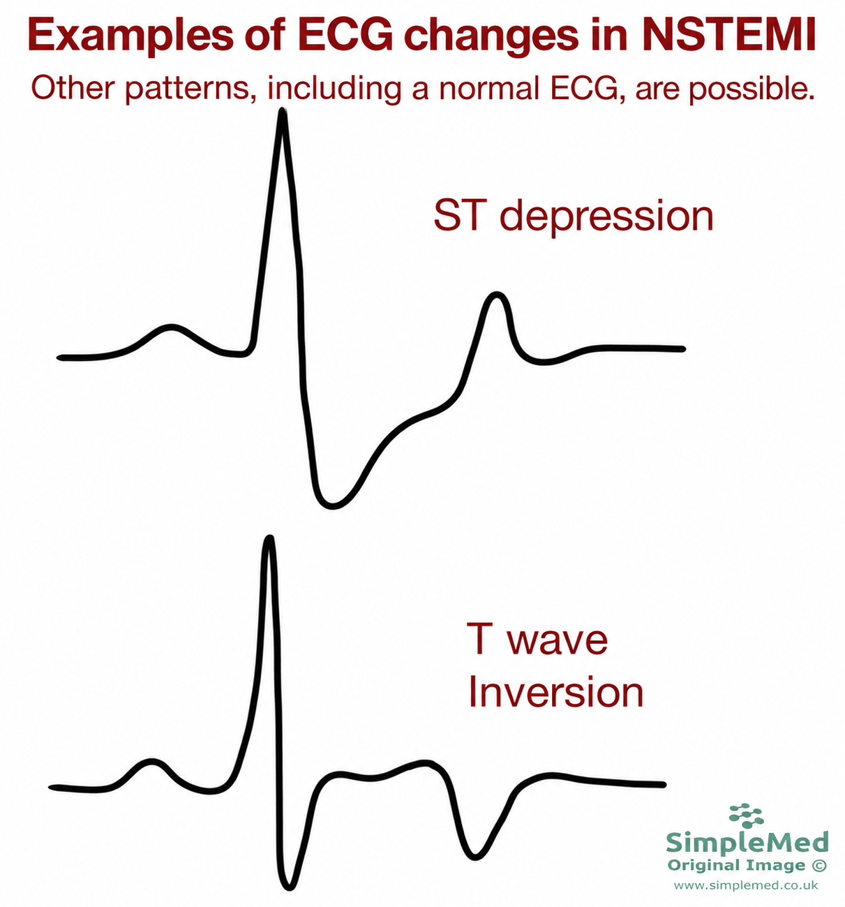
Diagram - Examples of ST depression and T wave inversion in NSTEMI. Other ECG patterns, including a normal tracing, are possible. The diagnosis also requires clinical assessment and evidence of acute myocardial injury; these patterns do not establish infarct depth.
Adapted from the SimpleMed original by Dr. Bethany Turner; revised diagram, 1 October 2026.
Hyperkalaemia makes the resting membrane potential less negative, depolarising it a little. This inactivates some voltage-gated sodium channels. The upstroke of the ventricular action potential is therefore slower, and cardiac conduction is slowed, which predisposes to arrhythmias.
In hyperkalaemia, the ECG changes tend to become more marked as the plasma potassium concentration rises. All values below relate to an approximate plasma [K+] in mmol/L (millimoles per litre). The ECG can be normal even when the potassium is high, and serious arrhythmias can occur without the classic changes. Do not rely on the ECG alone.
- 6.0 mmol/L - tall, peaked T wave.
- 7.5 mmol/L - tall, peaked T wave, flattened P wave and a prolonged PR interval.
- 8.5 mmol/L - tall, peaked T wave, absent P wave and a widened QRS.
- 9.0 mmol/L - widened QRS. The ST segment merges with the T wave, forming a sine-wave pattern (a smooth, wide wave like a sine curve). This is a pre-terminal rhythm and can progress to cardiac arrest.
Causes include: reduced renal function (such as acute kidney injury), medicines (ACE inhibitors and angiotensin receptor blockers, which treat high blood pressure and heart failure; mineralocorticoid receptor antagonists such as spironolactone; non-steroidal anti-inflammatory drugs such as ibuprofen; and the antibiotic trimethoprim), metabolic acidosis (too much acid in the blood) and cell lysis (cell breakdown, e.g. due to crush injury).

Diagram - The changes seen in hyperkalaemia on an ECG
SimpleMed original by Dr. Bethany Turner
Hypokalaemia is a plasma potassium concentration below 3.5 mmol/L. It results in a lengthened ventricular action potential and delays repolarisation. Longer action potentials can lead to early afterdepolarisations (explained below), which cause oscillations in the membrane potential. This increases the risk of ventricular arrhythmias, including ventricular tachycardia or fibrillation.
As the level of potassium in the plasma decreases, its appearance on an ECG changes:
- First there is a flattened T wave (a smaller T wave than normal).
- Then there is a flattened T wave and prominent U wave (a small extra wave after the T wave).
- Finally, there is a flattened T wave, a prominent U wave and ST depression (the ST segment sits below the baseline).
Causes include: diuretics (loop and thiazide diuretics are the most common medicines that cause it), severe diarrhoea and vomiting, metabolic alkalosis (too little acid in the blood), high urine output (e.g. in diabetes or with osmotic diuretics). Low magnesium (hypomagnesaemia) often occurs with hypokalaemia and increases the risk of dangerous ventricular arrhythmias.

Diagram - The changes seen in hypokalaemia on an ECG
SimpleMed original by Dr. Bethany Turner
Delayed afterdepolarisation (DAD)
DAD is when the membrane depolarises (without stimulation) soon after repolarisation following a contraction. DADs are more likely if intracellular calcium is high, because the extra calcium can trigger contractions when there should not be one. The depolarisation can reach threshold potential (the membrane voltage at which an action potential starts), initiating an action potential and causing the heart to contract.
Early afterdepolarisation (EAD)
EAD is when the membrane depolarises (without stimulation) during repolarisation following a contraction. This is more likely to occur if there is a long QT interval as the action potential is longer. Consecutive EADs lead to oscillations in the membrane potential which can trigger torsades de pointes, and this can degenerate into VF.

Diagram - The changes in an ECG in delayed and early afterdepolarisations
SimpleMed original by Dr. Bethany Turner
Edited by: Dr. Ben Appleby
Reviewed by: Dr. Marcus Judge
- 33947
